Cell
Historical document, translated for reference. It reflects medical knowledge of the 1920s–30s and is not medical advice.
Summary
This article from the 1928–1936 Soviet Medical Encyclopedia provides a historical and structural overview of the cell, tracing the development of cell theory from early microscopic observations by Robert Hooke to Max Schultze's definition of the cell as a lump of protoplasm with a nucleus, and details the morphological forms and components of cells.
Encyclopedia article (1928–1936)
CELL. Contents: Historical Outline............... 40 Structure of the C.................... 42 Shape and size of the C............. 42 Cell body................ 42 Nucleus...................... 52 Membrane.................... 55 Vital activity of the C.............. 56 Chemical composition of the C............. 56 Metabolism in the C............... 59 Energy metabolism in the C.............. 61 Reproduction and life cycle of the C........ 64 C. as a physicochemical system............ 65 Pathology of the C................... 71 Cell, corpuscle, loculus (Latin cellula, Greek kytos, German Zelle, French cellule), the simplest constituent part of a complex organism, possessing a definite structure (body, nucleus, membrane), endowed with the basic properties of a biosystem (metabolism, reproduction), and capable under certain conditions of existing outside the organism. The cell was defined as a lump of protoplasm with a nucleus (Max Schultze), an anatomical and physiological entity (Kolliker), an elementary organism (Brucke), a histomere (see), and a histo-system (M. Heidenhain). Homologous to the cells of a complex organism are single-cell (unicellular) organisms leading an independent existence. Historical Outline. The cell was first described by the English physicist R. Hooke (1667) in cork and other plant objects; upon microscopic observation, he found in them cavities separated by thin walls, resembling a honeycomb, and called them pores or cells. Several years later, scientists who first began to develop microscopic plant anatomy, N. Grew (1672) and M. Malpighi (1675), described among their composition, besides tubes and fibers, also cells ("pores" and vesicles of Grew, pouches, utriculi of Malpighi). In the 18th century, C. Fr. Wolff, studying plant development (1759), came to the conclusion that the vesicles forming the mass of parenchyma arise through the accumulation of liquid drops in a structureless organic substance. At the beginning of the 19th century, the plant cell began to attract special attention: the doctrine that cells represent pores or cavities hollowed out in the ground substance by an accumulation of fluid (Brisseau-Mirbel, Treviranus) was refuted (Moldenhawer, 1812); it was established that a cell is a vesicle or pouch with its own walls, that it is "a separate individual having its own special vital center of growth and displacement" (Turpin, 1826). At the same time, the term "cell" (cellule) came into scientific usage. In Meyen's textbook (1830), the cell theory of plants is outlined quite fully, finding its completion in the works of Schleiden (1838). In 1831, R. Brown described the nucleus (areola, nucleus) in plant cells, which had also been seen by some earlier investigators, and recognized it as a normal constituent part of the cell. In this way, the structural scheme of the plant cell was developed: membrane, contents, nucleus. Relying upon it, Th. Schwann proved in 1839 the presence of cells in the animal organism and established the cell theory of animals, which Dutrochet had spoken of even earlier (1824), whose works passed unnoticed. Of course, cells in animal tissues had been seen earlier, starting with Leeuwenhoek, but they were not compared with plant cells, not seeing a membrane in them, and were not attached any special significance. A major advance was the recognition within the cell contents of a special substance of a protein nature, transparent, viscous, capable of movement, which the botanist Mohl (1846) named protoplasm (the term had been proposed by Purkinje in 1840 for the formative substance of embryos). In the 1840s, a number of scientists (Bergmann, Bischoff, Kolliker) described formations in all respects similar to cells, but devoid of a membrane (cleavage spheres, embryonic elements); in view of the fact that they did not fit the concept of the cell, an essential part of which according to the views of that time was the membrane, Kolliker proposed for them a special name—protoblast (primary germ). He retained this name until the last edition of his manual (1889), one of the chapters of which is titled "Von den Protoblasten und Zellen." However, in the 1950s and early 1960s (Leydig, M. Schultze, Brucke, Beale), the membrane was recognized as an unessential part of the cell, a sign of cellular old age. Max Schultze (1861) formulated a new definition of the cell as follows: "A cell is a lump of protoplasm in which a nucleus lies." He also proved that sarcode—the main substance of protozoa (Foraminifera, Rhizopoda), studied by Dujardin as early as 1835—is identical with protoplasm. In the second half of the 1860s, Haeckel, based on the study of certain forms of protozoa (moners), argued that even the nucleus is not an essential constituent part of the cell; he called such primary life forms cytodes (cell-like) and united them with nucleated cells into a single group of plastids. In 1861, the well-known physiologist Brucke made an attempt to approach the cell from a physiological point of view as an elementary organism: the morphological data of that time seemed to him completely insufficient to understand the complex vital activity of the cell, and he postulated the presence in the cell of a definite organization and complex structure apart from the structure of organic molecules. This reasoning as if charted the path for the further development of the doctrine of the cell, and starting from the seventies, in-depth research of the constituent parts of the cell, first of all protoplasm and the nucleus, begins. Prerequisites for this were provided by the improvement of the microscope (immersion) and methods of histological research. The first monument of this new period, when the doctrine of the cell turned into a special discipline—cytology, is the classical work of Flemming (1881) (see also Histology, Blastema, Karyokinesis, Cell Theory). Structure of the cell. Shape and size of the cell. The shape of the cell is diverse (Fig. 1); the basic one is spherical (ova, leukocytes in the bloodstream) corresponding to the liquid state of the protoplasm. It can change under the influence of various
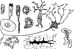
Figure 1. Various cell shapes (after Breyel).
moments: 1) movements of the cell itself (flattening, elongation, appearance of outgrowths of various shapes); 2) pressure of neighboring cells and tissues (cylindrical, conical and cubic, polyhedral, flat); in this case, cells round off upon the elimination of pressure; 3) uneven growth associated with the appearance of skeletal formations and a membrane (pear-shaped, spindle-shaped, ribbon-like, stellate, irregularly process-bearing).-The size of the human cell ranges from 4-5 µ (lymphocytes) to 40-80 µ (nerve cells) and even 0.2 mm (egg cells); the average size of the majority is 15-20 µ. Cells of various animals have different sizes: in tailed amphibians they are large, in tailless ones smaller; in birds and fish they are small; the size may differ in animals of close species and is not in direct connection with body size. The influence of the nucleus size on the cell size has been noted: cells containing a double number of chromosomes have a larger size. In the composition of the cell, 3 main constituent parts are currently distinguished: 1) the cell body, 2) the nucleus, 3) the membrane (Fig. 2). I. The cell body has a complex structure and consists of the main sub- Figure 2. Scheme of cell structure: 1-membrane; 2- protoplasm; 3-mitochondria; 4-chromatin clumps; 5-nuclear membrane; 6, 7 and 9-deutoplasm (inclusions); 8-nucleolus; 10- sphere with a centrosome in the middle; around it-the reticular Golgi apparatus. stance, protoplasm, in which are located 1) formations recognized as necessary constituent parts of any cell, or its organoids (centrosome with the sphere surrounding it, mitochondria, intracellular apparatuses, etc.); 2) inclusions of various kinds appearing in different cells and sometimes designated by the general name of deutoplasm. A. Protoplasm (cytoplasm, or simply plasma) is the main constituent part without which the cell as a living system cannot exist; it also gives rise to the majority of formed inclusions found in the cell body. Views on the morphological properties of protoplasm have changed since the 1860s. The first researchers (Mohl, Max Schultze, Kölliker) considered protoplasm to be a homogeneous transparent substance, which can become turbid due to the presence of small granules (microsomes); in young cells, protoplasm completely fills the entire cell body; subsequently, various inclusions appear in it, and it remains in the interstices. In the 1870s, protoplasm began to be viewed as a substance formed in the form of thin fibrils or threads intertwining into a network (Frommann, Heitzmann); this doctrine was developed in the 1880s by Flemming, Kupfer, Carnoy, Leydig and others and is known under the name of the fibrous-reticular theory of protoplasm. According to Flemming, protoplasm consists of fibrils, the combination of which forms a filamentous mass (Filarmasse), or mitome (Mitom); located between them is an aqueous interfilar mass (Interfilarmasse, Paramitom; Fig. 3). Kupfer called the threads protoplasm, the intermediate substance-paraplasm, Carnoy-reticulum-enchyleme, Leydig-spongioplasm-hyaloplasm and, unlike other authors, considered hyaloplasm to be the living substance, and the reticular spongi-Figure 3. cartilage hyoplasm-skeleton vro- "(nTSSSrT^ gybki. The doctrine of Flem" ming held on in science for a very long time and had supporters until recently. At the end of the 1880s, Altmann came forward with a theory according to which protoplasm consists of granules [granula-bioblasts (see)] (Fig. 4), possessing the ability to feed, grow, and multiply; threads arise from granules as a result of their growth or bead-like connection. At present, Flemming's fibers and Altmann's granules are considered mitochondria. At the beginning of the 1890s, the theory of foamy, or cellular structure appeared (Bütschli): protoplasm is two-phase and is constructed of two substances-viscous and highly light-refracting, which forms the walls of the cells, and the aqueous chyle enclosed in them (Fig. 5). The optical section of the thickened cell walls simulates fibers, and fluid flows in their corners give the picture of granules. A model of such protoplasm can be obtained by grinding provencal oil with potash and placing pieces of the mixture in water: a fine emulsion arises, forming a foam. Bütschli's theory met with a sympathetic reception from physiologists, since it made it possible to understand the simultaneous existence of various processes in a small space of the cell; subsequently, it was detailed by Rhumbler. While the proponents of all these theories argued, a conciliatory, eclectic trend began to arise in science
ruling (Waldeyer; 1895), according to which there is a grain of truth in every doctrine, and protoplasm, having no definite structure, can acquire a different appearance in different cases. On the other hand, a critical consideration of the question of the action
Figure 4.
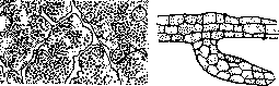
Figure 5. Figure 4. Section of the pancreas. Granular protoplasm (according to Altmann). Figure 5. Two protoplasmic strands from a mallow hair. Alveolar structure (according to Bütschli). of fixing reagents (A. Fischer; 1899) showed that fibers, granules, and foamy structure can arise as artifacts: they are easily obtained from solutions of various proteins upon fixation with appropriate liquids. Over the past decade, when scientists turned again to the intravital study of the cell, an impetus for which was given by tissue culture outside the organism, all these stated theories receded into the background, and the old doctrine of homogeneous protoplasm was again established in science (Chambers, M. and H. Lewis, Spek, and others). From this point of view, protoplasm represents a transparent mass of more or less liquid consistency, which can solidify and liquefy again, in other words, possesses the properties of a colloidal solution (sol), capable of turning into a gel. All formed structures, insofar as they are not artifacts, should be attributed to inclusions. The liquid properties of protoplasm, apart from its fluidity and the spherical shape of its liquid inclusions, are convinced by direct study of the cell body with a micro-needle using a micromanipulator; the solidification of individual sections with their subsequent liquefaction is most conveniently observed in the protoplasm of amoebas during the formation of pseudopodia (Chambers). Ultramicroscopic research shows that protoplasm can be optically empty (Haidukow), more often
sh ^W Figure 6. Granular disintegration of a protoplasm droplet (according to Verworn). in it finely dispersed granularity is visible, with individual particles being anisotropic, revealing a crystalline structure (Spek). By acting on the cell with various reagents (hypertonic solutions, formol, narcotics), one can induce coarser granularity, vacuolization, and further typical foamy structure (Giersberg, Spek, Herwerden); all these phenomena disappear when the cell is transferred to a normal environment. Upon death, protoplasm becomes coarsely granular and disintegrates into a pile of granules (Fig. 6). B. Centrosome and sphere. The centrosome (Boveri), or central corpuscle (van Beneden), was discovered by van Beneden first in dividing blastomeres of Ascaris (1876), then in resting ones (1887). Subsequent work quickly confirmed the existence of the centrosome in various cells: egg, seminal, pigment, wandering (Boveri, Henneguy, Hermann, Solger,
Figure 8. Diplosomes (under the membrane) in cells
Figure 7. Diagram illustrating various descriptions of centrosomes and spheres (according to Wilson). Flemming). M. Heidenhain (1891) developed a special method for staining centrosomes (iron hematoxylin with differentiation), with the help of which this formation could be detected in all cells, and produced the first detailed study of the centrosome in leukocytes. Since then, the centrosome has been recognized as the third component of the cell along with protoplasm and the nucleus and in the 1890s gave rise to an extensive literature. Recently, interest in the centrosome has declined partly because it is not visible in living cells with the exception of pigment cells. It was also not found in the cells of higher plants despite the initial assertions of Guignard. The centrosome is a formation pfi^eiffleHraHHy) of very small magnitude, and in various cells it has a different appearance. At first it was described as a light vesicle, then, after applying Heidenhain's method, attention began to be paid to the strongly staining granule in the middle-the centriole (Boveri), and most scientists consider this particular formation to be the centrosome (Fig. 7). In epithelial cells, the centrosome consists of two granules connected by a weakly colored bridge, the centrodesmose (diplosome, Zimmermann; Fig. 8); in large cells (megakaryocytes, corneal endothelium)- Figure 9. Centrosome and pure sphere v in a salamander leukocyte (according to Heidenhain), of a larger number of granules (from 3 to 100), which can be arranged in groups (micro- Heidenhain). Centrosomes have been described in the form of rods, straight or bent at an angle (Meves, Chasovnikov). The centrosome is usually located near the nucleus, very often in a special depression, but sometimes closer to the membrane; the assertion of some authors about the presence of the centrosome in the nucleus, whence it emerges

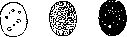
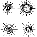


hod before division, was not confirmed. During mitosis, the centrosome becomes the center of the radiant figure, the aster (see Karyokinesis); this phenomenon is observed in some resting cells, mainly leukocytes (Fig. 9), pigment, and germ cells. It gave Heidenhain grounds to create the theory of "organic rays," elastic filaments running from the cell center to the membrane and determining the configuration of the cell's constituent parts, its movement and division. - Centroso-
Figure 10. Centrosome and aster in the spermatogonium of a salamander (after Duesberg).
ma often lies in the middle of a special spherical region of the protoplasm, which has received the names: attraction sphere (van Beneden), astrosphere (Strassburger), centrosphere (Wilson), centroplasm, archoplasm (Boveri). It has a different appearance under different fixations: sometimes as a light field penetrated by radially running rays and concentrically arranged clusters of granules (Figures 7 and 10), sometimes as simply a homogeneous or granular mass (Fig. 11). The achromatin spindle of karyokinesis is formed from and passes back into the substance of the sphere upon the completion of division. In life, the sphere is visible as a light spot without a special structure. - Closely related to the sphere is the yolk nucleus (vitelline body) of Balbiani. This formation, especially well developed in the eggs of spiders and myriapods and seen even by older authors, was described in detail by Balbiani (1864, 1893) and found by him in fishes, frogs, and mammals. It arises in the egg cell next to the nucleus in the form of a light vesicle, around which small granules accumulate. As it grows, its peripheral layers become compact and split into concentric zones consisting of yolk (Figure 12). Approximately in the same way arises the yolk nucleus, the vitellogen body, in mammals and man (Fig. 13); only concentric layers are not formed, but the arising yolk granules are located on the periphery (vitellogenic layer,
Figure 11. Sphere (idiosome) of a salamander spermatogonium (after Meves).
couche vitellogène). Balbiani derived the yolk nucleus from the cell nucleus by budding; later, the attraction sphere with the centrosome began to be seen in it (Mertens, van der Stricht). The newest researchers (Hirschberg) incline to the fact that the yolk nuclei of different animals have different origin and significance; mitochondria and the Golgi apparatus are also described in their composition.
B. Mitochondria, chromidia, trophoplasts. Mitochondria (thread-granules) were discovered Figure 12. Yolk nucleus
in germ cells by Benda (1897) using
in the egg cell of Tegenaria domestica (after Prenant).
a very complex method of elective staining [see separate table (vol. XII, pp. 583-584), Figs. 10 and 11] and studied in detail by Meves. The first decades of the 20th century, they stood at the center of attention of histologists and gave rise to a colossal literature and a large number of synonyms: chondriosomes (granular bodies), chondriomites (granular filaments), chondrioconts (rods), plastosomes (formative bodies, Meves), plastoconts; the totality of mitochondria is the chondriome. Mitochondria are found in all cells of the animal organism, as well as in plants and single-celled organisms, in the form of small granules, sometimes forming into chains, rods, and more or less long filaments, straight or curved (Fig. 14). In life, they have been observed in glandular cells (Michaelis), spermiocytes (Chambers), and mesenchymal cells of the chick embryo (Lewis, Carrel); they appear there in the form of pale threads and rods of semi-liquid consistency, sometimes curling into loops and rings and are very unstable; for better visibility, they are stained with a very dilute (1:50,000) solution of the dye Janus green [see separate table (vol. XII, pp. 583-584), Fig. 9]. When fixing mitochondria, one should avoid acetic acid and alcohol, which dissolve them, and use osmium acid, chromic acid, chromium salts, formalin, which are part of the most commonly used fixatives (Champy, Regaud, Altman). The complex method of staining with crystal violet according to Benda, which gives very beautiful pictures, is currently used rarely; usually, they are stained with iron hematoxylin or picrofuchsin according to Altman. - Views on the significance of mitochondria have changed. During the period of fascination with these formations, they were ascribed very great importance in the life of the cell, considering them permanent constituent parts of the cell, multiplying by division and
yolk nucleus in the egg cell of a woman (after van der Stricht).
transmitted from cell to cell; during fertilization, the sperm mitochondria mix with the mitochondria of the egg cell, which is why Meves considered them bearers of hereditary properties. Passing into the embryo cell, mitochondria accumulate various substances and give rise to all special inclusions: muscular, nervous, and connective tissue fibrils; pigment granules; secretion droplets, etc. (Meves, Duesberg, Hoven). As was justly pointed out (Retzius), the doctrine of mitochondria in this form resurrects the theories of protoplasm structure of Flemming and Altman. Most scientists, without going so far, were nevertheless inclined to consider mitochondria as cell organs that accumulate and process substances brought to the cell by the blood. Lately, the continuous succession of mitochondria has been disputed; intravital observations show that they can arise from protoplasm
. and disappear again; likewise, their participation in the formation of cell differentiations
Figure 14. Mitochondria: left - mouse liver cells; right - mouse intestinal epithelium. is called into question. Therefore, many consider them simply a specific substance that accumulates in the cell as a result of metabolism and precipitates in the form of special phases. When cell metabolism changes, for example, during starvation, the action of various poisons on the cell, X-rays, etc., mitochondria appear only in the form of granules or short rods instead of the normal form of threads; this makes it possible to judge the state of the cell at a given moment and makes clear the interest shown in mitochondria by pathologists. - Chromidia - granular or filamentous clusters lying near the nucleus, sometimes directly adjacent to the nuclear membrane and staining with the same dyes as the chromatin of the nucleus. They were described and studied by Richard Hertwig (R. Hertwig) in protozoa and represent, in his opinion, chromatin that has emerged from the nucleus. The existence of chromidia in the cells of complex organisms is disputed, and what was described in them as chromidia is considered mitochondria. - Trophoplasts, or plastids - inclusions in protoplasm characteristic of plant cells. These are small formations of rounded, oval, or irregular shape, colorless (leucoplasts) or colored green by the chlorophyll contained in them (chloroplasts or chlorophyll grains producing starch) or containing special pigments (chromoplasts). Their structure is little elucidated; they have the ability to multiply by division and are transmitted
from cell to cell. Some scientists (Levitsky, Maksimov) believe that trophoplasts are specially differentiated mitochondria. A certain analogy with plant trophoplasts reveals
Figure 15. Ergastoplasm (in
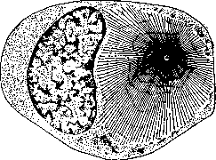
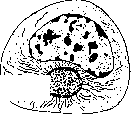
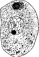

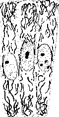
in the form of threads) reveal pigment granules in the cell of the mammary gland. Tonoplasts (de Vries) are formations that give rise to the vacuoles of plant cells; they are sometimes placed on a par with trophoplasts as autonomous inclusions. F. Ergastoplasm, accessory nuclei. Under the name of ergastoplasm (producing plasma), or higher protoplasm (protoplasma supérieur), French histologists (Garnier, Bouin, Prenant) described special fibrous or lamellar formations, usually convoluted or gathered into a tangle, found in the basal part of functioning glandular cells (Figure 15). They stain well with basic dyes, as well as with iron hematoxylin, and stand out clearly during the secretory activity of the cell, decreasing and disappearing during periods of rest. They were considered a special type of protoplasm performing special functions in the secretion process. Most authors, however, do not recognize the existence of ergastoplasm as a special formation, but consider it an accumulation of mitochondria or rod-like striation of the glandular epithelium altered by the action of fixatives.-Accessory nuclei have been described many times in sex and glandular cells. These formations have nothing in common with the nucleus, which is why they should be distinguished from true accessory (small) nuclei found alongside the main one in various tissues of amphibians (N. Rabl, Karpov). The accessory nuclei of authors are formations lying next to the nucleus and having very different structures: homogeneous or granular spheres, formations of irregular shape, bundles or tangles of fibrils. In sex cells, the following were described under this name: an accumulation of mitochondria (chondriome), a sphere, an idiosome, a yolk nucleus; in glandular cells, ergastoplasm and various artifacts.--Pseudochromosomes, rod-like, well-staining formations lying near the sphere in spermatocytes and oocytes (M. Heidenhain, van der Stricht), also have no independent significance; they are now considered mitochondria or are associated with the Golgi apparatus. D. Intracellular apparatuses. Under this name, a number of more complex formations in the form of networks, tubules, and vesicles can be combined, which arise in certain places of the cell body and are assumed to be related to the metabolism of the cell; their study is currently at the center of attention of cytologists. These include: 1. The internal reticular apparatus of Golgi (apparato reticolare interno Golgi), internal network (Binnennetz) (Kopsch), centroformin (Ballowitz), first discovered by Golgi (1898) using the silver-chromate method in nerve cells in the form of a reticular basket surrounding the nucleus (Fig. 16). Subsequently, through the works of his students and a number of researchers (Negri, Perroncito, Kopsch, R. Cajal, Sjövall, Hirschler, Dogel, Deineka, Kolachov, Nasonov), its ubiquitous distribution was proved, and more convenient methods for its detection were developed, which boil down to impregnation with silver or osmic acid. The apparatus is a network consisting of threads or trabeculae of various thicknesses that bend, wind, divide, and anastomose with each other (Fig. 16). In nerve cells, it surrounds the nucleus from all sides, but in most others it is adjacent to the nucleus on only one side; in glandular cells, it is placed between the nucleus and the free surface, sometimes giving off processes along the sides of the nucleus. In many cases, the location of the reticular apparatus along the periphery of the sphere was noted. Pieces of various sizes can separate from the network, and sometimes the entire reticular apparatus disintegrates into "separates" (nerve, egg cells). Separates arising
a
b
Figure 17.
Figure 16. Golgi reticular apparatus: a—nerve cell of the spinal ganglion of a rabbit; b—intestinal epithelium of a newt, I—leukocyte; c—spermatocyte of a newt; d—pancreatic cell of a newt, n—accessory nucleus; e—connective tissue cells of a newt. Figure 17. Trophospongium in a nerve cell of a rabbit. Processes of trophocytes forming a network in the protoplasm are visible (after Holmgren).
during karyokinesis, were named dictyosomes; they are distributed equally between the daughter cells (Perroncito, Deineka). At the site of the reticular apparatus, the accumulation of secretion takes place in glandular cells, dyes and other substances that have penetrated into the cell are released; in egg cells, its separates are associated with yolk granules; therefore, it was suggested that the reticular apparatus is an excretory and secretory organ of the cell (Nasonov). During life, the reticular apparatus cannot be detected.-2. Trophospongium, Holmgren's canaliculi (Holmgren). Almost simultaneously with Golgi, Holmgren described in nerve cells after special treatment and staining a system of canaliculi of various widths, connected in the form of a network and permeating the entire body of the cell. He suggested that the canaliculi arise from processes of special nutritive cells (trophocytes) that grow into the nerve cell, branch there, and then liquefy, supplying the cell with nutritive material, and named this network trophospongium (nutritive network) (Fig. 17).
Figure 18.
Figure 19.


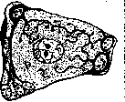
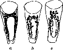
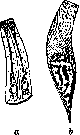
Figure 18. Holmgren canaliculi (a); Golgi apparatus; c-vacuome in the stomach epithelium of a newt (after Parat). Figure 19. Tonofibrils: a-in the intestinal epithelium of Ascaris ensicaudata; b-in a frog. Subsequently, Holmgren demonstrated the presence of canaliculi in all other cells in approximately the same places where the Golgi apparatus is located. The existence of trophocytes and trophospongia was not confirmed, and regarding the canaliculi, Holmgren himself expressed the assumption that they are identical with the trabeculae of the reticular apparatus (Golgi-Holmgren canaliculi), which, however, is far from shared by everyone (Dogel).-3. Recently, Parat and his pupils developed the doctrine of the v a c u o m e—the totality of special small vacuoles that appear as red vesicles when a living cell is immersed in a weak solution of Neutral Red [see separate table (vol. XII, pp. 583-584), Fig. 9] (see Vacuome). In his opinion, the Golgi apparatus and Holmgren canaliculi represent a vacuome modified by the action of reagents and have no independent significance; the locations of these formations coincide exactly (Fig. 18).-The study of intracellular apparatuses is still in full swing, and no definitive conclusions can be drawn about their mutual relations and significance. It should be noted that during cell poisoning and in a number of pathological processes, the typical appearance of the reticular apparatus changes; it disintegrates and may disappear entirely. E. Deutoplasm. In addition to the described formations, in which many scientists see permanent components of any cell, formed structures of various kinds can arise in the protoplasm of different cells, which can be grouped under the general name of deutoplasm (secondary plasma, van Beneden), metaplasma (Hanstein), or simply inclusions. By their appearance, they can be divided into 3 groups: 1) fibrous structures, 2) granules, droplets, and clumps, 3) crystals and crystalloids.-1. Fibrous, fibrillar structures belong to the category of vectorial formations, i.e., those whose physical and optical properties are unequal in different directions (longitudinal and transverse). Therefore, most deutoplasmic fibers possess optical anisotropy (birefringence); they are positive and uniaxial. At present, these properties are explained by the micellar structure, resurrecting the old doctrine of the botanist Nägeli on micelles, molecular complexes of a crystalline character, which, orienting themselves in the fiber in one direction, impart vectorial properties to it. Micelles arise in the protoplasm and can be detected by ultramicroscopic examination in polarized light. The character of fibers in different cells is various: in muscular and contractile cells in general, there are myofibrils; in nervous cells, neurofibrils, sometimes forming networks; in fibroblasts, collagen fibrils, which arise in the peripheral layers of the cell and then separate; in epithelium, supporting fibrils, tonofibrils; in sperms, skeletal formations (Fig. 19). M. Heidenhain considers fibrils to be histomeres, i.e., autonomous formations that can feed, grow, and multiply by longitudinal division. Vectorial structures probably also include the rod-like striation of glandular cells, which according to new data consists of lamellae.-2. Inclusions in the form of droplets, granules, and clumps are extremely diverse; various substances, accumulating in the protoplasm and overloading it, are secreted as special phases; their character is determined by the functional activity of the cell. These include watery inclusions in the form of vacuoles, fat droplets, glycogen clumps, protein granules, secretion droplets of various sizes and consistency, pigment granules, yolk spheres and plates, etc. Accumulating in a certain amount, they impart a definite physiognomy to the cell, making it possible to determine its character.-3. Crystals are often found in plant cells (crystals of calcium oxalate, protein), and rarely in animal cells (in the interstitial cells of the testis). P. The nucleus (nucleus) is currently considered a necessary component of the cell and unicellular organisms; the existence of non-nucleated protists (Haeckel's monera) was not confirmed, and non-nucleated cells encountered in a complex organism (erythrocytes, surface cells of the epidermis) originate from nucleated ones and do not possess full viability after the loss of the nucleus. Usually, there is a single nucleus in a cell, but cases of binucleated cells are not uncommon (glandular, nervous); a larger number is also encountered—dozens and even hundreds (striated muscle fibers, osteoclasts, syncytia). In such cases, a multinucleated formation corresponds to several cells, and the botanist Sachs even proposed for the designation of a nucleus with the adjacent portion of protoplasm the special term "energide," with which he thought to replace the term "cell."--The shape of nuclei is diverse (Fig. 20); the fundamental one is
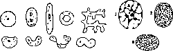
Figure 20.
Figure 21.
Figure 20. Various forms of the nucleus. Figure 21. Diagrams of the nuclear structure: 1-reticular framework immersed in the nuclear sap; 2-granular structure; 3-alveolar structure. spherical (egg cells), but in the majority of cells, adapting to their shape, the nucleus takes the form of an ellipsoid, a round or oval plate, and sometimes elongates into a rod. Due to special conditions, nuclei of a more complex shape may arise: horseshoe-shaped, annular, moniliform, lobular, lobed, branched; this phenomenon is known as nuclear polymorphism. The size of the nucleus is on average 4-9 µ, corresponding to the cell size; large nuclei are commonly found in large cells (giant bone marrow cells, egg cells, some glandular cells). According to R. Hertwig, there must be a definite ratio between the nuclear mass and the protoplasm (Kernplasma-relation). Structure of the nucleus. Views on the structure of the nucleus changed simultaneously with changes in views on the structure of protoplasm, and each cytologist described it in their own way: Flemming found in it a fibrous-reticular framework, Altmann a granular structure, Bütschli a foamed one (Fig. 21). Flemming's scheme received the greatest recognition and is placed at the basis of the description of the nucleus in textbooks. The nucleus is covered on the surface by a thin structureless membrane; it stains poorly and has been named achromatin. The membrane encloses the contents of the nucleus, the karyoplasm (nuclear plasma), which includes the fibrous-reticular framework (nuclear network), nucleoli, and nuclear sap. The main part is the framework, the basis of which consists of threads of various thicknesses penetrating the nucleus in all directions and attaching to the membrane; it stains poorly, and its substance has been named linin (linin, achromatin network). In the thickness of this basis and on its surface, grains and lumps of another substance are located—chromatin, which precipitates upon the action of acids and stains well (whence the name arose). There may be so many chromatin lumps that they mask the basis; some of them are located under the membrane, forming as if a second, chromatin membrane. Initially, a characteristic feature of chromatin was considered to be its stainability with basic (nuclear) dyes; but M. Heidenhain showed that along with basic chromatin (basichromatin), nuclei contain chromatin that stains with acidic dyes (oxy-chromatin), usually in the form of smaller grains; according to Heidenhain's opinion, it represents the first stage of basichromatin [see separate table (vol. XII, pp. 583-584), Fig. 8].-Nucleoli (nucleoli) in the nucleus are usually one or two: they lie at the nodal points of the framework and differ from chromatin in their appearance and reactions. These are formations of round or oval shape; they swell from the action of acids; they stain with acidic dyes, as well as safranin. Such are true nucleoli, or plasmosomes; false nucleoli (karyosomes), which are simply large, rounded lumps of chromatin, should be distinguished from them. The nuclear framework and nucleoli are immersed in the ground substance of the nucleus, the nuclear sap, which is described as a transparent liquid mass without special structures. Attempts to prove its granular structure (edematine grains of Reinke, cyaninophilic ones of Altmann) were not successful.-Such is the usual scheme of the nucleus established by studying fixed preparations, which include acids (Flemming's fluid) or sublimate (M. Heidenhain). But recently, researchers' attention has again turned to the study of the living nucleus and direct observation of the changes that reagents cause in it (Gross, American authors, Karpov). The nucleus appears in life in the form of a bright transparent vesicle, refracting light more weakly or strongly than the protoplasm surrounding it. It is demarcated by a smooth and even contour; the membrane as a special formation is not visible. The karyoplasm of many nuclei appears completely transparent, homogeneous, and optically empty even under ultramicroscopic investigation; only the nucleoli stand out clearly in it in the form of round or oval bodies, clearly contoured and more strongly refracting light [see separate table (vol. XII, pp. 583-584), Fig. 8]. In other nuclei, besides nucleoli, small grains, lumps, or blocks of chromatin are visible, paler, with an unclear contour; their number and size are different; uniformly granular nuclei are encountered (often in insects) or with large, elongated lumps (leukocytes). The consistency of nuclei is different: most often it is liquid (the microneedle moves freely, and the displaced nucleoli do not return to their former place); but there are nuclei of viscous and even gelatinous consistency. The nucleus reacts extremely rapidly to a change in protoplasm; upon injury of the cell, it acquires a distinct membrane, and the karyoplasm becomes filled with sharply outlined grains and lumps; weak acids cause the same effect. These changes are reversible, and after washing out the acid, the nucleus acquires its former appearance. Formalin and osmic acid preserve the vital picture best; acidic fixatives (Flemming's fluid), and especially sublimate, cause the coagulation of the karyoplasm and the appearance of a reticular framework in it. These data make one doubt the vital existence of the linin framework and the correctness of Flemming's scheme. The question of the fine structure of the nucleus becomes complicated if one takes into account its changes during karyokinesis (see), when chromosomes become visible in the karyoplasm, the number of which is constant for each species. The hypothesis of the preservation of chromosome individuality (Boveri), postulated by a vast number of facts, forces one to assume in the resting nucleus the presence of regions from which individual chromosomes arise, or special skeletal formations that collect scattered chromatin (Koltsov). But it is very difficult to discover the boundaries of individual sections in the nucleus; on the other hand, Tellyesniczky's hypothesis about the simple crystallization of chromosomes from the stock solution of the karyoplasm does not provide a satisfactory explanation of the principle of constancy. Thus, the question of the fine structure of the nucleus remains unresolved.-Nuclei of special structure. Deviations from the indicated scheme of nuclear structure can occur in two directions. 1. In the direction of condensation, when the liquid karyoplasm disappears and the nucleus turns into a compact lump of chromatin, with the volume of the nucleus decreasing and the shape changing; this takes place in the heads of sperm cells. 2. In the direction of swelling associated with the appearance of a special liquid phase in the nucleus. The nuclei of oocytes during the growth period greatly increase in volume; light fluid (nuclear sap) accumulates in them, and the karyoplasm together with chromatin lumps forms a network suspended in the fluid; along with this, enhanced multiplication of nucleoli occurs. Nuclei of this type correspond exactly to Flemming's scheme. Another example can serve as the nuclei of the salivary glands from the mosquito larvae Chironomus, first described by Balbiani (the same type was later found by Carnoy in many Diptera) (Figure 22). The nucleus is large, filled with light fluid; a thick curved vector is visible in it, at the ends and in the middle of which bumpy nucleoli are placed; the thread consists of chromatin disks separated by light intervals, and is covered on the surface by a thin membrane. III. Membrane. The protoplasm of any cell is demarcated from the environment by a special plasma membrane (Plasmahaut; Pfeffer), or surface film of a molecular nature, which determines the penetration of certain substances into the cell. As experiments show, injury to this film entails the breakdown of protoplasm, which stops only after the formation of a new film demarcating the broken-down area with a demarcation line. The plasma film in naked cells usually lies on a layer of surface-condensed protoplasm (ectoplasm), and such a membrane has been named the crust (crusta). Above the plasma membrane, a special membrane appears as a product of protoplasm secretion in plant cells always, and in animal cells comparatively rarely, which is easily seen in a microscope and possesses a certain degree of hardness (peel, pellicula). By placing a plant cell in hypertonic solutions causing compression of the cell body, it is possible to peel off the membrane in the form of an empty sac (plasmolysis); the plasma film remains on the body and as before regulates the intake of substances into it. Among animal cells, clearly segregated membranes are possessed by egg cells (membrana vitellina; another membrane—zona pellucida—is formed by cells surrounding the cell), muscle fibers (sarcolemma), long processes of nerve cells (neurilemma), fat cells. On the free surface of epithelial cells bordering the external or internal environment, a unilateral membrane always appears as a protective adaptation; it has been named the cuticle and can be very thin or reach enormous thickness (chitinous cover of arthropods). Vital activity of the cell. The starting point for cell physiology can be the fundamental proposition of the cell theory: the cell is an independent vital unit, a biosystem. Like any biosystem, the cell can exist only in an environment of a definite composition and definite properties, and its life proceeds in close interaction with the external environment.
This interaction reduces to the absorption by the cell of certain types of matter and energy, and, on the other hand, to the excretion of matter and energy in a processed form into the external environment; therefore, from a physiological point of view, the cell can be characterized as a transformer of matter and energy. Hence, the first vital property of the cell is metabolism and energy exchange. The second fundamental property of the cell, as of any biosystem, distinguishing it from a machine, is a definite life cycle: the cell arises, grows, undergoing certain changes, ages, and ends its existence. The third fundamental property is the ability to reproduce by division. Chemical composition of the cell. A necessary prerequisite for understanding metabolism is knowledge of the chemical composition of the cell and its constituent parts; in this regard, much of fundamental importance remains yet unclarified. Elemental analysis of organisms as a whole shows the presence of 28 elements in them; but the majority are present in very small quantities, and 12 are considered the principal ones (O, C, H, N, Ca, P, K, Na, Cl, S, Mg, Fe). Their compounds, which make up animal organisms, are as follows: 1) water (60-99%), 2) mineral salts (3%), carbohydrates (1%), fats (17%), proteins (19%) (Putter). Such is approximately the composition of the cell as a whole. - Composition of protoplasm. Analysis of cellular mass (pus corpuscles; Hoppe-Seyler) showed that per 100 parts of organic matter there are various proteins 13.762, nucleins 34.257, insoluble substances 20.566, lecithin and fats 14.383, cholesterol 7.400, cerebrin 5.199, extractive substances 4.433. In the ash, K, Na, Fe, Mg, PO3, and Cl were found. The composition of protoplasm according to the analysis of plasmodial masses (Reinke, Rohdewald) is as follows: water 71.6%, dry matter 28.4%. In the composition of dry matter: nitrogenous compounds 30%, ternary compounds 41%, mineral substances 29%. Among the nitrogenous compounds, plastin, vitellin, myosin, pepton, pepsin, leucine, guanine, hypoxanthine, xanthine, and ammonium carbonate were found; among the ternary: paracholesterol, resin, yellow coloring matter, amidodextrin, non-reducing sugar, fatty acids (oleic, stearic, palmitic), neutral fats; mineral substances: Ca in combination with fatty acids, as well as with acetic, formic, oxalic, phosphoric, sulfuric, phosphoric-acid salts of K and Mg, NaCl. Proteins are considered the main constituent parts of protoplasm (see); they are of extremely diverse composition, which explains the specific and individual differences of protoplasm. The presence of proteins determines a number of properties of protoplasm: swelling in water, coagulation upon heating, the action of alcohol, acids, and heavy metal salts. Older investigators (Reinke, Schwarz, Zacharias) particularly emphasized the significance of plastin (insoluble in ordinary reagents and during digestion). - Living protoplasm has a weakly alkaline or amphoteric reaction, easily passing into weakly acidic; it possesses the ability to reduce many substances (silver salts, indigo, methylene blue) and contains a number of oxidizing and reducing enzymes (which is disputed by some). - Regarding the fundamental question of what protoplasm constitutes in chemical terms, various opinions have been expressed. While the majority of scientists considered it a simple mixture of the indicated substances, others saw in it a special chemical compound, highly complex and unstable, which continuously decomposes and regenerates (living protein - Pflüger, biogenic molecules - Verworn). At the present time, the view of protoplasm as a colloidal solution containing electrolytes has been established; electron-ion reactions take place in it with the precipitation of inactive molecules, which can combine into complexes of various sizes; these complexes can again decompose and ionize. Under certain conditions, such a physical-chemical system is in a state of mobile equilibrium and maintains the constancy of composition characteristic of protoplasm. This view provides a synthesis of the two previous doctrines, retaining their positive aspects, but is in need of detailed elaboration. - The chemical composition of mitochondria has been little studied; based on the action of histological fixatives (dissolution in alcohol, chloroform, and acetic acid, darkening by osmic acid), it is hypothesized that they contain lipoids and proteins (lecithalbumin or phospho-lipin, Cowdry). The composition of the Golgi apparatus is even less known; the presence in it of an "osmiophilic substance," which makes it possible to stain the apparatus with osmic acid, suggests lipoids in its composition as well. The composition of deutoplasmic formations is more studied; accumulating in large quantities, they can be extracted from tissues and subjected to chemical analysis; they consist of inactive, stable molecules of all types of organic substances that make up protoplasm [simple and complex proteins (proteins), albumoids, fats, lipoids, carbohydrates, and products of their transformation]; their study forms the subject of biochemistry of tissues and organs. Some types of inclusions can be determined using microchemical reactions (glycogen, fats, lipoids) or specific stains. Composition of the nucleus. The names proposed by F. Schwarz (1887) to designate the substances making up the nucleus [chromatin, pyrenin (nucleolus), lines (achromatin network), paralinin (nuclear sap), amphipyrenin (membrane)] are morphological in nature and are now abandoned. Chemical analysis of nuclear substance from salmon sperm (Miescher) showed the following composition (in %): nucleic acids 48.68, protamines 26.76, other protein substances 10.32, lecithins 7.47, cholesterol 2.24, fat 4.53. A characteristic constituent part of the nucleus is nucleoproteins - complex protein compounds containing P and representing nucleic-acid salts of protein (for example, histone) (Altmann, Kossel); nucleic acids upon hydrolysis break down into phosphoric acid, purine or pyrimidine bases, and a carbohydrate. According to the scheme of Kossel and M. Heidenhain, nucleoproteins that have penetrated into the nucleus split off part of the protein and give rise to oxychromatin; the latter, splitting off another part of the protein, forms basichromatin (nuclein); the separated protein gives rise to the nucleolus. The specific reaction of nuclein was considered to be its stainability by basic aniline dyes, e.g., methyl green, characteristic of chromatin (Lilienfeld), and this was attributed to its phosphorus content; recently, a new color reaction (fuchsin- sulfurous acid; Feulgen) has been proposed, based on the splitting off of thymonucleic acid aldehyde, but it can hardly be considered specific, since it also stains elastic fibers. The composition of the nucleolus has not been precisely clarified; by its reactions, it differs from chromatin, since it is not soluble in water and swells under the action of acids; it is hypothesized that it consists of proteins. All these questions require further research. - Composition of the membrane. The plasma membrane, extremely thin and consisting of few layers of molecules, is formed mainly by lipoids, which is proven by the penetration into the cell of narcotic substances soluble in lipoids (theory of narcosis; Overton, Meyer). Recently, however, in order to explain the passage of other substances into the cell, the membrane is attributed a mosaic structure, partly of fats and lipoids, partly of proteins (Nathanson). A true isolated membrane in plant cells consists of a carbohydrate (cellulose), in animal cells - of nitrogenous compounds, most often albumoids. X-ray spectroscopic studies open the way to the chemical morphology of membranes (shape and arrangement of micelles, their molecular structure), and in this regard, significant results have already been achieved for plant membranes (Ambronn, Frey, and others). Metabolism of the cell. The metabolic process can be divided into three phases: 1) absorption of substances by the cell, 2) their processing inside the cell, and 3) excretion. - 1. The cell in a complex organism is supplied with all those substances from which it is built: water, salts, proteins, fats, carbohydrates, O in a dissolved state; in order to penetrate into the cell, they must pass through the plasma membrane, which is a semipermeable membrane, i.e., one that, while allowing water to pass in both directions, lets some substances through and holds others back. The amount of penetrated water depends on the osmotic pressure of the cell, which tends to equalize with the osmotic pressure of the surrounding environment (isotonia). The varying permeability of the cell membrane for different substances remains unexplained to this day despite a large number of works. To explain the passage of organic non-electrolyte compounds, three theories have been proposed: one regards the plasma membrane as a "molecular sieve" that lets through certain substances depending on the size and shape of the pores (M. Traube); the second sees in it a "solvent" that absorbs substances from the surrounding environment, dissolving them according to the law of distribution (Overton's lipoid theory); the third draws attention to adsorption phenomena, assuming the penetration of such substances that are capillary-active, i.e., accumulate on the surface of the cell, lowering its surface tension (J. Traube).
Also unclear remains the absorption by the cell of salts which dissociate into ions in solutions, whereas ions, as experiments show, cannot penetrate into the cell; the latter is explained by the fact that on the surfaces of the plasma membrane there are electrical charges (positive on the outside, negative on the inside) which impede the introduction of ions. The composition of the membrane and its charges are entirely determined by the composition of the protoplasm that generates it; this makes it possible to understand the selective absorption of many cells, which accumulate certain substances within themselves, as well as the change in the absorption capacity of one and the same cell—properties that in former times forced cells to be ascribed an activity of a vitalistic character (Bunge). Comparatively few cells can absorb solid substances and, if they are of organic origin, digest and absorb them (phagocytosis). In this case, the absorbed body turns out to lie in a vacuole, the walls of which possess the properties of a semipermeable membrane; it is digested by enzymes accumulating in the vacuole, and absorption proceeds in the same order as on the surface. 2. Substances that have penetrated into the protoplasm disrupt the mobile equilibrium established in it, increasing the amount of one or another component or introducing new compounds into the reaction. The cell, like any physico-chemical system, reacts to the occurred change according to the Le ChateliertBraun principle (Le Chatelier, Braun), i.e., reactions arise in it that proceed in the opposite direction, counteracting the change, as a result of which normally the system returns to its former state or differs from it as little as possible. This is achieved by various ways, and the fate of substances penetrating into the cell is diverse. Part of the substances is destroyed and, combining with O, burns into CO2 and H2O; in this way fats and carbohydrates can burn without a residue; proteins break down into the nitrogen-free part, which also burns, and the nitrogenous residue, which is removed from the cell in the form of urea. The destruction process is called dissimilation. The role of oxygen that has penetrated into the cell remains not entirely clear. The usual assumption that this specific O participates in the combustion process with the help of enzymes is opposed by another, according to which combustion occurs at the expense of intramolecular O, so to speak anaerobically, while free O destroys the side poisonous products developing in the cell (J. Loeb). Another part of the substances is eliminated from the protoplasm in the form of inactive molecules that aggregate into special phases. In this way deutoplasmic inclusions arise: drops of water, fat, secretion, lumps of glycogen, and along with them fibrillar formations of various character depending on the properties of the formed molecules. A comparatively small part of the substances goes to replenish or increase the protoplasm itself. If foreign fat or fat uncharacteristic of a given animal species is delivered to the cells, they undergo cleavage and restructuring, resulting in the formation of compounds characteristic of a given protoplasm (assimilation). 3. The excretion of substances from the cell represents a process inverse to absorption, and also occurs through their passage through the plasma membrane. In this way not only CO2 and products of reverse metamorphosis are removed, but in certain cases stores of fat and glycogen, which are previously mobilized by dissolution. Aside from such a process invisible to the eye, the cell can directly push drops or grains out of the protoplasm through the membrane covering the free surface, which is observed in glandular cells; sometimes special tubules serve this purpose (intracellular secretory capillaries). In protozoa, the excretion of products of reverse metamorphosis often occurs with the help of special organelles—contractile vacuoles. The role of the nucleus and nucleolus in the metabolic process. The nucleus represents an independent physico-chemical system separated from the protoplasm by a semipermeable nuclear membrane; it contains special compounds and, just like the plasma, is distinguished by the constancy of composition. Karyoplasm acts destructively on the cytoplasm; upon wounding the nucleus, the nearby plasma disintegrates; the same happens upon injection of the nuclear contents into the plasma (Chambers). Therefore, the nucleus has its own metabolism, with the cell body playing the role of the external environment for it. This metabolism is comparatively little studied (see composition of the nucleus), but it can be assumed that it proceeds similarly to the metabolism of protoplasm: part of the products «1»
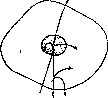
Figure 23. Arrows indicate the direction in which the movement of substances occurs.
is assimilated, part is excreted into the protoplasm, part precipitates in the form of inclusions; besides chromatin, lumps of glycogen, drops of fat are occasionally encountered in nuclei, and in plant cells—protein crystals. Experiments with enucleated parts of the cell body (merotomy of infusoria, obtaining enucleated algal cells) have shown that they can live for some time, but cannot assimilate substances and form a membrane. From this it follows that metabolism in protoplasm cannot occur in the absence of the nucleus. The role of the nucleolus in the metabolic process is depicted variously, depending on views on its nature, which have not yet been brought to unity. There are two main views: 1) the nucleolus is a "dead" formation, an inclusion analogous to the deutoplasmic inclusions of the cytoplasm, wherein according to some it represents a reserve substance consumed in case of need (A. Meyer), according to others (Hacker, M. Heidenhain) a side product of nuclear chemism, waste or secretion; 2) the nucleolus is a special organelle, a system with its own metabolism, as indicated by the formation of vacuoles and their effusion into the karyoplasm; it has significance not only on a nuclear, but also on a cellular scale (Born, Rohde, Dogel, Saguchi). Verworn's scheme. Bringing together all data concerning the metabolism of protoplasm and nucleus, Verworn proposed a scheme of cellular metabolism (Fig. 23), in which arrows designate substances entering the cell and their various fates; after all that has been said, it requires no explanation. If the nucleolus is recognized as an independent system, Verworn's scheme must be supplemented by the metabolism of the nucleolus. Energy metabolism in the cell, just like metabolism, can be divided into 3 phases: absorption, processing, and excretion. The cell absorbs various types of kinetic energy from the external environment: mechanical, or mass motion energy (shock, pressure), thermal, light; together with substance, it absorbs the potential energy of complex chemical compounds, which closely links energy metabolism with metabolism. The penetrated energy can pass through the cell without change only in rare cases (light through transparent bodies of the cell); usually it undergoes transformation. In this case, it is either deposited in the form of potential energy of the deutoplasm or excreted from the cell in one way or another: the cell comes into motion, emits heat, electricity, light, throws out substances containing energy in a latent form. The processing of energy perceived by the cell also occurs according to the Le Chatelier principle and leads to the unloading of the protoplasmic asset from the disturbance of equilibrium produced by energy. The presence of potential energy in the cell, passing into kinetic under the influence of small energy impacts, lies at the basis of the irritability (irritabilitas) or excitability (see) of the cell, a property that was considered specifically living and that is characterized first of all by the quantitative discrepancy between irritation and effect. With respect to energy metabolism as a whole, there are two main types of cells: some accumulate potential energy at the expense of absorbed kinetic energy (chlorophyll-bearing plant cells), others receive potential energy in ready-made form and translate it into kinetic in the process of work (all other cells). Of the types of energy manifested by the cell, movement must be put in first place. In the protoplasm of every cell, movement can occur in the form of the flow of its liquid parts, causing the displacement of inclusions, and sometimes a change in external shape; this movement is very slow and inaccessible to direct microscopic observation; it is successfully detected by applying microcinematography (fibroblasts; Carrel). Movements perceived by the microscope are divided into 4 main types: 1) flow of protoplasm, 2) amoeboid movement, 3) contraction, 4) ciliary movement. 1. Fluid movement of protoplasm is observed most often in plant cells, of animal cells—in hydroids. Such cells are covered with a dense membrane, the protoplasm forms a parietal layer, and the interior is filled with vacuoles of cell sap, through which protoplasmic crossbars in the form of networks can stretch. Rotational movement (rotation) is distinguished, when the parietal layer rotates in a definite direction, carrying inclusions with it (Elodea, internodes of Chara), and circulation; besides the parietal layer, protoplasm flows in crossbars in various directions with various speeds (hairs of Tradescantia). In this case, individual crossbars can tear and disappear, others form anew, so that the configuration of the protoplasm changes (Figure 24). The mechanism of the flow remains unclear; it can be assumed that it reduces to a change in surface tension.
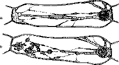
Figure 24. Cells from the hairs of Tradescantia virginica. The protoplasm forms cross-bars of various thicknesses; a - normal appearance of the cell; b - after irritation by an induction current; the protoplasm merges into drops. 2. Amoeboid movement is observed in cells without dense walls and which are free (amebae, leukocytes); in this movement, there is also a flow of protoplasm inside, but it is accompanied by a change in the shape of the cell due to the formation of outgrowths on its surface (false feet, or pseudopodia) and is associated with the movement of the cell in space, creeping (see Amoeboid movements). 3. Contraction is characterized by the fact that the cell changes its shape in a definite manner; it is observed in cells elongated in length (muscle fibers, infusoria), which become shorter and thicker upon contraction. In such cells, there are always fibrils running lengthwise (myofibrils, myonemes), which take part in the contraction process. 4. Ciliary movement is associated with the presence of special filamentous outgrowths on the cell surface, flagella or hairs, and consists in their oscillatory rhythmic movement (ciliated epithelium, infusoria, sperm). All these types of movement as manifestations of mechanical energy belong to the category of mass, ordered movements; but along with them, Brownian movement can be observed in the cell (see). It is found in watery vacuoles, in dead leukocytes (complex bodies), in some living cells with sufficiently liquid protoplasm. The generation of heat occurs in every cell in connection with metabolism, when complex organic compounds burn into simpler ones, releasing the energy contained in them in the form of heat. It increases during work, for example during muscle contraction, although it still remains very small and is accessible to measurement only by sensitive thermoelectric instruments (thousandths of a degree). Previously, the prevailing belief was that during cell work, heat generation is the primary moment, followed by the transition of heat into other types of energy, such as mechanical; in other words, that the cell works on the principle of a steam engine; it is now established that chemical energy is directly transformed into mechanical energy (Engelmann, Hill), and heat arises already in the recovery stage after work as a result of the combustion of residual products. The negligible amounts of heat released by individual cells, when summed up, create the temperature of a complex organism, always exceeding the temperature of the environment, and at the same time the conditions necessary for the life of the cell. The generation of electricity is also a constant moment in the life of the cell; the source is the charges of ions always present in the protoplasm. The concentration of ions of different signs in different places, for example, on both sides of a semipermeable membrane, creates a potential difference (concentration chains, Nernst) and is the cause of currents observed in muscles and nerves. Recently, attempts have been made using vital stains to determine the electrical charges and their location in the cell (Keller and his co-workers); theoretical considerations show that at small distances, such as those present in cells, potential differences can reach colossal sizes. The production of significant amounts of electricity, comparable to the charge of large Leyden jars, occurs in the electric organs of fish, which are modified muscles. The generation of light is characteristic of relatively few cells, concentrated usually in luminous organs (salps, fish, some insects). It is caused by a special substance produced by the cell—luciferin—which, under the influence of the enzyme luciferase, oxidizing in the presence of oxygen, begins to glow (see Luminescence). The generation of light is thus not directly connected with the life process and represents a special adaptation. Reproduction and life cycle of the cell. The doctrine of free cell formation prevailing in the era of Schwann (see Blastema) has long been abandoned; it was replaced by the proposition of R. Virchow (1855): every cell arises from a cell (omnis cellula e cellula); this occurs by the division of the mother cell into 2 or several parts (see Division, Karyokinesis, Amitosis). - Life cycle of the cell. The cell, like any biological system, goes through a definite life cycle, in which three sharply demarcated phases are distinguished: the period of growth and development, the period of maturity (acme), and the period of decline, or old age. The daughter cell resulting from division is smaller in size than the mother cell and in most cases is undifferentiated; its nucleus retains for some time an abundance of chromatin and its specific distribution. Then the period of assimilation and growth begins, upon the expiration of which the cell reaches the size of the mother; simultaneously with this or later, the cell may begin to differentiate, i.e., develop structural features characteristic of the cells of a certain tissue or organ (embryonic cells during histogenesis, replacement cells of the adult organism, germ cells). Having reached full development, the differentiated cell begins to function; its work may consist in contraction (muscle cells), transmission of excitation (nerve cells), mechanical and generally protective function (tegumentary epithelium), production of certain substances (glandular cells), etc. In such a developed state, various cells can exist for different times depending on the quantity and state of their protoplasm: if metabolism proceeds correctly, the protoplasm is cleansed and regenerated, cells live a long time and usually die after the death of the organism due to a radical change in their external environment (violent cell death, necrosis); such are nerve and muscle cells. In other cases, differentiation goes so far that the protoplasm undergoes changes: it degenerates and is spent on the formation of inclusions, or the cell loses its nucleus; such cells die relatively soon; they dry up or disintegrate (epidermal cells, some glands, mammalian erythrocytes); since this death is necessary for the whole organism and is not the result of pathological changes, it is called physiological. The totality of changes undergone by a cell during its life cycle is called cytomorphosis by Minot. In the described cases, the life cycle begins with the division of the mother cell and ends with death; but in embryonic cells and those that have not reached significant differentiation, it can also end in division: the cell does not die, but disappears as an individual, instead of which two new ones arise. In science, it is customary to call such cells "young" and to regard their division as a sign of increased vitality, but this view cannot be considered correct. In this case, too, the cell goes through the same three-phase life cycle; only the changes of a morphological character do not appear so sharply and are noticeable only upon careful comparison of cells of different ages. The aging of the cell occurs here as a result of incomplete cleansing of the protoplasm and its overloading with inactive molecules and molecular complexes, often of a microcrystalline character. It occurs quickly because these substances are not utilized for the construction of cellular structures. Usually, they accumulate near the nucleus, forming a sphere, and from them at the moment of division an achromatin figure arises; thus, division is the natural completion of cellular old age. Daughter cells each receive half of the material of the achromatin spindle and assimilate it during the growth period, increasing the amount of their protoplasm; they are rejuvenated compared to the mother cell and can thus begin a new life cycle.
B.
Karpov. The cell as a physical-chemical system has recently been subjected to detailed study by a number of scientists grouped around the journal Protoplasma. The question of the physical state of protoplasma can be considered one of the central problems of this discipline. Replacing the old morphological theories (Fromann, Altmann, Fleming) and others, which treated the question from the standpoint of protoplasmic structure, biological physical chemistry uses direct physical experiment for its purposes, in particular the determination of protoplasmic viscosity. The most widespread methods are: 1) centrifugation of the cell and study of the rate of movement of cell inclusions (granules, starch grains) returning to their places; 2) the magnetic method—introduction of iron and nickel particles into the cell and study of the rate of their movement toward an electromagnet brought close to the cell; 3) determination of viscosity by the rate of movement of cell inclusions in the fluid protoplasm of plant cells; 4) measurement of the rate of movement of small granules (Brownian motion) in the protoplasm. These methods are precise and make possible purely quantitative conclusions based on physical formulas and showing the magnitude of absolute viscosity. In addition, there are a number of methods making it possible to determine with less precision the r e l a t i v e (comparative) viscosity of protoplasm. These methods are: 1) the rate of outflow of protoplasm from a damaged cell, 2) the resistance offered by protoplasm to a micromanipulator needle, 3) the rate of fall of starch grains in the cell under the influence of gravity, 4) the shape of the plasmolyzed surface when hypertonic solutions act on a plant cell. Most investigators assert that the viscosity of the ground substance of protoplasm is small and this substance can be considered as a typical l i q u i d (see Aggregative state); in this ground substance inclusions of various sizes—micro- and macrosomes—are present in significant quantities.
The magnitude of the absolute viscosity of the ground substance of protoplasm is determined very differently by various authors; apparently, as precise studies by Heilbrunn show, it is equal to approximately 0.02-0.03 in absolute units, i.e., it exceeds the viscosity of water by only a few times. The viscosity of the entire protoplasm as a whole, which is a typical suspension in colloidal terms, is somewhat higher and exceeds the viscosity of water by 7-10 times. Yet another widespread method for studying the physical state of protoplasm is the study of its elasticity, i.e., its ability to resist external influences—pressure, tension, etc. The elasticity of a substance is inevitably connected with its internal structure, and therefore its excessive deformation, destroying the structure, leads to the disappearance of elasticity, which in turn should be expressed in a decrease in viscosity. In the case of protoplasm, enhanced centrifugation should lead to a new (decreased) value of viscosity. However, this cannot be established, and therefore a number of authors come to the view that the elasticity of the inner layer of protoplasm is negligible, i.e., again to the fact that protoplasm is a true liquid; this opinion, however, encounters objections from a number of authors (Seifriz and others) who studied protoplasm by the microdissection method and showed its some elasticity. The data just cited regarding the physical state of protoplasm should be considered only as a particular, although quite typical, case. The viscosity of protoplasm is subject to extremely sharp fluctuations both in different cells and in cells of the same type when the internal state and environmental conditions change. A certain "normal" temperature of about 15-17° (according to Heilbrunn) corresponds to maximum viscosity; a change in temperature in one direction or the other leads to liquefaction of the protoplasm. A further increase in temperature leads to a new, initially reversible increase in viscosity, which is then replaced (at 30-40°) by irreversible coagulation (thermal death of the cell). Upon a decrease in temperature relative to the optimum, following the liquefaction of protoplasm (for some objects around -3°), cell death ensues, accompanied by the formation of ice crystals and coagulation, apparently as a result of an increase in the concentration of salts entering the protoplasm. The action of the latter on the physical state of protoplasm is very significant: heavy metal salts cause coagulation even in negligible concentrations; monovalent ions K', Na", NH4" act similarly, but somewhat weaker; divalent and trivalent ions Ca", Mg"", Al''' possess the opposite action—they decrease the viscosity of protoplasm. According to the degree of liquefying action, ions can be arranged in the following series: Ca>Mg>K>Na>NH4; this series corresponds to the degree of adsorption of ions by protoplasm particles. Among anions, CNS' and I' cause rapid coagulation of protoplasm. Fat solvents exert a sharp influence on the physical state of protoplasm: ether, chloroform, etc. Their low concentrations cause a decrease in viscosity, high ones—an increase. The coagulation of protoplasm is caused not only by the action of hypertonic solutions (which is understandable from an osmotic point of view), but also of hypotonic ones. This latter circumstance is attributed by some authors to the partial dissolution of lipid substances of the cell by water (see above). Essential for the physical state of protoplasm is the active reaction (pH) of the medium. Acidity causes an increase in viscosity; alkalinization of the medium, on the contrary,—liquefaction. A number of diverse external influences—mechanical strokes, wounds, electric current—lead to an increase in viscosity and subsequent coagulation. Short ultraviolet rays, as well as X-rays and radium rays in small doses with short-term exposure cause a decrease, and with stronger exposure—an increase in viscosity. During cell aging, the viscosity of its protoplasm increases. All the cited data undoubtedly show that the physical state of protoplasm is labile and highly dependent on all processes occurring in the medium and inside the cell. When studying the physical chemistry of protoplasm, one must distinguish its inner layer from the outer, cortical one, which has a whole series of specific features. In terms of physical properties, the cortical layer possesses a high degree of viscosity and elasticity, being a typical gel. The specificity of the physical-chemical structure of the outer layer of protoplasm finds its most vivid reflection in semipermeability, first established by Pfeffer (1890) for the plant cell. Since then, the study of cell permeability, based on the special properties of the membrane or outer layer of the cell, has become one of the main tasks of physical-chemical biology, the cornerstone of modern cell physiology. Physiologically, the cortical layer also possesses some special, not entirely elucidated properties. In microdissection experiments on eggs upon separation of the inner and cortical layers, only the latter proved capable of development (Figs. 25 and 26). Cleavage of the inner
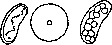
Figure 25. Isolation of the inner part (a) of the protoplasm from a crushed starfish egg; b and c—result of the action of sperm on the ecto- and endoplasmic fragments of the egg formed in the process.
layer is possible in the presence of at least a negligible fragment of the cortical layer. The latter also possesses a high degree of sensitivity to external influences. The touch of a micromanipulator needle causes the destruction of structure in a ciliated epithelial cell, the exit of hemoglobin from erythrocytes, the disintegration of protoplasm, etc. In the same way, a sharp effect upon action on the surface layer of the cell is caused by various chemical agents, a change in the reaction of the medium, etc. An important question of cytology, having fundamental significance also for genetics, is the question of the intravital structure of the nucleus. It must be noted that the physical properties of the nucleus have been elucidated to a significantly lesser extent than those
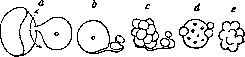
Figure 26. Inner part of the egg (a), connected with the region of its outer membrane; b—the same, treated with sperm; c, d, e—unsuccessful attempts at cleavage.
protoplasm. The study of the nucleus using an ultramicroscope reveals its optical emptiness, that is, the absence of any structures; similarly, the Brownian motion sometimes observed in the nucleus indicates that the nuclear contents are liquid; according to preliminary calculations by Heilbrunn, its viscosity is only twice that of water. On the other hand, however, there is a number of data indicating the existence of intranuclear structures, especially filaments lying on the surface of the nucleus. Thus, Zhivago, using an improved photomicrography method, discovered the presence of filamentous formations in the nucleus, which were moreover in motion. Similarly, Chambers, when damaging the nuclear membrane with a micromanipulator needle, obtained a structure of filaments in the nucleus that gradually changed and transformed into a typical chromosome figure. The later the nuclear damage was performed, the later the stage of the chromosomal tangle that could be obtained. Since all modern genetics is based on the premise that chromosomes are permanent formations, geneticists tend to explain the difficulties of direct observation of the structures of the living nucleus by the inadequacy of modern research methods, which do not allow seeing structures with almost the same refractive index. These data convincingly argue in favor of the presence of a nuclear structure in the form of a pre-existing chromosomal filament, invisible under normal conditions due to the equality of the refractive index of this filament with that of the main substance of the nucleus (nuclear sap). The physical state of cell organoids has been studied quite fully. Microdissection experiments show that the chromatin filament and individual chromosomes are dense formations that can be deformed and completely removed from the cell (Figs. 27 and 28). The spindle and asters in a dividing cell undergo a number of viscosity fluctuations depending on the various stages of karyokinesis. In the structure of cell stars—amphiasters—two parts should be distinguished: the liquid central astrosphere and the solid hyalinized rays, which apparently create the turgor of the dividing cell. These rays are very fragile formations that disappear, for example, upon touching with a micromanipulator needle. Permanent cell inclusions—mitochondria—are dense formations characterized by high stability in relation to various unfavorable external influences; in a dying cell, they persist longer than other parts of it. A number of processes studied by physical chemistry and colloid chemistry play a prominent role in the vital activity of the cell. Protoplasm is a suspension of proteins and lipids that are in complex and not entirely elucidated interrelations with each other. In addition to these substances, water and salts of alkali and alkaline earth metals are also particularly important. The composition of protoplasm thus includes various &«№ R ^>-7 /г Figure 27.
Figure 27. a - loop of the chromatin thread of a spermatocyte nucleus, stretched with a needle; b - stretched and unstretched parts of the thread. Figure 28. Chromosome from a nucleus in the metaphase stage, torn by needles. Both water-soluble and insoluble substances are present, and normal life of the cell is possible only with a certain equilibrium of all components of the physicochemical structure of the cell. This equilibrium leads to the normal course of processes both on the surface and inside the constituent parts of the cell, primarily oxidation and reduction processes. That the cell can exist only in the presence of certain strictly defined physicochemical conditions is shown by the fact that the concentration of hydrogen ions in it fluctuates very insignificantly in various cases (Chambers 6.7, Höber 5.2-5.7). An extremely essential role in the cell as a physicochemical system is played by electrical phenomena; according to some authors (e.g., Keller), generally all essential phenomena in the life of the cell, such as the course of assimilation and dissimilation processes, oxidative processes, neutralization of bacteria, copulation of sex products, etc., are determined by the charge of its various particles and the magnitude of the electrical potential. In proof of his position, Keller cites a number of facts based both on data of "electrohistology" (action of various dyes with different electrical charges) and direct measurements with a special cell galvanometer. He points out, for example, that plant pollen is charged negatively, while the egg cell is charged positively. If Keller's theory cannot be considered generally recognized at the present time, the significance for the cell of electrical phenomena, in particular cataphoresis, is beyond doubt. Rich factual material indicates that the particles of the inner layer of the protoplasm are charged positively, while the outer surface of the cell and the particles of the nucleus are charged negatively. The fluctuations in the physical state of the protoplasm considered above, especially those caused by the action of various ions, can be accounted for by changes in the electrical charge of the cell protoplasm. A number of observations are devoted to the question of the relationship between the work of the cell and the course of the physicochemical processes occurring in it. Thus, for example, the formation of pseudopodia in amoebae and wandering cells of higher animals can be regarded as the result of a local decrease in surface tension and fluctuations in the viscosity of the protoplasm in a segregated section of the cell, an alternation of liquefaction and subsequent condensation. This connection between the formation of pseudopodia and the physical state of the protoplasm is best illustrated by the circumstance that the shape of the pseudopodia essentially depends on environmental conditions, the presence in it of certain ions that act specifically on the viscosity of the protoplasm. Recently Heilbrunn put forward the assertion that a special reaction is specific for the protoplasm colloids, the essence of which reduces to the coagulation of colloids under the action of a special substance, cytotrombin, formed by the interaction of free calcium and certain organic (apparently lipoid) constituent parts of the cell. This reaction, which is extremely reminiscent of the blood-clotting process, in Heilbrunn's opinion not only causes the formation of a dense membrane around exposed parts of the protoplasm, but also underlies the widespread phenomenon of vacuolization, one of the most typical external manifestations of both the normal vital activity of the cell (the formation of vacuoles or granules close to them is associated with almost all types of secretions) and its reactive response to various external influences, insofar as this reaction can be detected morphologically. Continuing the chain of his proofs, Heilbrunn comes to the conviction that the work of the cell is inevitably associated with an increased viscosity of its protoplasm; in particular, for example, the old observations of Biedermann on the contraction of transparent muscles of certain invertebrates indicate that the work of a muscle cell is always associated with the condensation of its protoplasm. The anesthetic and narcotizing action of a number of fat-dissolving substances (ether, chloroform, etc.) reduces to the liquefaction of the protoplasm, which obviously terminates the processes of active functioning occurring in it. Despite the fact that Heilbrunn's generalizations must be accepted with a certain caution, the main proposition—the significance of a definite (condensed) phase of protoplasm for the course of physiological processes in the cell—is beyond doubt. This becomes especially clear when considering karyokinesis from a colloid-chemical point of view—a process studied most completely in this sense. The onset of karyokinesis is accompanied by a number of physicochemical changes in the cell. 1. Experiments with the penetration of vital stains into the interior of the egg (Herlant) show an increase in the permeability of fertilized eggs entering division; at the same time, detailed studies by Runnström elucidated that this process is associated with the transition of the envelope lipoids into a more dispersed phase. 2. Heilbrunn, using the centrifugation method, showed a sharp increase in the viscosity of the protoplasm after fertilization, during the extrusion of polar bodies, and at the beginning of egg segmentation. Stages of increased viscosity alternate with liquefaction of the protoplasm, which is especially noticeable at the moment of spindle formation. Thus, during karyokinesis, regularly alternating fluctuations in protoplasmic viscosity take place, well expressed on the attached curve. Viscosity fluctuations, in particular the presence of a condensed phase, are of decisive importance for the mechanism of karyokinesis. Thus, according to some authors (Chambers), the division process of the cell itself can be accomplished only under strictly defined conditions of equilibrium of the liquid and condensed phases in various parts of the cell (condensed middle part of the cell, liquid phase at the poles and in the equatorial zone). Karyokinesis ceases upon the destruction of these local viscosity fluctuations or a decrease in the viscosity of the entire protoplasm. Such, for example, is the action of small concentrations of fat-dissolving substances (ether) that stop karyokinesis. 3. The increase in oxidation processes in the protoplasm of dividing cells (according to Warburg's data, oxidation in the fertilized sea urchin egg is 6-7 times greater than in the unfertilized one). The data set forth above regarding changes in physicochemical processes in the dividing cell make the mechanism of external influences on the occurrence of karyokinesis more understandable (see Karyokinesis, physiology). Direct observations and precise measurements of viscosity, permeability, elasticity, and other physicochemical properties concern only individual constituent parts of the cell. The cell as a whole is a biological system consisting of the most diverse constituent parts; some of them have the properties of a liquid with greater or lesser viscosity, while others in the form of membranes, threads, networks, etc., possess more or less high elasticity and determine the often very stable shape of the cellular mechanism or individual cell organs—flagella, cilia, chromosomes, etc. (see Aggregate state).
S. Zalkind. The pathology of the cell is of enormous importance already because the basis of the majority of diseases ultimately lies in pathological changes in the cell. True, at present we no longer consider completely correct the basic dogma of Virchow's cellular pathology, which states that "all pathology is the pathology of the cell"; however, as applied to the pathogenesis of a significant majority of disease manifestations, cell changes undoubtedly have a dominant importance (see Virchow, Pathology). In general, pathological changes in cells are extremely diverse, and their classification is far from perfect. The division of all cellular changes that was very widespread in former times, based on whether the cell is in a state of decline or increase of vital activity—the division into regressive and progressive changes—is little used at present due to the circumstance that it is far from possible to judge the state of a cell's vital activity in all cellular changes. From the standpoint of pathogenesis, the greater part of pathological cell changes belongs to the manifestations of dystrophies (see), i.e., is based on certain disorders of cell nutrition and disturbances of tissue metabolism. As a result of the complete cessation of tissue nutrition, cell death ensues (see Necrosis). Inadequate nutrition of tissue in quantitative and qualitative terms, depending either on irregularities in the influx of nutrient material to the cell or on a violation of the assimilatory and dissimilatory capacity of the cell, leads either to a decrease in cell volume and a loss of their number, which is designated as atrophy, or to certain qualitative changes on the part of the cell protoplasm and nucleus, called transformations, degenerations; the latter, depending on which type of metabolic disorder underlies the given cell transformation, are subdivided into protein, fatty, carbohydrate degenerations, manifestations of impaired salt metabolism, and pigmentation disorders. Changes in the cell are expressed extremely diversely and generally represent violations (disorganizations) of the normal organization of the cell in morphological, physical, and chemical senses; at the same time, it should be noted that such a subdivision of cell changes is largely conventional, since many of them have a complex physical-chemical origin, and in addition, what relates to manifestations of morphological disorganization may be based on physical and chemical changes of the cell. Morphological disorganization of the cell manifests in changes in their sizes, as happens for example in atrophy and hypertrophy, in changes in the shape and appearance of the cell, for example from mechanical influences or during metaplasias. This also includes various irregularities in the volumetric ratios between the cell body and its nucleus (the nucleus turns out to be too large or small for a cell of the given type), changes in the nucleus in the sense of pyknosis, karyorrhexis, karyolysis, complete disappearance of the nucleus. In the protoplasmic body of the cell, various changes in the chondriosomal apparatus of the cell can be observed, phenomena of hyalinization and a sort of coagulation of the protoplasm, and sometimes a sort of liquefaction of it. The complete disintegration of the cell into granules, its transformation into detritus (see), as happens as a result of cell dying, represents the highest degree of morphological disorganization of the cell. Physical disorganization of the cell can be expressed in the loss of transparency by the protoplasm, apparently as a result of a change in state, possibly swelling of its colloids, in the precipitation of protoplasm colloids in the form of large aggregates, which yields pictures of droplet and granular degeneration of the protoplasm; in the appearance of moisture droplets in the protoplasm (see Vacuolar degeneration) or various protein inclusions in the form of grains, hyaline lumps, spheres. To the chemical disorganization of the cell can be attributed all cases of cell alteration in a chemical sense, both in the sense of the appearance in it of uncharacteristic
Related articles
Cite this page
“Cell.” Soviet Medical Encyclopedia. English translation of Bolshaya Meditsinskaya Entsiklopediya, 1st ed. (Moscow, 1928–1936), ed. N. A. Semashko. https://sovietmedicalencyclopedia.pages.dev/article/cell/