Karyokinesis
Historical document, translated for reference. It reflects medical knowledge of the 1920s–30s and is not medical advice.
Summary
Karyokinesis is the complex process of cell division, normal for cell reproduction in contrast to amitosis. It involves changes in the nucleus, particularly its chromatin, leading to the formation of chromosomes that split and distribute equally between daughter cells.
Encyclopedia article (1928–1936)
KARYOKINESIS, karyokinesis (Schleicher) (from Greek karyon-nucleus and kinesis-movement), synonym mitosis (Flemming), indirect nuclear division (indirekte Kernteilung; Flemming), a complex process of cell division that is the normal method of cell reproduction in contrast to amitosis (see), or direct division. History. Cell reproduction by division was established in the 1840s and 1850s by Naegeli for plants and Remak for animals. It was described simply: division of the nucleolus, strangulation of the nucleus into 2 parts with subsequent division of the cell body. In the 1870s, with the improvement of the microscope (immersion) and research methods, it became apparent that cell division is much more complex. The first detailed description of division was given for plant cells by Strassburger (1875) and the Russian botanist Chistyakov; for animals-in 1878 by Schleicher (cartilage cells), by the Kiev professor Peremeyko (triton larva), and mainly by Flemming (salamander larva). The latter in his classic works established the main phases of K. and developed a technique still used today. In the 1880s and 1890s, through the work of a number of outstanding researchers (Balbiani, van Beneden, Henneguy, Carnoy, C. Rabl, Hermann, Druner, Boveri, Kostanecki, M. Heidenhain), significant additions were made to Flemming's scheme; the universal distribution of K. was proven, and a number of important regularly recurring deviations from the general scheme were studied. In the 1890s, attempts began to explain the mechanism of K. Since the study of fixed preparations did not make it possible to resolve many problems associated with K., in the last decade, the study of living karyokinesis (mainly in tissue cultures) has resumed, supplemented by the application of microdissection (Chambers, Speck, Belar). Scheme of K. The changes in the cell during K. consist of 1) those changes observed in the nucleus, mainly its chromatin, which lead to the formation of chromatin structures, chromosomes, which then split along their length and are distributed equally between daughter cells (chromatin figure); 2) changes in the plasm: division of centrosomes, formation of the spindle and radiating rays, which play a role in the division of the cell body and distribution of chromosomes (achromatin figure). Both of these processes can proceed to a certain degree independently, but during normal K. they are strictly coordinated. K. in animal cells generally proceeds in the same way; the basis for the scheme usually presented in textbooks is
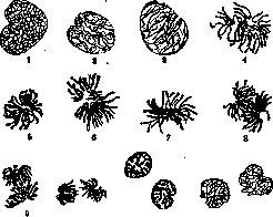
Figure 1. Karyokinesis in triton; chromatin figure: 1-resting nucleus; 2 and 3-coil stage; 4, 5 and 6-mother star stage; 7 and 8-metakinesis stage; 9 and 10-daughter star stage; 11-daughter coil stage; 12-resting nuclei. (According to Petersen.) the pictures observed in tailed amphibians (fig. 1) and during the division of the horse ascaris egg (for the achromatin figure) (fig. 2).- The process of K. is conventionally divided into 4 main phases: pro-, meta-, ana-, and telophase (Strassburger, M. Heidenhain) or into a series of successive figures (Flemming). Prophase begins with changes in the nucleus, which swells and rounds. The number of chromatin grains in it gradually increases; approaching each other, they begin to gather into threads, and in the nucleus a chromatin figure is formed, called a coil (spirema), in the form of a long, extremely convoluted thread, located mainly peripherically. Subsequently the thread becomes shorter and thicker, the coil as it were loosens, for which reason two stages are distinguished: dense coil (early prophase) and loose coil (late prophase). The coil further breaks down into separate segments, chromosomes (Waldeyer). It should be noted, however, that the latter often appear immediately as separate structures. Simultaneously with the changes in the nucleus, changes are observed in the protoplasm: the centrosome, which is not always visible in the resting cell, appears
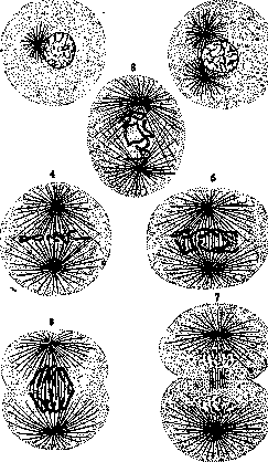
Figure 2. Semi-schematic representation of karyokinesis in Ascaris megalocephala: 1-resting cell; 2-division of centrosome; 3-prophase: centrosomes are located at the poles, rays are strongly developed; chromatin has broken down into chromosomes; 4-mother star; chromosomes are located at the equator; 5-metaphase: chromosomes, split along their length, move to the poles; 6-anaphase-beginning division of the cell body; 7-division of the cell body almost completed; nuclei pass into the coil stage. (According to Kastenetsky.) clearly as a small bubble, surrounded by a radiating glow, then divides, and between its receding halves a fibrous formation is stretched, having the appearance of a spindle (fig. 3). The combination of the spindle, centrosomes at its ends, and radiating glows (polar glows, asters, astrospheres) emanating from them, due to their weak staining in ordinary histological stains, is called the achromatin figure (otherwise-amphiaster). Once formed, it continues to grow throughout the prophase and comes into connection with the chromosomes. This occurs in such a way that at a certain moment the nucleus loses its membrane (it dissolves), the nucleoli also either dissolve or break down into small fragments, and in place of the nucleus a light nuclear cavity (Kernhohle) is formed, in which the chromosomes are located. Then between them and the centrosomes, thin connecting fibers growing from the centrosomes and connecting each chromosome with both centers (pulling fibers, "reins"; fig. 4) become noticeable. The spindle changes its position, turns, and is established in the middle of the cell, in the nuclear cavity, and the chromosomes are arranged along its equator at equal distances from the poles-equatorial plate, or mother star (monaster). With the establishment of the spindle, K. enters metaphase, during which the chromosomes of the mother nucleus split along their length into identical daughter chromosomes and begin gradually to move away from each other, remaining connected at their ends for some time (redistribution, or metakinesis). This phase without sharp boundaries passes into anaphase, when the chromosomes, finally separating from each other, continue to move toward the poles, sliding along the surface of the spindle (as was previously assumed, due to the contraction of the fibers going to them); reaching the poles, they stop, forming the figure of daughter stars (diaster). At this time, the division of the cell body begins: the polar glows grow strongly, reaching almost to the periphery, and at their crossing point, along the equator of the cell, a circular groove is outlined- strangulation. The final phase, telophase, is characterized by the fact that the daughter nuclei pass into a resting state, undergoing changes in reverse order to prophase (reconstruction of the nucleus). The chromosomes of the daughter stars approach each other, sometimes connect at their ends, forming figures of daughter coils (dispirema); a thin nuclear membrane appears around their periphery. It first tightly fits the ends of the chromosomes, then
distinctly. The chromatin grains gradually decrease in number, the threads become less distinct, and the nucleus acquires the appearance of a resting nucleus. The division of the cell body is completed, and two daughter cells are formed. In plant cells, the process of K. is complicated by the formation of a cell wall between the daughter cells. The division of the centrosome and the formation of the spindle occur in the same way as in animal cells. The main difference is in the division of the cell body: instead of strangulation, a cell plate is formed in the equatorial plane, which gradually grows and turns into a cell wall. The study of K. is of great importance for understanding the processes of growth, regeneration, and pathological changes in tissues. The knowledge of the laws of K. is the basis for the doctrine of heredity and variability.

Figure 3. Division of centrosome; Pfützen's grains; early splitting
chromosomes. (According to Hermann.)
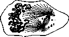
Figure 4. Formation of achro-
of the spindle (according to Hermann). As the nuclear sap accumulates, it lags behind them, and the nucleus becomes rounded. The chromosomes lose their smooth contours, become covered with knobs, connect with each other by bridges, and the chromatin is distributed throughout the resulting network in the form of clumps and grains. With the appearance of the nuclear membrane, nucleoli again appear in the nucleus—one or several. At the same time, division of the cell body continues, and the furrow finally separates the daughter cells, which remain connected for a certain time only by remnants of the spindle in the form of a narrow bridge. The polar rays and spindle gradually become less distinct and gradually disappear, sometimes forming a granular accumulation (archoplasm), while the centrosomes move from the polar part of the nucleus to the other side (telokinesis); sometimes this movement is accompanied by rotation of the nucleus (Heidenhain). Such is the picture of K. on fixed preparations; recent experimental observations in living organisms essentially confirm it, introducing only certain changes for the achromatic figure (see below).- The duration of K. is measured in hours (in the triton 1.5-2 hours, in the salamander 2-5 hours), in warm-blooded animals it is shorter (1/2 hour); heating within certain limits significantly accelerates the process. The chromatic figure of K. The number, relative size, and (with less strictness) absolute size, shape, and some individual features of chromosomes are by no means accidental; invariably recurring in all K. of a given species, they are characteristic and constant. However, even in closely related species, significant differences can sometimes be observed here, and with greater systematic distance, these differences are usually striking. Thus, the number of chromosomes varies in different species from two (Ascaris megalocephala, var. monovalens) to hundreds (e.g., in the brine shrimp Artemia salina 168 chromosomes); however, only a few tens are most common (60 in horses, 48 in humans and monkeys, 44 in rabbits, 40 in mice, 24 in lilies, 22-12 in different species of marsupials, etc.). The size of chromosomes can also vary considerably: the smallest are spheres of 0.3-0.4 μ in diameter, while large rod-shaped ones can reach several tens of μ in length. The set of chromosomes of a given species, or the chromosomal complex (chromosomal plate, karyogram), is a nuclear characteristic of the species, or the karyotype (see). In all somatic (body) cells of animals and plants [with the exception of haploid (see) generations of spores], the chromosomal plate consists of pairs of identical (homologous) chromosomes of paternal and maternal origin; only the sex chromosomes (hetero-, idio-, allo-somes, x-, y-, w-, and v-elements) may be unpaired, which in some species differ significantly from the others (autosomes) in size. It is interesting to note that the arrangement of chromosomes during K. is not random, but constant and regular, showing clear bipolarity; in connection with this, elements of constancy in arrangement can often be detected in other stages of K. The fact of the constancy of number, size, and distinctive features in chromosome sets is the main basis for the hypothesis of chromosome individuality (Boveri), the essence of which is that chromosomes continue to exist after reconstruction in resting nuclei in the form of separate nuclear regions, each of which, at the beginning of K., again gives rise to a chromosome. Experiments with crossing varieties with different numbers of chromosomes in the egg and sperm also speak in favor of this hypothesis: the number of chromosomes in the offspring is always equal to the sum of the maternal and paternal chromosomes (ascarid, sea urchins, plant hybrids); they can sometimes be distinguished by size. Finally, direct evidence is provided by the shape of some resting nuclei provided with finger-like projections (ascarid)—the number of projections corresponds to the number of chromosome ends; in crustaceans, the separation of maternal and paternal chromosomes is noticeable not only during cleavage but throughout life (gonomeria; Nägeli). New experimental evidence for individuality has recently been obtained under the action of large doses of X-rays on the chromosomes of the fruit fly Drosophila (Muller, Painter): various kinds of defects and anomalies caused by the experimental factor proved to be stably transmitted to offspring. The fine structure of chromosomes, despite all the efforts of researchers, cannot yet be considered established. According to one view, the chromosome is formed and consists of separate grains—chromomeres, or Pfitzner's grains; such a composition is clearly evident in some fixed preparations (Fig. 3), but was also observed in living organisms (Chambers). The splitting of the chromosomal thread then reduces to the division of chromomeres. Some authors (Eisen, M. Heidenhain) assume even smaller grains of chromatin, chromioles, from which chromomeres are composed. The chromomeres themselves, as many assume, consist of an achromatic basis (line) and the chromatin impregnating it; accordingly, the doctrine of chromosome individuality is based on the preservation of achromatic regions (Nägeli). According to another view, the chromosome is a morphologically integral formation consisting either of pure chromatin or of linin and chromatin; in this case, the chromatin is either distributed along the periphery in the form of a sheath or divided into two threads spirally winding around the axis (Bonnevie). Other authors, conversely, place the chromatin along the axis and assume on the surface an unstained sheath-like shell (Navashin) or assume that the chromosome maintains its shape due to a special skeleton in the form of threads (Koltsov). Chemically, chromosomes differ from the chromatin of the resting nucleus by a higher content of basichromatin rich in phosphorus (Lilienfeld, M. Heidenhain), due to which when stained with fuchsin-methyl green they give a purer green tint. In living organisms, chromosomes are homogeneous, strongly light-refracting formations that possess considerable density and elasticity; during movement they are easily deformed and can be stretched quite strongly with micro-needles before breaking. The achromatic figure of K. is usually studied in egg cells, where it reaches a large size. In fixed preparations, it consists of vesicular centrosomes with centrioles and distinct fibers extending from them. Some of them radiate to the periphery (polar rays, asters), some form a continuous connection between the centrosomes (passing fibers of the spindle), and some go to the chromosomes (pulling threads, mantle fibers). At first, all of them were attributed a contractile nature and even compared to muscle fibers (van Beneden, Kalb, Flemming, Boveri); with more detailed study, it became clear that in any case, not all spindle fibers can be attributed such a function—passing fibers, on the contrary, are solid, elastic formations that, as they grow, push the centrosomes apart and prevent their approach (Druner, Meves). Living research fully confirms this assumption (Chambers, Belar); microdissection shows that the spindle is a rather dense gelatinous formation. As for the fibers of the polar rays, they also show considerable hardness; when pressed with a needle, they bend; if the needle is moved strongly in the cell, the polar rays disappear, but after some time they reappear (Chambers). The place where the centrosome is located has a liquid consistency. The origin of the spindle has been the subject of long disputes: many thought that it is formed entirely or in part from the linin skeleton of the nucleus; but in a number of cases where its origin could be precisely traced, it undoubtedly arises from the protoplasm, according to some researchers—precisely from a special region of it lying near the nucleus (sphere, archoplasm); this question needs further development. The nucleolus during K., according to the usual view, disappears after dissolution of the nuclear membrane and reappears in the daughter nuclei; but in a number of cases, fragments of it were described throughout K.; sometimes it is stretched along the axis of the spindle and divides. Statistical studies on plant cells have shown that in a certain percentage of cases (for different species from 15 to 65), the nucleolus can be detected in metaphase, and in a small percentage, parts of it persist until anaphase and continuously pass into the daughter nuclei (Kotlyarevskaya). The mechanism of K. To explain the movements observed during K. (the wandering of chromosomes, the constriction of the cell body), many hypotheses were proposed in the 1890s and 1900s, which at present represent mostly only historical interest. The first researchers explained the divergence of chromosomes to the poles and the constriction of the cell body by the contraction of achromatic fibers. In detail, such a mechanical theory was developed by Heidenhain, who even constructed a special model of semicircular springs and rubber threads illustrating the constriction of the cell. Another series
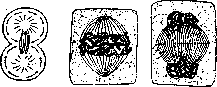
Figure 5.
Figure 6.
Figure 7.
Figure 5. Diagram of division of the cell body; flow of peripheral plasma toward the equator. Figure 6. Karyokinesis in plant cells - stages of the equatorial plate. Figure 7. Karyokinesis in plant cells - stages of daughter spirem formation - formation of the cell plate. Theories of a physical nature interpreted the achromatic figure differently: in it they saw the embodiment of force lines, and placed the source of force in the centrosomes (dynamic centers). At the same time, some scientists (N. E. Ziegler, Gallardo, Hartog) saw an analogy with the force lines of an electromagnetic field, which are revealed, for example, by iron filings around magnetic poles separated by a certain distance; others (Bütschli, Rhumbler) saw in the centrosome a section that absorbs liquid and thereby causes diffusion currents that produce deformation in the cellular plasma (hydrodynamic theories). Modern authors (Chambers, Heilbrunn), based on experimental data, tend to see in K. a complex process of solidification and liquefaction of colloidal plasma. The spindle, growing and hardening, provides points of support for the polar radiations (as Druner assumed). The fibers of the polar radiations cannot pull the cell membrane (as was previously assumed), since they liquefy toward the periphery and the entire peripheral layer of protoplasm becomes liquid at the beginning of division; in it there are currents flowing from the poles to the equator; here they bend according to the round shape of the asters and form a circular groove (Speck) (fig. 5). In the latest works conducted on living objects, Belarz (1929) makes the movement of chromosomes dependent on two factors: on the elongation of the middle segment of the central spindle (Stemm-körper) and on the contraction of the pulling threads, the origin of which, however, receives a completely new interpretation. Each chromosome, until it has finally come into contact with the central spindle, carries at a certain point (Insertionsstelle) a droplet of extremely viscous liquid, which, upon contact with the spindle fibers, spreads over it and gives rise to a contractile fiber sharply different from the others. These data coincide with the data of Nasonov, who managed to separately impregnate the pulling threads with osmium in a number of plant objects, and of S. Navashin, who long ago described on some large chromosomes apparently the above-mentioned droplets that, according to Belarz, give rise to the pulling fibers (Navashin's 'claspers'). Features of K. in higher plants (figs. 6 and 7) come down to the complete absence of centrosomes and polar radiations and to division of the body by formation of the cell plate. In the equatorial part of the barrel-shaped spindle, a series of grains forms, which, merging with each other, give rise to a partition, first simple, then splitting into 2 parts; the partition consists of cellulose. In some invertebrates (eggs of some mollusks and myriapods, hydras), the entire karyokinetic figure develops inside the nuclear membrane, which dissolves only very late or not at all - intranuclear K. Significant deviations toward simplification are characteristic of the nuclei of many protozoa, in which division proceeds in the form of promitosis (see). In the tissues of various vertebrates, under normal and pathological conditions, K. is encountered that does not reach completion due to dissolution of the achromatic figure, as a result of which binucleate cells are formed - abortive K. (glands, urinary bladder). The appearance of several centers in the cell at the beginning of K. leads to the formation of a complex achromatic figure from several spindles and the incorrect distribution of chromosomes between the individual poles - multipolar, or multipolar K. (megakaryocytes, cells of RIfig. 8. Chromo-some tumors). Division and here often does not reach completion. v In spermatocytes:
A special place among deviations is occupied by the reductional karyokinesis, which enters the cycle of maturation of the sexual elements (fig. 8) and leads to the formation of cells with a half (haploid) number of CHROMOSOMES.
V. Karpov. Physiology. Physiological research on karyokinesis starts from the fact that the rate of multiplication of biological objects, i.e., the number of cells appearing in a certain unit of time, is not constant, and the synchronism (simultaneity of all occurring divisions), characteristic of the early stages of egg cleavage, is lost quite soon (no later than the tenth division). The rhythm of multiplication of individual cells changes, which may depend either on an increase in the duration of karyokinesis or on an increase in the interval between two successive karyokineses. In addition to changes in the rhythm of karyokinesis of individual cells, one must observe general and local increases in the number of karyokineses both as a result of normal embryonic development (reaching a certain stage associated, for example, with the formation of a new organ) and under the action of extraordinary causes that obviously play the role of stimuli (cell losses, functional hypertrophy, regulation by the organism as a whole, etc.). Thus, upon extirpation of part of the rabbit's liver, Ponfick and others observed a large number of karyokineses that are normally absent in this organ. This example indicates the need to distinguish for each cell two separate concepts: the potential ability to divide and the 35» real possibility to divide. The realization of the latter necessarily requires the presence of certain changes in the cell plasma preceding the nuclear reaction, namely, an increase in its permeability and viscosity; only the presence of these conditions leads to the prophase of karyokinesis. The presence of a potential ability for karyokinesis is connected with the basic properties of cell plasma and is in antagonism with intensified metabolism, growth, and differentiation; an example may be the highly differentiated cells of the nervous system, sense organs, which under ordinary conditions apparently never enter into kariokinesis. According to a number of authors who have studied karyokinesis from a physiological point of view (Gurvich, Wassermann), when studying the causes of karyokinesis, one should strictly distinguish 1) factors affecting the ability, possibility of the cell to divide (Moglichkeitsfaktoren), and 2) factors determining, 'permitting' the very act of karyokinesis (Verwirklichkeitsfaktoren). The former in turn break down into stimulating and inhibiting. I. Factors stimulating readiness for karyokinesis. Oxygen stimulates the occurrence of karyokinesis, but stimulation of karyokinesis is sometimes observed also with a deficiency of O. In tumors, karyokinesis can proceed 'anoxiobiotically' (in the absence of free O). Enhanced nutrition stimulates karyokinesis, and its effect is manifested according to data from extensive research by Kornfeld only on the 6-7th day after the start of feeding. Starvation also causes a short-term stimulation of karyokinesis. The causes of this stimulation are not entirely clear; apparently, the increased expenditure of reserve substances in the body associated with lack of food leads to the fact that individual cells are temporarily supplied with them in increased amounts. Experiments with tissue cultures show the need for enhanced divisions of protein breakdown products (embryonic extract); ions, especially the Ca ion, as well as apparently K and Na, also play a significant role. For the normal occurrence of karyokinesis, a certain reaction of the medium is necessary. According to the data of Reding and Slosse, a certain alkalinity of the medium creates optimal conditions for karyokinesis, while an increase in acidity delays the onset of karyokinesis. Whether there are specific substances that create readiness of cells for division remains unclear. The repeatedly confirmed observation from the field of tissue cultivation and simplest organisms—an increase in the intensity of karyokinesis in large cell groups—can have various explanations (see also Mitogenetic rays). The significance of temperature for karyokinesis is undoubtedly clear, but it is difficult to fit into any general framework for all cases. The optimum of temperature effect on karyokinesis coincides with that for the basic physicochemical processes in protoplasm. The distribution of karyokinesis in different parts of a multicellular organism indicates the importance of the factor of place. Research by the school of Gurvich indicates a connection between an increase in the intensity of karyokinesis and subsequent morphogenesis. Research by Kornfeld on the cornea of salamanders showed that in paired organs not only the total number of mitoses, but also their distribution by stages in general coincides and is expressed by parallel curves. Here there is apparently a common regulating factor for both, as some think—of a hormonal nature. This latter assumption finds confirmation in the data of Romeis, who showed a sharp increase in the number of karyokinesis in the limb of tadpoles under the influence of the thyroid hormone. A significant amount of material has been accumulated indicating stimulation of karyokinesis under the influence of small doses of various salts and organic substances. Strong stimulants of karyokinesis are ethyl and butyl alcohol, as well as fat-soluble dyes Scharlachrot and Sudan III, whose introduction into the body causes, as a result of intensified karyokinesis, cancer-like tissue proliferations. Mechanical irritations (pressure) also stimulate karyokinesis. Small doses of radiant energy have a stimulating effect on karyokinesis: red and infrared rays, X-rays and Ra (radium) rays. Stimulation of karyokinesis has been obtained with prolonged (5-14 hour) exposure of galvanic current to the root of some plants; in the very recent time, a stimulating effect of short-term exposure of electromagnetic waves on the reproduction of yeast cells has been established. Many of the just-mentioned factors—organic compounds, hypertonic solutions, ultraviolet rays, injuries, etc.—have been successfully used by a number of researchers to stimulate the development of unfertilized eggs (artificial parthenogenesis). II. Inhibiting factors of karyokinesis. Among these, first of all, one should include violations of the usual metabolism: the lack of sufficient amount of O (for normal tissues), water, salts, and nutrients. Karyokinesis ceases with overheating, which can be accompanied by physicochemical changes in the structure of the cell: vacuolization, isolation of various phases in the protoplasm. This condition to a certain extent is reversible. To the inhibiting factors normally inherent in the organism should be attributed: 1. Aging of the cell, morphologically very close to the just-mentioned pictures of 'overheating'. The essence of the process of cell aging is little clarified; in recent times, by a number of works of the school of Ruzicka, the concept of hysteresis—an irreversible change in the colloids of protoplasm—has been introduced as the cause of aging. Hysteresis leads to a gradual fading of karyokinesis; 2. Differentiation. According to a number of authors, cell aging is associated with the formation of stable products of metamorphosis; perhaps differentiation and the formation of special cellular structures can be considered as premature aging and be compared with what (as was mentioned above) a differentiated cell usually does not divide, although it has not yet completely lost this ability, like an aged one; karyokinesis can appear in case of possible dedifferentiation. 3. A number of studies are devoted to the antagonism between the work of the cell and karyokinesis. Special investigations by Meves and especially Peter showed that during karyokinesis the special functions of various cells (renal, glandular, etc.) are significantly reduced. During karyokinesis in glandular cells, the disintegration of the Golgi apparatus into dictyosomes, the impoverishment of the cell in glycogen, a decrease in the content of Hb in erythrocytes, etc., are observed. Some data of Peter show the presence of the reverse relationship—work inhibits the course of karyokinesis. Thus, under the action of pilocarpine, which stimulates secretion, the number of mitoses in the glands sharply decreases. According to data from authors working with tissue cultures, a certain amount of substances inhibiting karyokinesis is normally present in the serum, apparently of a lipoid nature. The cause of cell aging and the associated cessation of karyokinesis these authors see in the action of the mentioned substances, along with some proteins of the blood that change over time. The effect of various poisons usually causes not cessation of karyokinesis, but more often— their abnormal course. Interesting are the data of Politzer, showing the inhibitory effect of the 'vital' dye Neutralrot even in significant dilution (1:150,000); in this case, both a distortion of the morphology of karyokinesis and their complete cessation are observed; restoration of the number of karyokinesis occurs after several days, and at first their number is far from normal. In contrast to small doses, the effect of radiant energy in large quantities leads to inhibition of karyokinesis. This effect can be obtained from both visible rays (electric lamp) and rays of Ra and X-rays; in the latter case, the effect resembles that just described for the effect of Neutralrot (distortion of the morphology of karyokinesis, their disappearance, and abnormalities in the course of newly appearing ones). Under the effect of X-rays on the testis, their inhibitory effect has been established only on typical karyokineses; maturation divisions prove to be much more resistant. A decrease in the number of karyokinesis in the skin is observed in female guinea pigs during the estrous period. According to the data of Stieve, the effect of heating the whole organism is manifested in a decrease in the number of karyokineses in the testis.
What the mechanism of these indirect effects is cannot be said at present; it is only obvious that here there is a very complex system of hormonal influences. Factors of realization. The data just considered referred to the cell's ability to divide. As for the immediate 'triggering' cause of K., understood by some authors under the term 'factors of realization,' there are currently 3 theories. 1. The intracellular theory (Kernplasmarelation; R. Hertwig) explains the onset of K. as a violation of the normal equilibrium between the amount of nuclear and plasmatic substances. This theory finds confirmation in a number of facts, mainly from the field of embryology. However, its generalization seems difficult for now; likewise, the concept of Kernplasmarelation as the sole factor realizing K. requires special proof. -2. Haberlandt's hormone theory is based on a series of established facts in plants regarding the influence on K. of decay products arising from injury (wound hormones). Haberlandt generalized these data and created a theory considering each K. as a result of the action of necrohormones. In recent times, Haberlandt's views were adopted and generalized by Guthehrz, who uses the term 'metabolin' for such a division hormone; at the same time, he points out that even in normal metabolism lie the causes of K. In support of his idea, Guthehrz cites a number of data regarding the stimulating effect of autolysis products (parenteral administration of autolysate, nonspecific protein therapy, etc.). However, the specificity of Haberlandt's hormones as a 'factor of realization' is far from established, primarily in the sense of their universality. Sometimes when injuries are inflicted, K. cannot be detected, and the defect is replaced, at least initially, through cellular movements. Independently of this, a number of data forces one to assume that Haberlandt's hormones should be classified as 'readiness factors,' not 'factors of realization.' - 3. Mitogenetic rays. In 1922, Gurvich discovered the presence in plant and animal organisms of ultraviolet rays, about 2,000 Å in length, which he named mitogenetic and which, in his opinion, are a specific stimulus for K. In recent years, mitogenetic rays have been studied in detail (see Mitogenetic rays). Their universality, proven for a significant number of cases of K. in animals, plants, and in protozoa, both by the works of Gurvich himself and his school, as well as by a number of other scientists, makes it very probable their importance as a necessary factor in each karyokinesis. The question, however, cannot at present be considered finally resolved.
S. Zalkind.

Chromatolysis of cell nuclei: A - sperm cell from the testis of Salamandra maculosa; B - prometaphase cell from the testis of Ascaris megalocephalus.
Related articles
Mentioned in
Cite this page
“Karyokinesis.” Soviet Medical Encyclopedia. English translation of Bolshaya Meditsinskaya Entsiklopediya, 1st ed. (Moscow, 1928–1936), ed. N. A. Semashko. https://sovietmedicalencyclopedia.pages.dev/article/karyokinesis/