Immersion
Historical document, translated for reference. It reflects medical knowledge of the 1920s–30s and is not medical advice.
Summary
This article explains the optical principle of immersion microscopy, where the air gap between the microscope objective and the specimen is replaced by a liquid medium. It details how water or cedar oil immersion increases the numerical aperture and resolving power of the microscope compared to dry objectives.
Encyclopedia article (1928–1936)
IMMERSION (from Latin immersio—dipping), a common designation for immersion objectives. The difference between these objectives is as follows: the layer of air that is located between the coverslip and the objective when viewing a specimen is replaced by a layer of water or cedar oil (less frequently by a layer of monobromonaphthalene). In the first case, we have an objective with water immersion, or water-immersion objective; in the second, with oil immersion, or a homogeneous objective. Immersion objectives possess a significantly greater resolving power compared to dry objectives of the same focal length, which is explained quite simply by the Abbe theory of secondary image formation (see Microscope), since their numerical aperture is always greater. For greater clarity, let us trace the path of light rays from the illuminator to the front lens of the objective. As is known, a light ray, when passing from a medium with a higher refractive index to a medium with a lower refractive index (if the angle of incidence is less than a right angle), is refracted, and the more so, the greater the difference in the refractive indices of the media. In a microscope with a dry system, the bundle of rays, upon exiting the upper lens of the illuminator, passes into a layer of air and is refracted; then, within the thickness of the slide, it assumes a direction parallel to the original, i.e., it is displaced; having passed through the thickness of the specimen, upon exiting the coverslip into the air, it is refracted again (and upon reaching the critical angle, it undergoes total internal reflection). When a drop of water (refractive index 1.33) is placed between the specimen and the objective, this refraction is significantly reduced; in the presence of a layer of cedar oil, there will be no refraction at all, since cedar oil and the coverslip have the same refractive index (1.515). A medium that is homogeneous in an optical sense is obtained, which is why such a system is called homogeneous (see the figure, where the path of rays from the specimen into a dry system is depicted on the left, into water immersion in the middle, and into homogeneous immersion on the right: the letter O denotes the illuminator, P—the slide, d—the coverslip, l—the objective).
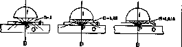
According to the Abbe theory of secondary image formation, light, when passing through the apparatus, produces a series of diffraction spectra, which are deflected from the axis of the bundle more strongly the smaller the interval between the elements of the specimen's structure. Consequently, for the path of these spectra (secondary maxima), the scheme of the drawings provided above remains valid, and a greater number of spectra will pass into immersion systems, and this determines the resolving power of the objective. The same follows from an analysis of the formula for numerical
aperture Ap = n sin a; i.e., by increasing the refractive index of the medium between the specimen and the objective, we increase the numerical value of the aperture, and consequently, the resolving power of the objective. When using immersion objectives with a high aperture (homogeneous), to fully utilize the aperture and resolving power of the objective, it is necessary to use a three-lens condenser (aperture 1.40) and to place a layer of cedar oil between its upper lens and the slide in order to avoid the displacement of the ray parallel to its original direction. The first immersion objectives (water) were introduced by Amici in 1850, and in 1878, according to the instructions of Abbe and Stephenson, the first objective with oil immersion was manufactured at the Carl Zeiss factory. Immersion objectives are usually designated by the length of their focal distance, expressed in fractions of an English inch (1/7, 1/12, 1/16) or in millimeters (3.0; 2.0; 1.5), less frequently by letters (J, D*, PL); recently, the Carl Zeiss factory has been designating all objectives by the number of their own magnification (40, 60, 90).
Related articles
Mentioned in
Cite this page
“Immersion.” Soviet Medical Encyclopedia. English translation of Bolshaya Meditsinskaya Entsiklopediya, 1st ed. (Moscow, 1928–1936), ed. N. A. Semashko. https://sovietmedicalencyclopedia.pages.dev/article/immersion/