ULTRAMICROSCOPY
Historical document, translated for reference. It reflects medical knowledge of the 1920s–30s and is not medical advice.
Summary
Ultramicroscopy is a technique for observing objects smaller than the resolution limit of ordinary microscopes by using dark field illumination and intense lighting to make particles visible as points of light against a dark background.
Encyclopedia article (1928–1936)
ULTRAMICROSCOPY, ULTRAMICROSCOPE. The main task of ultramicroscopy is to create conditions of observation under which it becomes possible to detect objects that lie beyond the limits of visibility in the microscope. The visibility of an object in the microscope (apart from other conditions) depends on its size and the resolving power of the microscope (see). This limiting value of the resolving power is equal to 0.15 μ. From this it follows that it is impossible to construct an objective lens with which, using visible light, one could observe individual objects in the field of view of the microscope at a distance less than 0.15 μ. The largest colloidal particles have a size of 0.1 μ. Thus, the extreme points of the contour of such a particle will be at a distance less than the limiting resolving power, and therefore they will no longer be visible separately, but will merge into one, and the entire contour of the given object contracts into a single point. From this it follows that it is no longer possible to "see" a colloidal particle, i.e., to observe its shape, its contours. Instead of a colloidal particle, one can only hope to see a point without a visible diameter - a signal of the presence of the particle. This is the first principle of ultramicroscopy. Magnification in ultramicroscopy plays no role. In ..... '*' reality, if there is an object ab (fig. 1), then when viewed with the naked eye it will be visible at an angle α; in the microscope its enlarged image a'b' is observed at an angle β. As a first approximation, it can be assumed that the magnification is equal to β/α, but if the distance between points a and b is less than 0.15 μ, then the images of these points a' and b' merge for us into one, i.e., the angle β turns into 0, and the magnification β/α = 0/α = 0 (the physical meaning of this is that magnification in this case has no significance). In an ordinary microscope, it is not even possible to see signals of the presence of particles, individual points, because by the very construction of the microscope, objects in it are viewed against a light background. To make the weak signals of the presence of colloidal particles visible in the microscope, it is necessary to create the same conditions of observation as exist on the night sky, i.e., to make the field of view of the microscope dark. This is the second condition of ultramicroscopy. But many colloidal particles scatter so little light that even on a dark field of view they will not be noticeable. Therefore, it is necessary to illuminate them with the brightest possible light, since with an increase in the intensity of the latter, the brightness of the particles also increases. The brightness of illumination is the third condition of ultramicroscopy.
These three conditions of ultramicroscopy are implemented in practice as follows. Although individual colloidal particles are visible only as points, for ultramicroscopy it is necessary to use objectives with high resolving power so that with a sufficient concentration of the colloidal solution, the images of several particles do not merge together. Dark field of view is achieved in two ways: with the help of "slit" ultramicroscopes and by means of "dark field condensers". In slit ultramicroscopes, the light beam falls on the preparation not from below, as in ordinary microscopes, but from the side (fig. Light is concentrated on the object G with the help of the illuminating objective B and then passes by the observing objective M, the axis of which (and of the microscope tube) is perpendicular to the direction of the illuminating I).
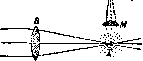
Figure 2.
beam. Thus, the latter does not enter the microscope, and the field of view remains dark. But the beam emerging from objective B illuminates the colloidal particle G, scattering light in all directions (dotted lines), which falls on the front lens of the microscope M. As a result of all this, colloidal particles will appear as bright points on a black background. It should be noted that due to diffraction, the particles appear not as separate bright points, but as a system of concentric black and bright rings, the area of which is many times larger than the actual size of the particle. - If in ultramicroscopes with dark field condensers ^ the third condition - possibly intensive illumination - is fully implemented due to the construction of these illuminating devices, then in slit microscopes the use of bright illumination encounters great difficulty. Indeed, the intensity of illumination of the object depends not only on the brightness of the light source used, but also on the numerical apertures of the illuminating and observing objectives, namely - the intensity of illumination is directly proportional to the product of the squares of the numerical apertures of both objectives. When the numerical apertures of the objectives are not high, then the distance of the object (fig. 3) from the observing objective B2 will be sufficiently large, similarly the focus (intersection of rays) of objective B1 will also be sufficiently far away, and both objectives can be moved so far that the point of intersection of the rays emerging from B1 coincides with the point where the object is located. For the reason mentioned above, it is of course extremely necessary to increase as much as possible the numerical apertures of both the observing and illuminating objectives, but such an increase in numerical apertures entails a decrease in focal lengths, a decrease so
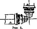

Figure 4.
strong that the frames of the objectives collide before it is possible to make the foci of both objectives coincide. To eliminate this inconvenience, Zsigmondy in 1913 proposed to bevel not only the frames of both objectives, but also their front lenses. This allowed, using objectives even with very high numerical apertures, to move them so close that their foci coincided (fig. 4). With such a construction, the light-gathering power is increased 21 times compared to that of the old slit ultramicroscope. A slit ultramicroscope equipped with such beveled objectives received the name "immersion", since both of its objectives are water immersions. Fig. 5 shows the general appearance of an ordinary (1903) slit ultramicroscope of Siedentopf and Zsigmondy. On a rail (10), mounted
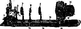
Figure 5.
on a board (15), are installed: an arc lamp (11), an illuminating lens(#), the so-called "slit" diaphragm (8), intended for quantitative measurements, then a second illuminating lens (7), then a stand (1), to which the microscope (5) and cross-shaped slides (6) are screwed, carrying the illuminating objective (2). The microscope has neither a stage nor a mirror. Observation is carried out in a special cuvette (fig. 6), consisting of a glass tube, square in cross-section (1-2), having a funnel (3) on one side and a stopcock on the other. The tube is equipped with two quartz windows: one (2), facing the observing objective, and another, through which a horizontal beam of light from the illuminating objective falls into the cuvette. In the area of the windows, the cuvette tube is blackened in order to absorb direct transmitted rays. Quartz windows are used to eliminate possible illumination of the field of view due to fluorescence, which glass gives. The arrangement of the individual parts of the instrument in im
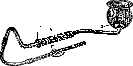
Figure 6.
mersion ultramicroscope is essentially the same. The cardioid ultramicroscope is shown in fig. 7. Light from the arc lamp, before reaching the mirror of the microscope, passes through a special porcelain cuvette with flat parallel glass walls, containing either a solution of Mor's salt or a 0.5% solution of copper sulfate. The purpose of this cuvette is to absorb thermal rays. After passing through the cuvette, the light beam falls
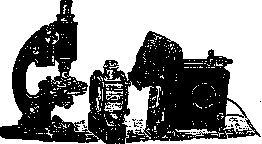
Fig. 7.
on the mirror of the microscope and, after reflecting from it, enters the cardioid condenser, located in the sleeve under the stage of the microscope. From here the light beam enters a special quartz cuvette enclosed in a metal holder and illuminates the preparation. For biological research, such a cuvette is often not used, but ordinary slide and cover glasses are used, but on the condition that the slide glass has a thickness of 1.2 mm.
Related articles
Mentioned in
Cite this page
“ULTRAMICROSCOPY.” Soviet Medical Encyclopedia. English translation of Bolshaya Meditsinskaya Entsiklopediya, 1st ed. (Moscow, 1928–1936), ed. N. A. Semashko. https://sovietmedicalencyclopedia.pages.dev/article/ultramicroscopy/