Tissue Cultures
Historical document, translated for reference. It reflects medical knowledge of the 1920s–30s and is not medical advice.
Summary
This 1930s article from the Great Medical Encyclopedia covers the history, methodology, and applications of tissue cultures, detailing methods for growing living tissues outside the organism in vitro and highlighting key pioneers such as Harrison, Carrel, and Fischer.
Encyclopedia article (1928–1936)
TISSUE CULTURES. Contents: History..................... 74 Methodology and technique.............. 75 General biology of tissue cultures...... 82 Application of the tissue culture method in various fields of biology and medicine . . 90 Tissue cultures (syn.: tissue explantation, tissue cultures), the cultivation of tissues outside the organism, in vitro. The term tissue culture method refers to the methodology and technique whose essence is as follows: a small piece of tissue or organ, freshly excised from the organism, is placed in a suitable medium under conditions such that not only is the life of the tissue preserved (simple "survival"), but true tissue growth (cell multiplication) outside the organism is obtained. Such a method of experimentation, combined with various ways of studying the "tissue cultures" obtained in this way, has proved very fruitful and has found wide application in various fields of cytology, histology, embryology, biochemistry, physiology, pathology, pharmacology, bacteriology, immunology, hematology, radiology, etc. Just as the transplantation of a part of one organism to another (or to another site of the same organism) is designated by the term "transplantation," so the placement of a part of an organism into any other medium, but not into a living organism, is called (according to the term proposed by Roux and introduced into use by Oppel) explantation. Consequently, tissue cultures represent such tissue explants which, in contrast to various other explants, exhibit tissue growth in one form or another. Although explantation of an entire embryo (Totalexplantation), an entire embryonic heart, etc. is possible, in experiments with tissue cultures one ordinarily deals with the explantation of small parts (Teilexplantat, Teilzuchtung). For the isolation and cultivation of tissues, other methods can also be used: one can, according to the method of Ekman, Stöhr, Bautzmann, and others, place a piece of an embryo, a heart rudiment, etc. into a pouch excised from the ectoderm (ordinarily of the amphibian larva Bombinator); in this case, various embryonic tissues grow and develop inside the ectodermal pouch. The isolation and cultivation in vivo of embryonic tissues ("in vivo tissue cultures") is achieved by transplanting a piece of an embryo onto the chorioallantois of a chicken embryo developing in the egg (Murphy, Danchakoff, and others), by implantation into the abdominal cavity, the orbital cavity, etc. (see Interplantation). In what follows, data are presented only on in vitro tissue cultures in the sense indicated above. History. Although individual attempts at cultivating tissues outside the organism were undertaken at various times by many authors (Roux, Born, L. Loeb, Haberlandt, and others), nevertheless R. Harrison should be recognized as the founder of the modern methodology of tissue cultures. He was the first to correctly establish the basic principles of cultivating tissues outside the organism (the necessity of support for cell movement and growth, the small size of the explanted tissues to facilitate metabolism, the necessity of medium sterility and asepsis of all manipulations, etc.) and first (1907) published successful experiments on the cultivation of frog embryo tissues in a drop of coagulated frog lymph. Burrows, working in Harrison's laboratory, improved the method: he began using blood plasma instead of lymph. Subsequent works by Carrel and Burrows (1910), carried out already at the Rockefeller Institute in New York, described tissue cultures from various tissues and organs of various animals, as well as humans. To prolong the growth of tissue cultures, which in initial cultures continued for only 3–15 days, Carrel and Burrows began performing "passages," i.e., they washed the explanted pieces of tissue and then placed them in a new medium. Of particular importance for the further development of successes in this field was Carrel's discovery (1913) of the fact that embryonic extract possesses the ability to enhance tissue growth. The use of blood plasma with the addition of embryonic extract as a medium (which is recognized as the best medium to this day) and the systematic execution of "passages" made it possible for Carrel and others to establish the fundamentally important biological fact that somatic cells of various animals and humans are capable of growing outside the organism for an indefinitely long time. Thus, modern methodology and technique can be called the "Harrison-Burrows-Carrel method" (it is less correct to call them the "Carrel method"). With the help of the passage method developed by Carrel, which are performed every 2–3 days (see below), the growth of tissue cultures prepared at the Rockefeller Institute on January 17, 1912, from the heart of a 7-day-old chicken embryo has been maintained to the present day. During the first fifteen years, about 3,000 passages were performed with these cultures of embryonic fibroblasts. At present, these cultures, living outside the organism for 18 years, show former good growth without the slightest signs of aging and can be considered "immortal."—Albert Fischer in 1921 succeeded for the first time in obtaining a pure culture of epithelium from the iris of a chicken embryo; then pure strains of thyroid epithelium, chondroblasts, monocytes, and some other cells were obtained. A particularly important success was the acquisition of pure strains of malignant tumors: chicken sarcoma (by Albert Fischer in 1924), a pure culture of cancer cells from transplantable Ehrlich mouse adenocarcinoma (by him in 1927), sarcomatous cells in pure culture from 2 rat sarcomas (by Carrel in 1927). The ability to experiment with pure strains of tissues and to perform quantitative accounting of experimental results by measuring the growth rate (Carrel, Ebeling) or by analyzing metabolism (Krontovsky and his co-workers) has made tissue cultivation in vitro a more precise method of scientific research and has contributed to a wider and more fruitful application of the method of such cultures in various fields. Improvement of methodology and technique took place in other directions as well: for physiological studies, Carrel (1923) proposed a new method according to which a piece of tissue is explanted into a two-layer medium (see below) in such a way that tissue growth continuously proceeds in the solid layer of the medium, while the upper liquid layer of the medium is periodically renewed. Maximov (1916) thoroughly developed the histological technique of tissue culture research; Krontovsky (1925) introduced physicochemical and microchemical methods for studying the vital activity and chemical dynamics of tissue cultures, including non-growing ones (e.g., explants from various parts of the brain of adult animals, etc.). Experiments with cultures from tissues and organs of various animals and humans have recently been so widely and so frequently and systematically applied to the study of various issues of biology and medicine that in 1925, with the participation of specialists from various countries, a special international journal was founded, «Arch. f. exper. Zellforschung besonders Gewebezüchtung», and in September 1927, during the 10th International Congress of Zoologists in Budapest, a special section was created dedicated to work in this field. In various states, there are specially equipped laboratories adapted for work with pure strains of tissues (with passage cultures): the laboratories of A. Carrel at the Rockefeller Institute in New York, the department of A. Fischer at the Biological Institute in Berlin-Dahlem, the laboratories (headed by A. A. Krontovsky) at the Bacteriological Institute and the Roentgenological and Radiological Institute in Kiev, the histological laboratories of G. Levi at the Anatomical Institute in Turin. Recently, abroad and in the USSR, similar laboratories are being established at various institutes. Methodology and technique. The equipment of special laboratories and the general setup of work with tissue cultures are carried out either 1) according to the principles of surgical asepsis (a room analogous to a surgical operating room, sterile gowns, caps, masks on the face of the experimenter, a large sterile piece of black fabric on the table as a background, etc.), and then certain manipulations, such as cutting out pieces from the tissue culture for subsequent passage, are performed without any protection against contamination through the air, etc., or 2) in accordance with the principles of experimental bacteriology, when the preparation of tissue cultures, passages, etc., are carried out in an ordinary laboratory room, sufficiently clean (the experimenter can work without a sterile gown, mask, etc.), but all manipulations are performed with strict adherence to the rules of bacteriological cleanliness: cultures are cut under the protection (from airborne contamination) of a Petri dish lid, slightly raised on one side to introduce instruments; liquids are taken according to bacteriological rules, etc. With sufficient skill, good results are obtained when organizing work according to both the first and the second type. In all work with tissue cultures, all glassware, solutions, instruments, etc., must be sterilized, and all manipulations performed aseptically. 1. Basic types of preparation of tissue cultures.
Although tissue cultures can be prepared in various vessels and by different methods, recently two main types of tissue culture preparation have crystallized, which should be used (whenever possible in identical "standardized" conditions, unless the nature of the given case requires special technique): 1) tissue cultures (or explants in general) by the hanging drop type (see figure in vol. V, article 89); 2) tissue cultures in a Carrel flask in a double-layer medium [see figure 1 and separate table (articles 79-80), figures 1 and 2]. These types can be combined. For g
rowth and continuous circulation, in which grow Figure 1. Scheme of a tissue culture in a dish.
Carrel. The tissue is constantly washed by a flowing fluid: some physiological solution, serum, exudate fluid, etc. (apparatuses of Burrow, Romeis, Vasilyev, de Haan, etc.); however, by means of the passage method (see below) or by changing the liquid part of the medium in Carrel dishes, prolongation of life is achieved more successfully, for which reason the aforementioned apparatuses have not found practical application. For the preparation of tissue cultures according to the first type, it is best to constantly use special thick glass slides with a depression of the same size (to facilitate the comparison of results with data from various authors, etc.), namely: 26x76 mm with a round depression of a certain depth (mostly 3-4 mm) with a diameter of 20-21 mm and coverslips or mica plates (which tolerate sterilization better) 24x40 mm. Recently, Carrel proposed preparing simultaneously 4 tissue cultures according to the hanging drop type on a round (5 cm in diameter) mica plate, which is then fastened as a lid on a metal ring (rim), wherein the bottom of such a dish also represents a mica plate (see below). It is possible to place coverslips with cultures in Petri dishes, prepare explants in Gabrichevsky dishes, etc. For cultures of the 2nd type, Carrel dishes (see separate plate, Fig. 1 and 2) with a single neck, with a dish diameter of 3 cm (type D1) or 5 cm (D6), are used as a rule. The solid part of the medium (Fig. 1-a), in which the explanted tissue piece is located (Fig. 1-e), consists of blood plasma mixed with diluted embryonic extract; the liquid part of the medium (Figure 1-b) usually consists of Tyrode's fluid with 5-10% embryonic extract (or of heparin plasma, sometimes of serum). To obtain very slow growth (which contributes to the development of certain histological structures), Fischer sometimes applies a new modification—adds a negligible amount of diluted embryonic extract to the Carrel dish, needed only for better clotting of the plasma, and completely aspirates the liquid part of the medium after washing. In addition to the main types of tissue cultures, a multitude of modifications have also been proposed: growing tissues in test tubes according to the method of Carrel, Rivers, Haagen and Muckenfuss [Figure 2 (a and b—two Рисунок 2. layers of the medium; c—tissue culture)], according to Champy, in a Borrel flask, in special chambers, vessels, etc. 2. Media for tissue cultures. The best medium for tissue cultures up to the present time is considered to be coagulated blood plasma (in which the fibrin network is a good support for moving and multiplying cells), usually mixed with embryonic extract (or with diluted embryonic extract), sometimes with proteoses, etc. Blood plasma is obtained in the following way. Blood is taken by means of an oiled cannula from an animal (most often from a chicken) from the carotid artery or by means of a syringe (lubricated on the inside with vaseline) from the heart of small animals or from a vein in the human arm. Blood is collected into paraffin-coated centrifuge tubes (or under a layer of liquid paraffin) standing on ice. The blood is centrifuged for several minutes in an electric centrifuge (a hand one can be used). The transparent plasma, collected above the settled erythrocytes and leukocytes, is removed and used immediately for preparing cultures or kept in reserve on ice. If working with "heparin plasma" (see Heparin), one can dispense with paraffin coating and cooling. Usually, plasma from an animal of the same species as the cultured tissue is used. However, one can also use heterologous plasmas, successfully growing axolotl tissues, for example, in rabbit plasma (Krontovski and Shustova, Khlopin), or mouse tissues in chicken plasma (Fischer), etc. In general, chicken plasma, according to Carrel's conclusion, is the best medium for growing tissues of various animal species, including mammals (including humans). Since heterologous embryonic extracts can also be used (Carrel), a mixture of chicken plasma with chicken embryonic extract is especially often used for cultivating tissues of various animals and humans. Apparently, the latter can be replaced by a solution of "proteoses" or products (like Witte's peptone) of brief peptic digestion of fibrin or of tissues of various organs (see below). Frequently, plasma alone is also used as a medium, usually diluted, with Tyrode's or Ringer-Locke's solution, or with the addition of tissue extracts from bone marrow (Maksimov), from the spleen (Krontovski), from tumors (Drew), from yeast (Heaton). It must be kept in mind that fibroblasts and normal epithelium can grow indefinitely (with cell multiplication) only in the presence of embryonic extract (even if diluted), whereas monocytes and tissues of certain tumors can grow without it (e.g., in plasma with serum). In certain cases, combined media according to Krontovski can be used. If, according to the research objectives, it is desirable to have tissue cultures in liquid media (since plasma often strongly impedes special investigations, e.g., under dark-field illumination, with various micro-chemical tests, etc.), then Locke-Lewis's medium, embryonic extract alone, hemolymph (Goldschmidt), Drew's fluid (see Drew's solution), a mixture of serum with Tyrode's solution, etc., are used; but to obtain growth, the liquid medium must be distributed in a very thin layer so that the tissue and cells do not float in a drop of liquid, but are pressed against the glass, which will serve as a support; in the indicated liquid media, usually only embryonic tissues grow (from embryos of early stages, e.g., chicken embryos from 5 to 11 days) and only for a short time. The addition of fibrinogen (Ebeling) proved impractical; instead, Carrel has recently been using coagulated plasma (in a Carrel dish) from which the serum has been removed by washing. Embryonic extract is prepared fresh each time: most often a 7-10-day chicken embryo is taken, crushed into pieces, and centrifuged; the obtained embryonic extract is usually diluted with Tyrode's or Ringer-Locke's solution. When preparing media, attention is paid to ensuring that the medium is isotonic (или slightly hypotonic), has an appropriate reaction, etc. 3. Preparation of tissue cultures. Tissues for explantation are prepared under strict aseptic conditions in Tyrode's or Ringer-Locke's solution with sharp instruments (Graefe's cataract knife or small scissors) and cut into small pieces with a diameter most often of approximately 1 mm (0.5-2.0 mm). Depending on various conditions, the dimensions of the explant may vary. For the explantation of blood leukocytes, the procedure is usually as follows: normal or leukemic blood of man or animals is strongly centrifuged, the plasma is aspirated, several drops of embryonic extract are dropped onto the remaining leukocyte layer (located above the erythrocytes); when the leukocyte layer coagulates in the form of a film, it is washed and cut into pieces and explanted, just like pieces of tissue. Cells of exudates can also be used for explantation. Tissue cultures from transplantable animal tumors are relatively easy to succeed with. If the tumor liquefies plasma, it is preferable to cultivate it according to Fischer's method, placing a piece of muscle (or some organ) in the medium next to the tumor pieces. When explanting plant tissues, the material is obtained either mechanically by cutting out small pieces of tissue, or by isolation using plasmolysis. The medium is most often Knop's solution with the addition of glucose, Witte's peptone, etc., to which a little agar is sometimes added: in such a medium Kotte, Shustova, and others observed the growth of pieces of pea rootlets grown sterilely using special techniques. Many authors (especially Haberlandt and his students) explanted pieces of potato tubers, pieces of leaves, etc. 4. The passage method, developed by Carrel, is used to maintain the long-term (unlimited) life of tissue strains, especially in pure culture. Passage is performed in such a way that from a well-grown (usually 48-hour) tissue culture (Figure 3-a), the peripheral parts of the newly grown zone are cut off with four straight incisions, and the remaining middle part (the former piece with a border of new tissue) (Figure 3-b) is washed in Tyrode's fluid, and a new tissue culture (subculture) is prepared from it, which during 48 hours of growth grows again to its previous size (Figure 3-c); then the passage is performed again, and so on. A well-grown culture can be cut into 2 parts (Figure 4-1) and then half of each piece (Figure 4-2) can be explanted.
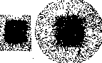

Figure 3. Passage tissue cultures.
tigate in the form of a separate culture (Fig. 4-3); such cultures usually exhibit almost identical growth (Figure 4, growth curves), which makes them a very precise biological reagent for studying all kinds of influences to which one of them is subjected (the other is left as a control). Sometimes it is possible to excise a part of the newly formed zone and obtain further growth from it. Passages are usually made every two to three days; when working with cold-blooded tissues growing at room temperature, significantly less often, for example every 2 to 21/2 weeks. A passage tissue culture that has grown for 24 hours in a thermostat can then be left at room temperature or in the cold for many days (up to 25 days according to Meyer's experiments), and then successfully passaged. This circumstance made it possible to transport or send such cultures by mail over very great distances. 5. Investigation of tissue cultures. a) Observation of living cultures. Microscopic observation of living tissue cultures is of particularly great importance: brief examination is performed simply at room temperature; for prolonged observation of amoeboid motility, cell division, rhythmic contraction of tissue cultures from the heart, etc., the explants are placed on a warming stage, or the microscope along with the culture is placed in a special thermostat box. Observation under dark-field illumination provides an invaluable service, because mitochondria (chondriosomes) are visible in the living cell under it, and the appearance of various changes in the structure of the cytoplasm and nucleus is noticeable, etc. [see separate plate (Vol. XIV, Art. 375-376), Figures 6 and 7]. To obtain clear images (under dark-field illumination), it is necessary to mount the preparation in a special way or to use Peterfi's Preparier-Wechsel-Condensor (Zeiss). Excellent images are obtained using vital staining methods. Drawing and microphotography are generally performed in the usual manner. For microcinematography, which makes it possible to trace the finest details of the movement of normal and cancer cells, their reproduction, etc., relatively simple equipment has recently been proposed. Tissue cultures are also particularly suitable for various microoperations on cells, for micrurgy (see), because during the operation the cells are, so to speak, in more "physiological" conditions than with other methods, and after the operation the cell can be followed in tissue culture conditions for a long time; for recording the microoperation process and its consequences, a photographic eyepiece (for example, the Zeiss "Phoku") is particularly suitable. To carry out micrurgy (see) in tissue cultures, some special techniques and devices developed by Peterfi, Krontowski, and others are necessary. b) Fixing and staining of tissue cultures for cytological and histological purposes. To obtain microscopic (whole-mount) preparations from entire tissue cultures [which, to obtain good preparations, must be prepared beforehand (during the preparation of tissue cultures) sufficiently thin, not containing a thick layer of plasma, or prepared in liquid media], Maksimov recommends fixing the cultures in Bouin's fluid (for thin preparations - Zenker's fluid) and staining with Delafield's haematoxylin. Carrel and his pupils usually use formalin. Fischer fixes tissue cultures in 2% formalin (in Ringer's solution) or 2% formalin-alcohol, stains with haematoxylin, washes, and then brings them to Canada balsam via alcohol, acetone-xylene, and xylene. To obtain stained sections, Maksimov applies fixation of tissue cultures almost exclusively in Zenker-Formol (15-20 min.) and rapidly embeds them in celloidin. Levi particularly recommends Maksimov's fluid of the following composition for fixing tissue cultures: to 8 cm3 of Zenker's stock solution, add 1 cm3 of pure formalin and 1 cm3 of 2% osmic acid. Fix for 2-6 min. After fixation, embedding in celloidin or celloidin-paraffin according to Peterfi (in paraffin for no more than 15 minutes) and staining with Heidenhain's iron haematoxylin. Based on careful studies by Strangeways and Canti (see below) devoted to the study of changes in living cells upon fixation, Niven preliminarily exposes tissue cultures to the action of a 2% osmic acid solution, then fixes them in Zenker's fluid (without acetic acid) and stains with iron haematoxylin. For special purposes, appropriate fixation and staining methods are used. c) Biological and physiological methods. When investigating the vital activity of tissue cultures and applying them to the study of various questions of biology and medicine, quantitative methods are of particularly great importance. The vital activity of tissue cultures can be measured in various ways: by the intensity of metabolism, the rate of growth, the number of mitoses, etc. Since the increase in tissue mass of a growing culture cannot be measured directly (due to the difficulty of weighing), it is judged by the increase in the surface area of the tissue culture, and the growth rate is usually expressed according to Ebeling's method: the contours of the just-explanted piece are drawn (using a projection drawing apparatus) (Fig. 4-2) and the surface area (A0) is determined using a planimeter after 48 hours of growth (or generally during time t), the contours of the entire expanded culture are drawn again (Fig. 4-3) and the total area (At) is determined, so that the newly formed zone will equal At - A0. Then the Ebeling index ("relative increase" of the tissue culture) = (At - A0) / A0. When studying various influences, the ratio of the growth index in a given experiment (E) to the growth index of the control culture (K) quantitatively expresses the effect of the studied influences on growth. Instead of the "relative increase" according to Ebeling, according to Buch-Andersen and Fischer's calculations, it is more correct to use \u221aA or log \u221aA, where A is the surface area of the grown culture. To judge the intensity of metabolism, either the consumption of certain substances—sugar, oxygen, etc.—or the formation of certain products—lactic acid, an increase in medium acidity, a change in pH (Krontowski)—is determined. Sugar consumption (preferably together with the determination of lactic acid formation) proved to be a particularly convenient indicator of metabolic intensity in explants from various tissues, growing and non-growing (e.g., from different parts of the brain), an indicator of changes in vital activity under the influence of radium rays (Krontowski), changes in K and Ca content (Yatsimirskaya-Krontowskaja), etc. To determine the vital activity of explants according to Krontowski's method, substances (e.g., glucose and other carbohydrates, tributyrin, etc.) are added to the medium which, under the influence of tissue vital activity, are capable of decomposing with the formation of products causing a change in the reaction of the medium, and after a certain period, the change in pH is measured. To study the effect of different tissues on growth ("blastotropic" influences), on growth polarity, etc., Centanni applies the method of opposed ("affronted") explants: in a single drop of medium at a distance of approximately 1 mm, 2 pieces of identical or different tissues are placed. Another method of Centanni: at the same distance from the explanted piece, a small glass capillary filled with the test liquid is fixed. Among physiological methods, one can note Krontowski's method of recording contractions of heart explants and movements of ciliated epithelium on a kymograph tape, the standardization of pharmacological substances by the cessation of contractions of cardiac explants, peristaltic movements of intestinal pieces (Fischer), etc.
In general, modern technique provides the experimenter with the most diverse opportunities, makes it possible to prepare tissue cultures from various animal and human tissues, to obtain very intensive uniform growth (e.g., in passage tissue cultures or in a Carrel flask) or cultures with very slow growth (according to Fischer's modification), to obtain proliferation of a comparatively simple homogeneous ("pure") tissue (so-called cytotypic or histotypic growth) or the growth and development of complex, organ-like or even organism-like explants (see below) and to investigate tissue cultures by various morphological and physiological methods. When working with the tissue culture method, a critical attitude toward the studies performed and caution in conclusions are required, since some phenomena proceed both in tissue cultures and in the organism in generally identical ways (manifestations of vital staining, behavior of connective tissue and blood cells and their further transformation, and so on), while others proceed differently (e.g., the realization of the effect of X-rays in the organism compared to tissue cultures, the not always identical reversibility of damage inflicted on cells in the organism during transplantation and outside the organism in explants, and so on). Both for the proper setup of experiments and for obtaining correct conclusions, appropriate knowledge of the features of tissue cultures, a correct assessment of the advantages and disadvantages of the technique, and knowledge of the limits of application of the method are necessary. In appropriate cases, it is desirable to introduce other research methods in parallel. General biology of tissue cultures. — 1. General morphology. If a piece of a chicken embryo is explanted, then already after a few hours at the edges of the piece into the surrounding medium in large numbers (mostly radially) individual connective tissue spindle-shaped or process-bearing cells begin to grow in ("connective tissue type of growth"), so that already after 48 hours around the piece there is usually formed a wide (see figure 3-a and c and sep. tab., fig. 3) newly formed zone, the so-called "growth zone" (or "invasion zone" — zone of penetration). Epithelium usually grows in the form of a continuous thin and wide sheet, a membrane ("epithelial type of growth" — see sep. tab., fig. 6), consisting of epithelial cells (see separate table, fig. 4). Epithelium sometimes forms cords, lines the walls of a cavity, often formed in plasma. According to Rubashkin and Shakhov, growth in the form of a symplast (continuous layer) is characteristic of epithelium in general, and syncytial growth is characteristic of connective tissue; in both the one and the other, individual cells can become isolated. Mixed growth frequently takes place in cultures. Sometimes the outgrowth of muscle fibers with a multinuclear expansion at the end (as during regeneration) and of nerve fibers, the peripheral end of which is expanded and provided with the finest, thread-like mobile amoeboid processes, is observed. Upon explantation of pieces of the spleen, lymph nodes, bone marrow, leukocyte film, and the like, already after a few hours various wandering cells — lymphocytes, leukocytes, reticular cells, and the like — emigrate from the piece into the surrounding environment, so that at first the "growth zone" consists only of emigrated cells; only later do spindle-shaped fibroblasts begin to grow from the piece, and then the actual multiplication of cells sets in, mitoses appear in hypertrophied reticular cells (macrophages), in fibroblasts, and in other cells. In general, one should distinguish between more complex phenomena developing inside the explanted piece itself (in the case of a gland — phenomena in glandular parts, excretory ducts, proliferation of interstitial tissue, processes of progressive and regressive character, reaching necrosis in the center of the piece, and the like) and external proliferation (in the medium) around the piece of a simpler epithelial sheet (see sep. tab., figs. 4 and 6). In certain cases (for example, in the cultivation of early-stage rabbit embryos), the usual growth of cultures — the ingrowth of tissues into the surrounding environment — does not occur at all, but the pieces of the embryo round off, their surface becomes overgrown by epithelium (see sep. tab., figure 5, upper and side parts), internal tissues sometimes reveal organoid development; a peculiar growing organism is obtained as it were (often resembling a teratoma). Maximow proposed to call such explants "organotypic" or "organospecific" in contrast to ordinary ones characterized by "histotypic" or "cytotypic" growth. 2. Tissue culture as a biological system. A tissue culture is not a simple aggregate of individual independent cells, but represents a peculiar biological system. Individual (isolated) fibroblasts, for example, and epithelial cells, judging by the experiments of Fischer, Policard, and others, cannot multiply: for their proliferation a certain combination, a certain minimum number of cells is necessary [Fischer connects this circumstance with the presence of hypothetical desmons (see), Burrows with a certain concentration of hypothetical substances — "archuxy" and "erguxy", and so on]. Upon the fusion of two tissue cultures from pieces of the heart with two different contraction rhythms, a culture with one common rhythm is obtained (experiments of Fischer, Olivo), so that a unified system is formed not only in morphological, but also in functional relation. Regarding the question of the connection of cells with each other in tissue cultures, it should be noted that many authors have described syncytial formations in them and pointed out that from these syncytia under certain conditions individual cells easily become isolated, liberated, and emigrate into the medium. The opinions of authors on the nature of intercellular connections diverge: some believe that the cell processes only tightly adhere (stick) to one another (Lewis), so that different granules, for example, during their movement, do not pass from cell to cell (de Garis), whereas according to others there is a direct protoplasmic connection between cells (Fischer). The results of experiments with cell damage by a micromanipulator needle in some cases (e.g., epidermis epithelium) speak in favor of a direct connection (Chambers), in others against this (Kredel). In those cases when it is impossible to notice boundaries between neighboring cells, the individuality of the cells in the biological (physiological) sense, according to Levi, is nevertheless preserved, since each nucleus usually has its own sphere of influence. In complex tissue cultures containing different tissues, as well as upon the fusion of pure cultures of different tissues (for example, epithelium and connective tissue, and the like), the influence of tissues on each other (intertissue correlations) is expressed quite clearly. In organotypic explants, a number of phenomena having the character of regulation (organization) are observed: Maximow described the delimitation of the surface of embryonic explants from the surrounding environment by an epithelial cover (see sep. tab., fig. 5), tissue transformation, elimination by the epithelium of degenerated cells with subsequent restoration of the defect, and so on. Rounded, epithelialized, self-contained explants were observed by Smirnova upon explantation of axolotl digits, Timofeevsky and Benevolenskaya — of the terminal parts of human embryo extremities, Fischer, Khlopin — of pieces of the embryonic intestine, mucous membranes of the urinary bladder, Umela, Gasul, and others — of pieces of the frog pharyngeal mucosa, Murray — upon explantation of pieces of planarians, and so on. Upon the cultivation of blood leukocytes of various animals (vertebrates, as well as invertebrates) and man — monocytes (amoebocytes and the like) and a part of lymphocytes (granular leukocytes quickly perish) transform into macrophages (see), ultimately even into fibroblasts, and in cultures of guinea pig leukocytes Maximow described the formation of true connective tissue with argyrophilic and collagenous fibers (experimental synthesis of tissue from individual cells). In other (lower) animals, not only tissues, but even an entire organism can be obtained from individual separated cells: in the experiments of Galtsoff and others, rounded agglomerates formed from individually artificially dissociated cells of sponges (Microciona prolifera and others), which developed into real small sponges, sometimes (upon the destruction of the surface membrane) becoming similar to ordinary tissue cultures. By means of the joint cultivation of pure strains of different tissues, biological formations (units) of a higher order are obtained, for example, resembling glands (Fischer, Parker, and others) and the like (see sep. tab., fig. 7). In tissue cultures from embryos, it is also possible to observe the development of a neural sympathetic network encompassing entire cellular areas (Maximow), and the attachment of grown nerve fibers to the elements of a neighboring piece of muscle tissue (Lavrentiev, Grigoriev). Thus, the method of tissue cultures makes it possible not only to dissect the living organism — to obtain and analyze the pure races of cells making up the organism, but also to experiment in the synthetic direction (in the sense of the experimental "synthesiology" of Heidenhain).
As regards the problem of differentiation and dedifferentiation (retrograde development), Champy advanced the proposition that in tissue cultures the epithelium from various organs undergoes dedifferentiation, turns into indifferent epithelium, and subsequently the epithelial cells become indistinguishable from cells of connective tissue origin. This proposition is disputed by many authors (Maksimov, Khlopin, Fischer, and others). At the present time it has been established beyond doubt that in tissue cultures from embryonic fragments, whole limb rudiments, eye rudiments, the auditory vesicle, and the like, differentiation is observed: the ectoderm exhibits keratinization, the formation of skin papillae, rudimentary mammary glands, and abortive hair follicles (Maksimov); the formation of cartilage and bone in vitro has been established (Fell), as well as of various parts of the eye (Fell, Filatov), functional (the appearance of rhythmic contractions) and subsequently histological differentiation of the cardiac muscle (Olivo), and so on. Biochemical differentiation has also been discovered: when explanting embryonic thighs that do not contain phosphatase, along with the normal development in vitro of typical zones of cartilage, upon the formation of hypertrophied cells, phosphatase also appears in large quantities (Fell and Robinson), an enzyme participating apparently in the process of ossification. Of course, the liver rudiment, for example, merely by virtue of spatial, mechanical, and other conditions, does not develop into a real liver—only the proliferation of embryonic liver cells, blood capillaries, and the like is observed (Maksimov, Benevolenskaya). It has also been established that pure strains of epithelium retain their basic epithelial properties for an indefinitely long time, furthermore, the epithelium of the thyroid gland, for example, can produce colloid for more than 4 months of life and growth outside the organism; the iris epithelium can form pigment, and so on. On the other hand, morphological dedifferentiation, a known simplification of structure, is also observed in tissue cultures: epithelial cells of the prostate with special secretion granules (Champy) or the structure of glandular cells characteristic of different sections of the pancreas (Khlopin) gradually lose their organspecific glandular structure in tissue cultures, and the epithelial sheets grown from different parts of the gland yield an identical indifferent epithelium (Khlopin). Dedifferentiation is also observed in explants from the heart (Olivo and others). It must, however, be borne in mind that various changes (transformations) in the shape and character of cells in many cases may not pertain properly either to differentiation or to dedifferentiation, but depend on surrounding conditions, on the properties of the medium (Lewis, Barta, and others), on enhanced proliferation, and the like. Upon a change in conditions—upon combination in vitro with another tissue and the like—the epithelium, as indicated, again exhibits a polar, characteristic structure, and so forth (Maksimov, Drew, Fischer, and others). The addition of connective tissue promotes the differentiation of epithelium (Drew), neuroblasts (Lavrentiev, Grigoriev). The fusion of cultures of a strain of embryonic fibroblasts and epithelium cultured separately for a long time, with the epithelium growing out in the form of an indifferent epithelial sheet, yielded formation resembling a gland (see sep. tab., fig. 7), with the typical arrangement of epithelium and connective tissue (Fischer and Ebeling). Moreover, obtaining more differentiated structures is also successful in experiments with a pure strain: Fischer and Parker, with intensive growth of cultures obtained from the perichondrium of a chicken embryo, observed morphological dedifferentiation, and with artificially slowed growth, the appearance of more differentiated tissue with intercellular substance. Obviously, enhanced proliferation hinders differentiation, and this is also observed in the organism: during embryonic development, periods of enhanced growth alternate with periods of differentiation (Schmalhausen and Stepanova). The conditions of life for cells and tissues in explants are peculiar, but in many respects similar to those that take place in the organism during regeneration, wound healing, tissue new formation, inflammation, and the like (Strangeways, Maksimov). In accordance with this, in explants (as in the organism under various conditions) there occurs an energetic mobilization of resting wandering cells (histiocytes), reticular cells that become free, blood monocytes, hypertrophy of all these cells, and their transformation into "macrophages" ("polyblasts" of Maksimov). The most diverse cells—including pigment cells (in explants from organs of the frog and axolotl), chloragogen cells (in cultures from earthworm tissues), neuroblasts (in embryonic cultures), and the like—acquire amoeboid mobility (it is clearly noticeable also at the edge of the growing epithelial sheet, at the terminal expansion of the growing nerve fiber, and so on) and emigrate into the medium. Division is undergone by various tissue cells, leukocytes of normal and leukemic blood; sometimes even cells that normally do not exhibit division in the organism begin to multiply, for example, Müller cells of the retina (Champy). Myasoedov observed the cleavage of the egg in explanted ovarian follicles of rabbit ovaries, which Champy later described under the name of "experimental parthenogenesis in the rabbit." 3. General type of chemical dynamics of tissue cultures. Diverse cells and tissues in explants (as in the organism during regeneration, wound healing, productive proliferative inflammation, and the like) exhibit (as indicated above) high activity. The source of energy necessary for all this serves mainly sugar (glucose), as shown by the studies of Krontovsky and his associates Bronstein, Yatsimirskaya-Krontovskaya, Kolomiyets, and others (confirmed now already by many other authors). The consumption of sugar in tissue cultures is so significant that a small piece of tissue (the surface of which is approximately equal to 1 mm2), constituting by weight only about 1/500–1/1000 of the entire medium, absorbs up to 60% (or even more) of all the sugar contained in the medium within 48 hours (fig. 5). If the sugar content in the medium is artificially (e.g., by dialysis) lowered below a certain level (0.005% according to Wind), tissue cultures do not exhibit growth; the addition of sugar to such a (dialyzed) medium makes it suitable for growth again. In the most diverse tissue cultures (both on mica and in Carrel flasks, even in an atmosphere of oxygen), along with the disappearance of a large amount of sugar, a significant increase in the amount of lactic acid is always discovered. Thus, normal tissue growing well in vitro covers a significant part of its energy requirement through fermentation (which, according to Warburg's theory, is exclusively characteristic of the malignant growth of carcinomas and sarcomas). Since upon muscle excitation (and in some other cases) the anaerobic phase of carbohydrate splitting also appears first, and excitability (irritability) represents a general elementary property of living protoplasm, Krontovsky put forward a theory according to which, upon the excitation of various tissues, first of all the indicated general energy mechanism is set in motion, a shift is observed toward the intensification of fermentation processes, i.e., processes that easily yield energy, while energy, depending on various conditions, internal structure, and so on, goes either to muscle contraction or to processes of enhanced activity of cells and tissues during regenerative processes, in tissue cultures in vitro, during wound healing in the organism, during inflammation, and so forth. In various experiments with tissue cultures, for the correct understanding of many phenomena, it must be borne in mind that the medium does not remain constant: already during the 2-day growth of embryonic cultures, the sugar content drops sharply (fig. 5), the amount of lactic acid increases, CO2 accumulates, a pH shift toward the acidic side occurs (in hanging-drop cultures, the medium pH is initially around 8.6, then over the course of 2 days it moves to 7.0–6.8 and further to the acidic side; in Carrel flasks, when measured with a special electrode, the pH of the tissue piece initially = 7.2, then 7.0–6.8, and of the medium—8.1–8.3), and so on. The accumulation of CO2 occurs partly due to tissue respiration, but mainly as a result of the displacement of CO2 from bicarbonates by the formed lactic acid (as shown by Magath in direct experiments with the addition of a corresponding amount of lactic acid to a freshly prepared, hermetically sealed culture). By means of ventilating cultures in a new (metal) Carrel flask with 2 side tubes (see above) or by simply temporarily lifting the mica with the culture of the hanging-drop type (Magath), the reaction can be shifted back to the initial, more alkaline point, and blowing air with an admixture of 5% CO2 into the Carrel flask can slightly acidify the fresh, somewhat alkaline medium (pH around 8.0); in this way it is possible to a certain extent to make the medium more stable in the indicated respect. 4. Analysis of cell growth and nutrition.
The phenomena observed in tissue cultures—enhanced sugar decomposition with the formation of lactic acid in the presence of respiratory insufficiency, the displacement of CO2 by lactic acid from bicarbonate, leading to acidification when CO2 retention occurs—can serve as a prototype, easily accessible to study, of a number of phenomena in the organism: during ischemia, inflammation, regeneration, wound healing, transplantation, and the like, and help to understand their mechanism. Cultures of a pure strain of "fibroblasts" (mesenchyme cells) and other cells proved to be an excellent biological (physiological) reagent, allowing the discovery and study of a number of factors promoting or inhibiting tissue growth. Prolonged, unlimited growth of fibroblasts and epithelium is impossible (according to Carrel's experiments) in plasma or serum alone; however, in the presence of embryonic extract in the medium, these tissues can feed and synthesize their protoplasm for an indefinitely long time. Long-term reproduction of monocytes, lymphocytes, and cells of certain tumors is also possible in serum. Growth-promoting substances are contained not only in embryonic extract, but also in other tissue extracts (from the spleen, from Rous sarcoma, etc.), but only in smaller quantities. According to Carrel, special nitrogenous nutrients, which he called "trephones," are present in the protein fraction of embryonic extract. Carrel, Baker, Fischer, Wright, Heaton, and others thoroughly investigated their nature. It turned out that the initial stages ("proteoses") of pepsin digestion (incomplete hydrolysis) of various proteins—fibrin, pure crystalline albumin, crystalline edestin, various organs (liver, pituitary gland, etc.)—significantly enhance tissue growth in vitro, sometimes no worse (according to Carrel, even better) than embryonic extract. According to Carrel, the effect of embryonic extract depends on the fact that the splitting of the extract's proteins into proteoses easily occurs within it through cellular enzymes; according to Fischer, the effect depends not on chemical substances, but rather on a special physicochemical state. If the medium is composed of digestion products of pure substances (crystalline albumin or casein), the addition of glycine and nucleic acid, which are missing in this mixture, improves growth (Carrel, Baker, Ebeling). When studying the significance of the oxidation-reduction potential for tissue cultures, Baker found that cultivation in a synthetic medium (a mixture of casein products, glycine, and nucleic acid) yields better results upon the addition of glutathione and hemoglobin (or liver ash). Serum exerts an inhibitory effect on cultures of fibroblasts and epithelium and shortens their life (Carrel). Further research led Carrel to the conclusion that a certain amount of an antagonistic principle promoting growth is also present in serum. The inhibitory effect of serum increases with the age of the animal (Carrel and Ebeling). According to Carrel's theory, cell nutrition and growth depend, on the one hand, on the presence in the fluid surrounding them of two groups of antagonistic substances (growth-promoting—"trephones"—and growth-inhibiting), and on the other hand, on the intrinsic energy of the given tissue, which is greater in tissues of embryos and young animals and less in old ones. An idea of the intrinsic energy of tissue growth can be obtained (according to Carrel) by measuring the so-called "residual growth energy," i.e., the duration and rate of culture growth in a medium lacking nutrients. According to Carrel, the growth curve of fibroblasts and epithelium (Fig. 6) in a nutrient medium containing embryonic extract is similar to a parabola; in a medium inadequate for nutrition, it has an S-shape. Based on mathematical analysis of various actually obtained curves, Buch-Andersen and Fischer believe that growth curves of the first type were not observed at all; a greater or lesser successive deceleration of growth is always noted. Since, according to Carrel's experiments, leukocytes in vitro can produce growth-promoting substances from serum ("leukocytic trephones"), leukocyte cultures are capable of exerting a rejuvenating effect on neighboring, degenerating fibroblast cultures, etc., and since similar products are apparently also formed in the organism (they were found in extracts from inflamed tissues, in peritoneal exudate with macrophages, etc.), Carrel put forward a hypothesis according to which white blood elements, especially lymphocytes and monocytes as he elucidated later, act in the organism as mobile unicellular glands capable of nourishing fibroblasts and epithelium. And as a result, it is precisely these cells that play a major role in wound healing, inflammatory proliferation, and so on, since tissue growth depends on a certain concentration in the surrounding environment of "trephones," which are secreted by white blood corpuscles and also released from dead tissues (muscles, etc.). Many other factors are also necessary for tissue growth: for example, a specific reaction of the medium (for fibroblasts, the optimum pH, according to Fischer, is 7.4–7.8), a specific combination of electrolytes [changes in the content of K and Ca sharply affect both growth and metabolism of explanted tissues (Yatsimirskaya-Krontovskaya)], a known osmotic pressure, a specific temperature, sugar (glucose) as an energy source, and so on. 5. Main types of tissue cultures and their life cycle. Based on living conditions and their life cycle, 3 main types of tissue cultures can be distinguished. 1) Tissue cultures (explants) with a simple elementary life cycle: a small piece of tissue (about 1 mm across) is placed in a drop of medium (e.g., plasma with embryonic extract); in this case, neither washing of the tissue cultures to remove metabolic products nor the addition of new medium is performed. Under these conditions, tissue cultures grow (at body temperature) usually for 5–15 days (longer depending on various conditions; tissues of cold-blooded animals, for example, at room temperature for more than a month). Then the cells degenerate, and the culture dies. 2) Tissue cultures whose life is prolonged by renewing the medium (or upon explantation in a two-layered medium in a Carrel flask by renewing its liquid part), while the growing part itself remains untouched (Figure 1). The growth of fibroblasts continues for about 15 days, with their colonies reaching large sizes—2–3 cm in diameter; the growth of monocytes (macrophages)—thirty days or more (Carrel). The growth curve of the epithelium is depicted in Fig. 6. 3) Tissue cultures whose life is maintained by constant passages for an indefinitely long time (passaged, "eternal" cultures). During passages, not only is the explant washed and placed in a new medium, but in addition, during each passage, the peripheral parts are cut off (Figure 3); consequently, a constantly renewed regeneration is observed, which, in Krontovsky's opinion, is of fundamental importance. This circumstance was compared by Krontovsky with some analogous phenomena: repeated amputation in Child's experiments with planarians, and Hartmann's with amoebas and other protozoa, acted in a rejuvenating manner, and the experiments could be continued for an indefinitely long time without aging of the individuals. In connection with this, Krontovsky also explained the fact that an 18-year-old Carrel tissue culture of fibroblasts still grows with its former activity, appearing as if forever young; in reality, during 2–3 days of growth it ages somewhat, but with each passage it artificially returns to its initial state again, which can be repeated for an indefinitely long time. The circumstance that speaks in favor of such an explanation is that if passages with the cutting off of peripheral parts are not performed, the growth of the culture gradually slows down, the cells transform into more mature forms, and special histological differentiated structures appear—various kinds of fibers and the like (Maximow, Fischer, Parker). The growth curve of "passaged cultures" is given in Figure 4, sugar consumption in Figure 5. Application of the tissue culture method in various fields of biology and medicine. The method of tissue cultures is successfully applied in the most diverse fields of biology and medicine. 1. Cytology, histology, pathology. The trends of modern cytology—first of all, to study objects whenever possible in a living and least altered form [fixing, embedding, and the like inevitably cause substantial changes in the complex colloidal structure of living protoplasm (see Histological technique)]—are best met precisely by the method of tissue cultures, since the most reliable proof of a living (undamaged) state is tissue growth and cell proliferation (Péterfi). The structure of various living cells in tissue cultures has been studied in great detail by many authors (under ordinary and dark-field illumination) [see separate table (vol.
XIV, col. 375-376], figure 6], changes in the cytoplasm and nucleus of cells that easily occur under any unfavorable conditions (the appearance of a granular or other structure in the nucleus, which is initially "optically empty," a change in mitochondria and the appearance of various granules in the cytoplasm, etc.), upon a change in the reaction of the medium (Lewis), osmotic pressure (Hogue), under the action of X-rays and radium (Strangeways, Canti, Donaldson, Spear), etc. In living active cells, systematic recording revealed (Levy, Strangeways, Canti, and others) the motility of mitochondria (chondriosomes), constant changes in their size and shape (Figure 7); peculiar "vermiform" movements of mitochondria are clearly visible in the remarkable film by Canti demonstrated by Krontovsky at the Congress of Zoologists, Anatomists, and Histologists in Kiev in 1930; various details of cell division under different conditions have been studied, etc.; in the aforementioned film, the stage of mitotic cell division characterized by a sort of "bubbling," the formation of pulsating bubble-like protoplasmic protrusions, and other details of cellular dynamics is particularly well noticeable. Observations of living cells at the moment of action of fixing liquids on them made it possible to study in detail in what respects and to what extent the microscopic picture of the cell changes under various fixations. On a separate plate (vol. XIV, col. 375-376, Fig. 6), it can be seen, for example, how an "optically empty" cell nucleus (even under dark-field illumination), in which only the nucleolus is noticeable, acquires a peculiar structure upon the action of Zenker's mixture, becoming matt-granular [see separate plate (vol. XIV, col. 375-376), Figs. 6 and 7], how the initially structureless background of the cytoplasm changes, in which fat droplets and filamentous chondriosomes are clearly visible in the living cell, how the general contours of the cell are smoothed out after fixation, the periphery of which in the living state possessed a mass of the finest amoeboid, motile pseudopodia, etc. (Strangeways and Canti).
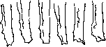
Figure 7. Movement and change of mitochondria in a cell process (under dark-field illumination) over the course of 5 minutes.
Thanks to the tissue culture method and "pure strains," individual cell types are currently receiving not only a morphological characterization (which formed the basis of "classical cytology"), but also a physiological, dynamic one—according to the peculiarities of their movement, growth rate, nutrition, reactions to influences, and behavior under different conditions, etc., which, according to Carrel, is the distinguishing feature of "new cytology." Studies on cell morphology in tissue cultures under various conditions have necessitated a critical attitude toward the habitual notion that cell shape is something frozen, unchanging, and characteristic of cells of a given species: a change in the consistency of the medium (Uhlenhuth), tension (Weiss), etc., is sufficient for a typical epithelial cell to turn into a spindle-shaped or stellate one (Uhlenhuth, Khlopin), making it look like a fibroblast ("morphological convergence"); however, it retains its nature in the process and, placed in other conditions, acquires the typical epithelial shape again (see above the experiments of Fischer and Ebeling, Drew, etc.), which is why great caution is necessary when judging (on the basis of microscopic preparations) the conversion (transformation) of cells of one type into another. In tissue cultures, it is possible to systematically study changes in living cells during their differentiation and to trace the colloid-chemical changes of the protoplasm occurring in the process. In experiments on spermatogenesis in vitro (with cultures from the testes of the butterfly Samia cecropia), Goldschmidt, as shown in Fig. 8, observed step by step the development in vitro of all stages of sperm formation and was able to experimentally study certain factors of spermatogenesis. As was established, the formation of the axial filament begins with the appearance of many protoplasmic protrusions, peculiar pseudopodia, on the surface of the cell facing the lumen of the follicle (Figure 8 a-b). Then one of these protrusions (Figure 8 b) grows and after some time suddenly turns into a fixed filament (Fig. 8 c) with a thickening at the end, which is caused by a rapid change in the colloid-chemical structure, which Goldschmidt also managed to induce artificially. Then a second filament is formed (Fig. 8 c), and further...
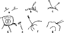
Figure 8. Spermatogenesis in vitro.
further development occurs, the course of which can be sharply influenced by changes in osmotic pressure and the like (Goldschmidt). The tissue culture method has also yielded very valuable results in clarifying the problem of the transformation of connective tissue and blood cells, which is of special interest to a whole series of disciplines: cytology, histology, developmental mechanics, pathology, and clinical hematology. The changes in histiocytes (wandering cells) and their transformation into hypertrophied, energetically phagocytosing polyblasts (macrophages), as well as the transformation of blood monocytes and part of the lymphocytes into such macrophages (part of the lymphocytes perish), have been traced, which is observed both in cultures from blood leukocytes (Avrorov and Timofeevsky, Maksimov) and in cultures from lymphocytes obtained from the thoracic duct (Bloom). At first, hematogenous polyblasts (macrophages) differ from polyblasts of tissue origin (both in explants and in the organism in inflammatory foci); then they become completely similar (Maksimov, Zilberberg). Later, during the formation of scar tissue, macrophages of both origins transform into fibroblast-like cells and finally into true fibroblasts (fibrocytes), while in vitro and in vivo true connective tissue (argyrophil and collagen) fibers are formed, as Maksimov, Bloom, and others have shown. These observations also clarified the controversial question of the origin of fibers. Granular leukocytes, emigrating from an explant fragment or having emigrated from vessels during inflammation, are incapable of further development and gradually die. The reverse transition of fibroblasts into histiocytes has been described by some authors (Carrel and Ebeling, Fischer), but this referred to embryonic fibroblasts, and these cases were not subjected to thorough cytological analysis. Immature mesenchymal cells (resembling fibroblasts) can undergo further changes, whereas fibroblasts represent a mature form incapable of further transformations (Maksimov). The endothelium, according to Maksimov, behaves similarly to fibroblasts; according to Lewis, it forms a reticular tissue similar to mesenchymal cells. Analogous changes in wandering cells—histiocytes, monocytes, and lymphocytes—were established in tissue cultures from various organs and blood leukocytes even during the formation of a tubercular tubercle in vitro (Maksimov, Timofeevsky and Benevolenskaya, and others). Such experiments were carried out both with virulent tubercle bacteria and with the low-virulence BCG strain. At the same time, Lewis and others managed to observe directly under the microscope step by step many controversial phenomena, for example, the formation of multinucleated giant cells by the fusion of polyblasts (Fig. 9—sketches every 10 min.). Timofeevsky described a similar behavior of cells in tissue cultures from human leprous tissues, and he had the opportunity to observe the formation in vitro of typical "leprosy cells" packed with leprosy bacilli. Granulopoiesis and erythropoiesis in tissue cultures were described by Maksimov, Benevolenskaya, and others. Systematic studies of the aforementioned cycle of questions made it possible for Maksimov to create a general doctrine on the morphology of mesenchymal defense reactions of the organism against local and general harmful agents and to give a detailed outline of the histogenesis of various inflammatory reactions. The tissue culture method makes it possible to study the basic properties of blood cells, their prospective potencies, etc., in various forms of leukemias (Avrorov, Timofeevsky, Hirschfeld, and others). The epithelium of various organs (Champy, Khlopin) and mesothelium (Myasoedov) have been less studied. According to Khlopin, epithelial tissues can be divided into 3 groups: 1) epithelium of the stomach, intestine, pancreas, which is characterized by single-layer sheets; 2) epithelium of the skin, oral cavity, etc., with a tendency to form multi-layer structures, cell complexes of the "pearl" type, etc.; 3) various epithelia of mesodermal origin (kidneys, serous membranes, etc.). The development of smooth muscle fibers (Lewis), nerve fibers (Levi, Olivo, Grigoriev, and others), as well as the reaction and regeneration of nerve fibers upon their damage by a micromanipulator needle (Levi), etc., has been investigated in tissue cultures. 2. Physiological and pharmacological data. The speed and character of amoeboid movement of various animal and human cells—lymphocytes, monocytes, histiocytes, etc.—have been carefully studied, as well as the peculiar, gliding movement of fibroblasts, in which, according to Carrel and Ebeling, usually one end of their spindle-shaped body proves to be active (the character of their movement is clearly visible in Canti's motion picture). In active monocytes and wandering cells (histiocytes), it was possible to discover a very thin, mobile, undulating membrane, the movement of which has also been captured cinematographically (Carrel and Ebeling), resembling the hyaline membrane of certain amoebocytes ("choanoleukocytes") of invertebrates (Faure-Fremiet). Data on the analysis of muscular movement are of particular value. The so-called "myogenic origin" of rhythmic contractions of the heart muscle has been proved by many authors, since in tissue cultures from the heart and other objects from embryos and newborn animals it was possible to observe rhythmic contractions of individual isolated cells (Burrows, M. Lewis, Olivo, and others). The details of contractions of individual cells, their reaction to a needle prick of a micromanipulator (M. Lewis), rhythmic contractions of explants from various parts of the heart (Cohn), etc., have been studied. The rhythm of contractions is usually quite regular. Fig. 10 shows the contractions of 2 parts of the same chick embryo heart explant registered by Krontovsky (on a kymograph tape): a—136 contractions in 1 min. at 39°, b—104. Under unfavorable conditions, the second part showed a disorder of rhythmic activity—bigeminy (Fig. 10—b). Contractions continue even with a significant decrease in t°, for example at 22° 11–20 contractions in 1 min., but already with an irregular rhythm (Fig. 10aa). Peristalsis is observed in explants from pieces of the intestine. Carbohydrate metabolism was studied in tissue cultures (see above—chemical dynamics). Microchemically, a strong consumption of sugar and the formation of lactic acid were established, as well as protein metabolism (see above), respiration of explants (Erdmann) and release of CO2 (de Haan), fat and lipid metabolism, with a tendency of various cells to accumulate a significant amount of neutral fat droplets in tissue cultures (Krontovsky and Popyev), formation and fate of pigment (Smith, Luna, and others), and the like. The direct action of various pharmacological substances on living cells and tissues has been studied: quinine (Partashnikov), arsenic (Wilson, Rybinsky), potassium cyanide (Olivo), lead, mercury (Bianchini), and many others, and the reversibility of certain changes was clarified, the adaptation of tissue to arsenic was established (Wilson), and so on. Lambert subjected the action of a number of disinfecting substances on bacteria and tissue cells to a parallel study. Kavetsky made observations on the action of hydrogen sulfide water from the Matsesta springs and Sergievsk mineral waters on the growth of tissue cultures. 3. External factors—radiobiology and roentgenobiology. With the help of the tissue culture method, the influence on living cells of various physical factors was investigated—temperature, tissue drying, medium consistency, and the like; physico-chemical—medium reaction (pH), osmotic pressure, surface tension; chemical—various salts, narcotics, quinine, arsenic, and the like; biological factors—extracts from various organs, including endocrine glands, etc. Tissue cultures provide an opportunity to judge the direct action (in the organism there is always a summed effect) of X-rays and radium on cells and tissues (Amato, Wood, Gasul, Krontovsky, and others); in particular—to study the effect of various doses of β- and γ-rays of radium on mitoses (Strangeways, Canti, Donaldson, Spear, and others), on the growth and carbohydrate metabolism of explants (Krontovsky, Kolomiets, Lebenzon, and others). By means of appropriate combination of experiments in vivo and in vitro, it was possible to demonstrate the participation in the realization of the effect, besides direct action, of various secondary factors (experiments of Krontovsky, confirmed by Schubert, Strangeways and Fell, and others). Klauber quantitatively studied the influence of ultraviolet rays on the growth rate of a fibroblast strain; no direct stimulating effect was observed in this process. 4. Oncological data. Regarding the study of oncology problems, the tissue culture method has yielded extremely valuable, and sometimes decisive, results.
Already the mere fact that it is possible to obtain in the form of a pure culture in vitro strains of mouse carcinoma (Fischer) and rat sarcoma (Carrel), which retain their malignancy and virulence under various conditions (independently of the organism), proved that special malignant "cancerous" and "sarcomatous" cells exist, and forced the abandonment of a number of previous hypotheses and theories (in particular, the position of many prominent pathomorphologists that there is no cancer cell, but only "malignant proliferation of the epithelium"). It made it possible to subject cancer cells to a detailed and comprehensive study under various conditions necessary for analysis, and moreover directly, whereas within the organism the biology of cancer cells is usually accessible only to indirect research methods. By means of tissue cultures, it is possible to isolate individual types of cells that make up a tumor and study their properties individually, which of them possess malignancy, etc. (Carrel). The tissue culture method for the first time made it possible to precisely clarify the question of the rate of proliferation of cancer and normal cells under identical conditions (in the organism the conditions are not equal: the growth of normal tissues is more or less inhibited by normal tissue correlations, whereas malignant cells do not succumb to these regulatory influences). It turned out that cancer and sarcoma cultures grow more slower than normal tissues (Carrel, Fischer), which speaks against the frequently given explanation according to which greater growth energy is inherent in malignant tumors (in the organism under appropriate conditions, normal cells can also exhibit very rapid proliferation during regeneration, wound healing, etc.). On the other hand, normally growing tissue cultures in vitro (so to speak, in freedom), when implanted into the organism, immediately fall under the action of regulatory factors, and their growth is delayed (Kontovsky), while cancer cultures grafted into the organism continue to grow boundlessly in it as well, having apparently lost the ability to submit to the regulatory influences of normal tissue correlations. Such studies provide quite definite indications of the exact respect in which cancer cells biologically differ from normal ones. Tissue cultures also provide an important indication that atypical proliferation with invasion into the underlying tissue, etc., on a histological preparation sometimes morphologically completely indistinguishable from true cancer (Maximow), can occur in cultures from normal tissues and organs, for example, from the mammary gland (Maximow), ureter, and many others. Morphological studies alone turn out to be insufficient here as well; a biological understanding of the cancer cell and a careful study of its physiological properties are necessary. If pieces from various normal tissues and organs are added in vitro to cultures from tumors, the tumor cells grow through and destroy the latter (Fischer, Sharavsky). According to Fischer, normal epithelium can exhibit growth (regeneration) in vitro only in the presence of a relatively large conglomerate of epithelial cells (see above), fibroblasts at a somewhat smaller number of cells; cancer cells need only a small accumulation of cells for their growth, Rous sarcoma (see Chicken sarcomas) is capable of growing from a single cell. Cancer cells, according to Fischer, are distinguished by short life and lesser stability in relation to various external influences (for example, to an increase in the partial pressure of oxygen, etc.). The growth of cultures of mouse cancer cells can take place in vitro in a medium such as, for example, serum without embryonic extract, in which normal tissues (epithelium and fibroblasts) do not grow (Fischer); however, rat sarcoma cells in this respect are completely similar to a culture of normal fibroblasts (Carrel), in view of which further research is necessary. Fischer explains the main characteristic feature of cancer cells to grow boundlessly in the organism among normal tissues on the basis of his experiments by the fact that 1) cancer cells for the construction of their protoplasm can indefinitely use (like leukocytes) serum (plasma) and 2) that for this purpose they can also use the substances of neighboring normal cells coming into contact with them. Grafting into the organism tissue cultures exhibiting intensive, atypical growth in vitro as a rule does not yield a positive result (Kontovskiy et al.), even if cultures that have grown for a long time in vitro (with repeated regeneration) are grafted directly into the brain (Kolesnikov). This speaks in favor of the fact that separation from the maternal tissue and prolonged autonomous proliferation of tissue in themselves are insufficient (contrary to some theories) for the transformation of normal cells into malignant ones. By means of prolonged cultivation of cultures from embryonic spleen with the addition of very insignificant amounts of arsenic (in the experiments of Laser—when cultivating in a medium with coal tar), Fischer managed, after grafting such a culture to a chicken, to cause the development of a true sarcoma. The evaluation of this experiment requires caution, since the positive experiment remained singular despite further subsequent attempts by Fischer himself and other authors (Carrel et al.). The transformation of a normal cell into a malignant one is apparently an irreversible process: under no conditions was it possible to transform a culture of tumor cells into normal ones (Carrel, Fischer). Microbiology and the doctrine of immunity. Of the numerous studies related hereto, only a few heterogeneous examples are given here. Regarding the study using tissue cultures of various filtering viruses, which are generally distinguished by a particularly intimate connection with the cells of the host organism, it can be indicated that in tissue cultures (infected in vivo or in vitro) various viruses can be detected: of vaccinia, poliomyelitis, herpes, rabies, typhus, vesicular stomatitis of horses, etc., and that already now, despite the imperfection of technique and ignorance of the required conditions, some of them have been successfully cultivated in this way for a long time, whereas by means of ordinary bacteriological methods their cultures have generally not been obtained: the virus of vaccinia in the experiments of Parker and Nye with in vitro infected testis explants by means of passages was successfully cultivated for 132 days, and the strength of the virus, according to their calculations, increased (in the 11th passage) by 51,000 times. The multiplication of the virus is also supported by the interesting fact that if a culture is prepared from monocytes in rabbit plasma containing negligible traces of virus, then already after a few days of growth the virus can be easily detected by ordinary methods (inoculation on the cornea, etc.). In the experiments of Carrel and Rivers with embryonic cultures in Carrel dishes, the indicated virus multiplies to such an extent that, according to the aforementioned researchers, from a single chicken embryo dissected into separate tissue cultures, one could obtain approximately as much vaccinia virus as can be had from a whole calf.—The herpes virus is successfully cultivated by Haagen in tissue cultures already for 10 months (50 passages). In cultures of the rabbit cornea infected with the smallpox virus, the development of typical Guarnieri bodies can be observed, and upon infection with the herpes virus, the presence of characteristic intranuclear oxyphilic inclusions, and both are observed in cells newly formed during the proliferation of epithelium in vitro. The virulence of microorganisms in tissue cultures has been almost unstudied: according to Bacher, the virulence of attenuated pneumococcus strains increases, so that in his opinion tissue cultures can be used for this purpose instead of animal inoculations. According to Erdmann's experiments with fowl plague, virulence weakens (further research is needed with new technique, paying attention to the bactericidal properties of plasma, etc.). In tissue cultures from testis infected with herpes virus, Gildemeister, Haagen, and others observed a weakening of virulence after the 7th passage; but it was only necessary to introduce a small technical improvement—to make passages every 3–4 days instead of 6—so that in 15–16 passages no weakening of virulence was noticed (experiments are continuing further). There are interesting observations on the behavior of amoebae, various species of Trichomonas (Hogue) among tissue cells in vitro, bacteriophage (Dresel and M. Lewis), etc. From the field of immunity studies, it can be noted that the formation of specific antibodies was observed in tissue cultures: hemolysins, bacteriolysins, agglutinins, precipitins. With modern technique, the effect of successively decreasing doses of various toxins can be quantitatively studied on tissue cultures. By the difference in the growth rate of halves of cultures of a pure strain of embryonic fibroblasts, of which one half was subjected to the action of the toxin, the effect of diphtheria toxin at a dilution of 1:100,000, 1:1 million (Kontovskiy) and the like (upon addition to the medium) can be easily detected.
Even the simple addition of diphtheria toxin to the liquid part of the medium above cultures growing in a Carrel flask has a distinct effect on growth (Crontowski and Yatsimirskaya-Crontowska); this is best seen from the growth curves (Fig. 11). The effect of the toxin can be neutralized by antitoxic serum (Levaditi). The possibility has also emerged to delve more deeply into the mechanism of action of diphtheria toxin on vital processes: experiments by Crontowski and Yatsimirskaya-Crontowska demonstrated a clear effect of diphtheria toxin on the chemical dynamics of tissue cultures, on sugar consumption, and on the formation of lactic acid (see table). MM3 1/ / /, -""y. loooo зфт.то Д н и l 2 3 4 Figure 11. Influence of diphtheria toxin on the growth of fibroblast cultures. Tissue cultures in Carrel flasks Surface of the explanted piece in mm%..........:
i,o !
i,o Surface of the entire 3-day tissue .
8,5 |
1,0 Sugar consumption in % .... '
73,0 '
49,5 Formation of lactic acid in mg %.
23,1 !
10,8 a-tissue cultures control b- »
» +0.15% diphtheria toxin Experiments with tissue cultures are undoubtedly a very valuable method for a more precise analysis of humoral and tissue factors of immunity, as can be seen from a number of works by Lambert, Rivers, Haagen, Muckenfuss, and others on immunity in smallpox, Crontowski and Hach on typhus fever, and many others.
Related articles
Mentioned in
- Adenoid Tissue
- Carrel
- Chicken Sarcomas
- Desmons
- Drew Solution
- Extracts
- Foot And Mouth Disease
- Growth
- Heparin
- Karyokinesis
- Krontovsky, Alexey Antoninovich
- List of Scientific Research Institutes of the USSR (Medicine and Related Fields)
- Mechanics of Development
- Microscope Slide
- MICROSURGERY
- Moist Chamber
- Obliteration
- Organism
- Physiological Solutions
- Polyblasts
Cite this page
“Tissue Cultures.” Soviet Medical Encyclopedia. English translation of Bolshaya Meditsinskaya Entsiklopediya, 1st ed. (Moscow, 1928–1936), ed. N. A. Semashko. https://sovietmedicalencyclopedia.pages.dev/article/tissue-cultures/