Thyroid Gland
Historical document, translated for reference. It reflects medical knowledge of the 1920s–30s and is not medical advice.
Summary
The thyroid gland is one of the most important endocrine glands in vertebrates, developing from the epithelium of the pharyngeal region of the intestine. It consists of follicles containing colloid, which produces hormones that regulate metabolism, growth, and development.
Encyclopedia article (1928–1936)
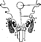
THYROID GLAND (gl. thyreoidea, syn. corpus thyreoideum), one of the most important endocrine glands of vertebrate animals. In embryonic development, the thyroid gland arises from the epithelium of the lower wall of the pharyngeal part of the intestine; in the larvae of cyclostomes, it still appears as a groove-like depression of the anterior intestine at the level of the II-IV gill arches, and only in the adult lamprey it separates off and acquires typical follicular structure (see below). In higher vertebrates, the formation of the thyroid gland proceeds in a similar manner, but at earlier stages of development (Fig. 1). The structure of the thyroid gland in various representatives of vertebrates is subject to significant variations: in amphibians and birds, it is associated with other glands of branchiogenic origin: I and II - the first and second entodermal pockets: 1-tuberculum impar (of the tongue); 2-ductus thyreoglossus; 3-gl. thyreoidea; 4-trachea; 5-esophagus; 6-gl. thymus; 7-gl. parathyreoidea sup.; 8-postbranchial body; 9-gl. parathyreoidea inf. (According to Tourneux).
In contrast to the deep location in the chest cavity in birds and lower mammals, the thyroid gland in humans is located in the area of the laryngeal cartilages. Nearby are usually the parathyroid glands, sometimes penetrating into the tissue of the thyroid gland, which in some cases contains areas of lymphoid tissue. According to histological structure, the thyroid gland represents a collection of a large number of vesicles, so-called follicles; their walls consist of one or several layers of epithelial cells, and the internal cavity is filled with a glassy viscous mass, colloid, which is a derivative of the secreting epithelium. The size of the cells is subject to significant fluctuations (height from 1 μ to 10 μ and more), their structure to a certain degree reflects the functional state of the gland: a high cylindrical epithelium is considered a sign of hyperfunction, low, flattened - hypofunction. Colloid, according to its ability to take dyes, is divided into chromophobic and chromophilic; the latter in turn is stained by both acidic and basic dyes. These phenomena should be regarded as different phases of the formation and subsequent transformation of the colloid substance. The latter is the main carrier of the hormonal principles of the thyroid gland, although in some cases the so-called basal secretion is probable, i.e., the release of an active substance by cells directly into the bloodstream, bypassing the follicular cavity. The cytological relationships in the process of formation of the thyroid gland's secretion are not yet fully clarified, just as the interpretation of the staining reactions of the colloid.
The follicles are covered on the surface with connective tissue cells; the latter acquire relatively strong development in mammals and humans, thanks to which the thyroid gland is divided into a number of lobules (sometimes with different structures). Between the follicles, compact islands of the same epithelial cells are often found, which apparently should be regarded as the initial phase of follicle formation. Colloid either overflows and greatly stretches individual vesicles or is almost absent; the latter may occur either at the initial stages of gland formation or in cases of complete pathological inactivation of the gland, sometimes such a picture indicates a rapid release of colloid into the bloodstream. The chemical nature of the thyroid gland hormone is relatively well understood. Developing previous views of Baumann and Oswald on the secretion of the thyroid gland as an iodine-containing protein compound, Kendall (1919-20) and later Harrington (1926-27) elucidated the structure of this substance, named by Kendall thyroxine. However, the views of both authors on the chemical nature of thyroxine do not coincide: Harrington considers it a derivative of tyrosine with the empirical formula C15H11O4NI4 and has achieved the synthesis of this compound in the laboratory. In any case, thyroxine in extremely small concentrations reproduces the physiological and morphogenetic effect of the thyroid gland introduced into the organism by one means or another.
The thyroid gland has an exceptionally strong physiological effect, extending to various aspects of the body's life activity. The thyroid hormone activates gas exchange, which sharply decreases upon extirpation of the organ (oxidative processes are reduced by half). Protein metabolism is also in close dependence on the function of the thyroid gland, prompting the body to enhanced protein breakdown; with hypofunction or removal of the thyroid gland, glycogen accumulates in the liver and fat is deposited, with hyperfunction - the opposite phenomena. The morphogenetic manifestations of the thyroid gland function are just as diverse. The overall growth of the organism and the associated phenomena of differentiation at late stages of development normally proceed only in the presence of a functioning thyroid gland. Insufficient secretion or experimental disruption of the integrity of the organ leads either to a cessation or at least to a sharp inhibition of the growth and development of a young animal. The earlier the stage of development at the time of the operation, the more pronounced are the phenomena of thyroid infantilism. Developmental disorders also occur in hyperthyroidism. The most well-known phenomenon is the accelerated metamorphosis (transformation) of amphibian larvae, which is caused by feeding them the thyroid gland or its preparations (Gudernatsch and others). That the transformation of a tadpole into a frog really depends on the activity of the thyroid gland is proven by experiments of thyroidectomy, as in this case metamorphosis does not occur. Under the control of the thyroid gland is a whole complex of form-building phenomena, including sexual differentiation, development of the skin cover and its derivatives (feathers, hair), general body proportions, regeneration. The greater the importance of the doctrine of the thyroid gland as a constitutional factor.
V. Larionov. On living subjects, upon inspection of the anterior region of the neck, in the triangle bounded on the sides by the relief of the sternocleidomastoid muscles, somewhat below the protrusion of the cricoid cartilage of the larynx, one can notice to a greater or lesser degree a convexity caused by the thyroid gland located here. The thickness of the subcutaneous fat layer, the degree of development of the muscles covering the thyroid gland, the length of the neck and finally the size of the organ itself determine the character of the relief of the thyroid gland area, which changes with movements of the head and cervical region.
Figure 2.
Figure 3.
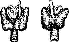
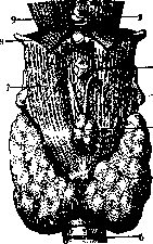
Variants of pyramidalis gl. thyreoideae (thyroid gland of the newborn). Figure 2. Pyramidalis, originating from the junction of the left lobe with the isthmus. (After Lucien, Parisot and Richard.) Figure 3. Pyramidalis, originating from the isthmus. (After Lucien, Parisot and Richard.) of the spine, as well as during swallowing. Tightly connected with neighboring organs and in particular with the larynx, the thyroid gland changes its position, rising or falling (during swallowing). The nature of the relief presents a number of features, depending on age, sex, body type and individuality. Enlargement of the thyroid gland (goiter) gives particularly sharp changes in the relief of the described area of the neck. Structure. The thyroid gland has a horseshoe or semi-lunar shape; it has three main parts: two lateral lobes, lobi lat. (lobus dext. et lobus sin.) and one middle unpaired part, the isthmus (isthmus), also called the middle lobe. To these three constant lobes of the thyroid gland should be added a fourth, inconstant lobe, which is called the pyramidal process (processus pyramidalis) or lobule (lobus pyramidalis, Lalouette's pyramid) (figs. 2 and 3). The shape and size of the individual lobes and of the gland as a whole are subject to significant variations. The lateral lobes are usually asymmetrical both in shape and in their size. The shape of each of the lateral lobes can be compared to a pyramid with rounded faces (edges), the base of which is turned downward and the apex upward. The middle unpaired part of the thyroid gland (isthmus) is most variable in form, size, and position; it connects the lower parts of the lateral lobes. The position of the isthmus can give the thyroid gland the appearance of a horseshoe with a low-lying transverse and obliquely directed vertical lines). The isthmus has an anterior convex surface and a posterior concave surface, by which it is closely applied to the anterior surface of the trachea. The upper edge of the isthmus is concave and lies at the level of the first tracheal cartilage (rarely the second or third); in children it is located somewhat higher, touching the cricoid cartilage. The lower edge of the isthmus is also concave, but less so than the upper one. The isthmus covers the first 2-3 tracheal cartilages and without sharp boundaries passes into the lateral lobes. The pyramidal process has the appearance of a flat triangular lobule with a base connected to the upper edge of the isthmus or to the anterior edge of one of the lateral lobes; with its apex this process reaches the upper edge of the thyroid cartilage or the body of the hyoid bone or rises even higher. The pyramidal process is considered as a lobe of the thyroid gland, which developed in place of the thyroglossal duct that existed in the embryonic period (ductus thyreo-glossus) (figs. 1 and 11). The varying degree of preservation of this duct gives various variations in the pyramidal process (see below). The size of the thyroid gland as a whole and of its individual lobes is subject to significant fluctuations; even in the same individual the size of the thyroid gland can change depending on its functional state at a given period, on the degree of blood filling, etc. From this point of view the thyroid gland is compared with the spleen-an organ that is also very unstable in terms of size,
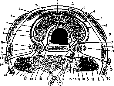
Figure 4. Horizontal section of the neck in the area of the thyroid gland (diagram): 1-deep leaf of fascia colli; 2-m. sterno-thyreoideus; 3-fascia colli media; 4-superficial leaf of fascia colli; 5-outer capsule of the thyroid gland, connected with the fascial sheath of the vascular-nerve bundle; 6-space between the outer and inner capsules of the thyroid gland; 7-n. recurrentes (dext. et sin.); 8-lymph node; 9-v. jugularis ext. with its accompanying fascial plate; 10-m. trapezius; 11-fascia colli profunda; 12-m. scalenus ant.; 13-n. vagus; 14-truncus sympathicus; 15-fascia praevertebralis; 16-a. vertebralis; 17-fascial plate; 18-spatium retropharyngeum. OF DEVELOPMENT AND WITH EACH OTHER [DEPENDING ON THE SIDE OF DEVELOPMENT AND WITH EACH OTHER]. In relation to the development of the lateral lobes, according to Vierordt, the average length of the lateral lobes is 5-6 cm, width 2-3 cm, thickness 1-2 cm; according to Henle, length 5-7 cm, width 3-4 cm, thickness 1.5-2.5 cm. The right lobe is more voluminous than the left both in adults and in children. According to Sappey, the width of the isthmus is 0.5-1.5 cm, height 4-20 mm, thickness 6-8 mm (according to Testut, 4-6 mm). The isthmus is less pronounced in a child than in an adult. Weight. With average development, the thyroid gland of a newborn weighs 2-3 g, of an adult 25-30 g (according to Rauber-Kopsch 30-60 g): in tables 16-. According to Vierordt, the weight of the thyroid gland of a newborn is given as 0.16% of the total body weight, while in adults only 0.05%. The relative and absolute weight of the organ, according to observations by Gubobin, fluctuates greatly; the relative weight of the gland in newborns is the same as in adults. Data regarding age variations of the thyroid gland are very contradictory: some indicate that the thyroid gland in a child is less voluminous than in an adult; others, on the contrary, believe that it is more developed in the fetus and in the child. Based on data from Parsky, Gubobin makes the following conclusions: 1) all dimensions of the gland double by 12 years; 2) in an adult it is 2-3 times larger than in a newborn; 3) the growth of the gland is especially energetic during the period of puberty (12-15 years); 4) the isthmus in a newborn, although about 2 times smaller than in an adult, this is not characteristic for the child's gland and one cannot speak, as Tillaux does, that the isthmus is less developed in children, since its relative size strictly corresponds to the other dimensions of the thyroid gland; 5) in old age the thyroid gland decreases in size and volume (as well as in weight). The specific gravity of the thyroid gland, according to Krause, ranges from 1.0381 to 1.0655 (Testut). Individual variations in the size of the thyroid gland are so significant that it is difficult to establish even a conditional norm. As for sexual differences, it is noted that in women the thyroid gland is more voluminous than in men. During pregnancy and during menstruation it increases in volume; the same phenomenon is observed in girls during the onset of sexual maturity. In addition to the above-mentioned variations in the size of the thyroid gland, one must take into account fluctuations in size in connection with the prevalence in a number of populated areas of the so-called endemic goiter (see Goiter).
The color of the gland is usually described as grayish-red with a yellowish tint, sometimes darker, sometimes lighter. The color depends to a large extent on the degree of blood filling: it sometimes appears more red, sometimes, for example, in venous congestion, it becomes almost purple. The consistency of the organ is described by some authors as soft, others as dense; it depends on a number of factors: physiological state, degree of blood filling, amount of colloid, degree of development of connective tissue, etc. Topography. The thyroid gland lies in the cervical triangle, which in topographic anatomy is called trigonum colli infrahyoideum (or trigonum colli medium), bounded above by the body of the hyoid bone, below by the jugular notch of the sternum, on the sides by the sternocleidomastoid muscles. The area occupied by the thyroid gland itself is called regio thyreoidea (the pyramidal process extends into the area called regio laryngea). The isthmus of the thyroid gland is covered in front by the sternohyoid muscles, then by the superficial and middle cervical fasciae (fascia colli superficialis et fascia colli media) and finally by the skin. Along the midline of the neck, corresponding to the interval between the medial edges of the sternohyoid muscles, the isthmus is covered only by the fascial sheets fused in this place and by the skin. The lateral lobes are covered in front and partly on the outside by mm. sterno-hyoideus, sterno-thyreoideus, and on the side by m. omohyoideus with their covering fascia. More superficial structures covering the lateral lobes are m. sternocleidomastoideus, fascia colli superf., m. platysma and skin. The isthmus embraces the trachea in front; the lateral lobes of the thyroid gland are applied to the lateral surfaces of the trachea, the cricoid and thyroid cartilages of the larynx and the posteromedial surface to the lower part of the pharynx (m. constrictor pharyngis inf.) and to the very upper part of the esophagus; on the posterior surface-to the a. carotis communis. The thyroid gland covers the upper part of the groove between the esophagus and the trachea, and therefore the recurrent branches of the vagus nerve lying in these grooves, along with the lymph nodes located along their course (lgl. paratracheales) (see.
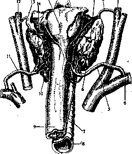
Figure 6. Artery of the thyroid and parathyroid glands (posterior view): 1-gl. parathyreoidea sup. dext.; 2-a. thyr. sup. dext.; 3-a. carotis ext. dext.; 4-a. subclavia dext.; 5-a. thyr. inf. dext.; 6-gl. parathyreoidea int. dext.; 7-n. recurrens dext.; 8-trachea; 9-esophagus; 10-gl. parathyreoidea inf. sin.; 11-a. thyr. inf. sin.; 12-a. carotis ext. sin.; 13-a. thyr. sup. sin.; 14-gl. thyreoidea; 15-posterior wall of the pharynx (Vagus nerve). Of the organs that are in the closest topographical relationships with the Thyroid gland, the parathyroid glands should also be mentioned, which are sometimes included even in the parenchyma of the Thyroid gland (see Parathyroid glands). The Thyroid gland is covered by two connective tissue capsules, one of which is called the internal capsule, capsula interna, s. propria (tunica fibrosa, s. albuginea), and the other the external, capsula externa, s. tunica vaginalis. The internal capsule directly covers the surface of the gland, tightly fusing with its parenchyma and sending processes that penetrate into the organ and form the interstitial tissue (septa) between the lobules. The external capsule is formed by the compacted cellular tissue of the neck, the so-called fascia endocervicalis, which provides fascial coverings for the organs of the neck and begins at the external base of the skull (according to other indications, from fascia colli media). The fascial plates forming the external capsule of the Thyroid gland separate from the fascial sheath of the neck vascular-nervous bundle, surround the Thyroid gland, and from behind pass to the esophagus, meeting there (along the midline) with each other. Between the external and internal capsules of the Thyroid gland there remains a slit-like space filled with loose cellular tissue and veins. From the external capsule extend well-defined connective tissue bundles, which connect the Thyroid gland with the larynx and contribute to its fixation; these strands are called the ligaments of the Thyroid gland, lig. gland, thyreoideae (Henle) or lig. thyreoidea (Gruber) (fig. 4 and 5). Arteries. The constant arteries of the Thyroid gland are considered to be: two superior thyroid arteries (aa. thyr. sup. dext. et sin.) and two inferior (aa. thyr. inf. dext. et sin.) (fig. 6). A. thyreoidea sup. is usually the first branch of a. carotis ext.; it goes in a descending direction, and, approaching the apex of the lateral lobe, divides into three main branches: anterior, lateral, and posterior. The posterior branch of a. thyr. sup. passes between the Thyroid gland and the trachea, where it anastomoses with a. thyr. inf. of the corresponding side. The anterior branch at the upper edge of the isthmus connects by an anastomosis with its counterpart. The superior thyroid artery is distributed in the upper and lateral-anterior part of the Thyroid gland (Latarjet et Alamartine). A. thyreoidea inf. is one of the branches of truncus thyrocervicalis. It initially goes along the anterior surface of m. scaleni ant., passes between a. carotis comm. on one side and a. vertebralis and truncus sympathicus on the other; sharply bending, it approaches the thyroid gland from behind. A. thyr. inf. divides into three terminal branches: 1) the inferior one, going along the lower edge of the isthmus to connect with the corresponding branch of a. thyr. inf. of the opposite side,
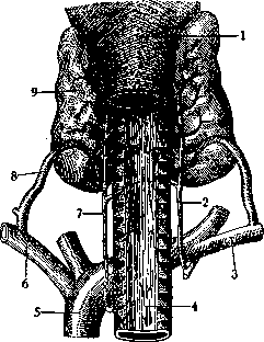
Figure 7. Relationships of the recurrent nerves and inferior thyroid arteries (aa. thyreoideae inf.): 1-pharynx; 2-n. recurrens dext.; 3-a. subclavia dext.; 4-trachea; 5-aorta; 6-a. subclavia sin.; 7-n. recurrens sin.; 8-a. thyr. int. sin.; 9-gl. thyreoidea. 2) the posterior one, passing along the posterior edge of the lateral lobe, anastomosing with the corresponding branch of a. thyr. sup., and 3) the deep one, lying on the inner surface of the Thyroid gland. From the terminal branches of a. thyr. inf., a thin branch almost constantly departs, penetrating into the apex of the corresponding lobe of gl. thymus. A. thyr. inf. with its branches is distributed mainly in the lower and posterior-inner parts of the Thyroid gland. From a topographical point of view, the relationships between a. thyr. inf. and ramus recurrens n. vagi are of considerable interest (fig. 7). According to Dwight, in 51.7% n. recurrens lies behind a. thyr. inf., in 35.9% in front of it; according to Taguchi, n. recurrens is located in front of the artery in 27%, behind the artery in 36%, and between its branches in 37%. Behind a. thyr.
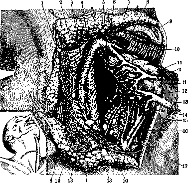
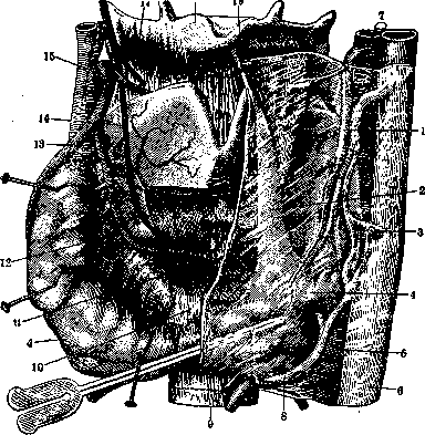
Figure 8. Topography of the thyroid and parathyroid glands (the diagram shows lines of surgical incisions in the area of the thyroid gland and incisions made in the preparation of the specimen presented in the figure): 1-2. sternocleidomastoideus; 2-3. thyroidea media (cut and retracted to the left); 3-4. jugularis int. dext.; 4-5. prevertebral fascia and muscles; 5-6. thyroidea sup.; 6-7. thyroidea sup. dext.; 7-8. lateral wall of the pharynx; 8-9. omo-hyoideus (cut and retracted upward); 9-10. incisura thyroidea cartilaginis; 10-11. stemo-thyreoideus (cut and retracted upward); 11-12. right lobe of gl. thyreoideae (retracted forward and to the right); 12-13. parathyreoidea inf. dext.; 13-14. thyroidea media (cut and retracted to the left); 14-15. thyr. inf. dext.; 15-16. trachea; 16-17. thyr. inf.; 17-18. recurrens dext.; 18-19. carotis comm. dext.; 19-20. vagus dext. inf. is located (if only it is developed) the middle cervical sympathetic ganglion (thyroid ganglion of Haller). The fifth artery of the thyroid gland is the inconstant unpaired artery bearing the name a. thyroidea ima (Neubauer's); according to the indications of most authors, it occurs in 10% of cases, according to Gruber, in 11-13%. All the described arteries of the thyroid gland anastomose with each other both inside the organ and outside it (on the surface of the gland). Some anastomoses have a transverse direction, connecting arteries of the same name of one and the other side; others - longitudinal, connecting the upper and lower thyroid arteries of the corresponding side (Latarjet et Alamartine). Veins. The thyroid gland is very rich in veins. On the surface of the gland, branches of the thyroid veins form plexuses, from which arise common venous trunks, flowing into the large cervical veins. The following veins of the thyroid gland are distinguished (fig. 8): 1) vv. thyr. sup., 2) vv. thyr. inf., 3) vv. thyr. mediae (one on each side), going from the anterolateral surface of the lateral lobes, at the middle of their course they unite into common trunks, opening into v. jugul. int., 4) v. thyr. ima, s. impar, well-developed, constant unpaired vein, going independently from the arteries. Here these veins form a richly looped plexus, called plexus thyroideus impar, s. imus. V. thyr. ima most often flows into v. anonyma sin., more rarely into angulus venosus dexter. Anastomoses of the thyroid veins with other veins of the neck are also observed, e.g. with v. jugularis ant. All veins of the thyroid gland are devoid of valves. The blood vessels of the thyroid gland are located between its outer and inner capsules; large arteries and venous trunks pierce capsula externa.
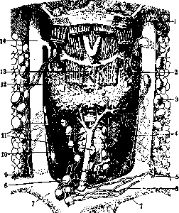
Lymphatic vessels of the thyroid gland begin from the intralobular networks; in the interlobular spaces vasa lymphatica interlobularia are formed, which, following the course of the blood vessels, come to the surface of the gland, where from them the outer network of lymph vessels is formed. From this network depart larger efferent lymph vessels-vasa efferentia. According to observations of Bartels in humans, when injecting lymph vessels of the thyroid gland of one side, it is difficult to fill the vessels of the other side, while in animals (e.g. in a cat, rabbit) unilateral injection easily fills the entire network of lymph vessels of the thyroid gland. All lymph vessels are divided, based on the direction of lymph flow, into ascending, or upper, and descending, or lower (fig. 9). Nerves. The thyroid gland is innervated from the vegetative nervous system (see), from which it (from nn. sympathic. and vagus) and receives vasoconstrictor, secretory and trophic nerves. Braeucker indicates that sympathetic branches depart from the cervical sympathetic ganglia (upper, middle and lower) or from the interganglionic branches of the cervical part of the trunk and can be designated as nn. thyreoidei sup., medii et inf. These branches arise mainly together with the cardiac branches of the sympathetic nerve or else separate from the trunk as independent branches reaching the thyroid gland. Sympathetic nerves go to the thyroid gland also together with the arteries, forming on them plexuses: plexus thyroideus sup. et inf. Branches of the vagus nerve, according to Braeucker, depart from the upper, middle and lower segments of the cervical part of n. vagi and are designated by him as rami thyreoidei sup., medii et inf.; these branches go to the gland through the medium of ramus ext. n. laryngei sup., rami cardiaci et n. recurrens vagi. In rare cases, participation in the innervation of the thyroid gland of branches from ansa hypoglossi, fibers of which come from the roots of the cervical plexus (Vishnevsky, Braeucker), is noted; some authors also point to the participation of n. glossopharyngei. Nerve branches, according to the opinion of most authors, end partially in the walls of blood vessels, partially at the cells of the follicles of the thyroid gland. Variants of innervation of the thyroid gland according to data of typical anatomy (Vishnevsky) can be reduced to three types: sympathetic, vagal and mixed, middle, i.e. in some cases a predominance of sympathetic branches is observed, in others - the main supply by the vagus nerve, in the third - uniform supply by both nerves. It is noteworthy the commonality of innervation of the thyroid gland and heart, which gives some authors reason to explain this by the appearance of certain functional disorders from the heart in diseases of the thyroid gland.
Anomalies and variants. Attention should be paid to the variants of the thyroid gland associated with a violation of the correct course of its development. From this point of view, more often than others, the pyramidal lobe gives various kinds of deviations in terms of size, position, degree of development, etc. It may be absent (in approximately 1/4 of cases); different degrees of development of this lobe may be observed, which determines its length and form. Thus, in some cases it is barely outlined, short; in others it reaches the upper edge of the thyroid cartilage or even the hyoid bone. In terms of its place of origin and position, it is noted that the pyramidal lobe may separate either from the isthmus, and then it is located strictly along the median line, or it departs from one of the lateral lobes (more often the left), and then lies obliquely. Cases of doubling of the pyramidal lobe are observed, with a double pyramid
GLAND
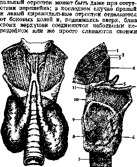
Figure 10.
Figure 11.
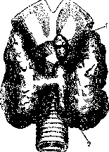
Figure 10. Absence of the isthmus of the gl. thyreoideae and doubling of the proc. pyramidalis. (After Lucien, Parisot and Richard.) Figure P. Tractus thyreo-glossus (in the adult): 1-root of the tongue; 2-papilla vallata; 3-foramen caecum; 4-part of the tractus thyreo-glossus lying above the hyoid bone; 5-os hyoideum; 6-gl. thyreoidea; 7-trachea; 8-proc. pyramidalis of the gl. thyr.; 9-thyroid cartilage; 10-part of the tr. thyreo-glossus lying below the hyoid bone; 11-part of the tr. thyreo-glossus lying behind the hyoid bone. The pyramidal lobe usually consists entirely of glandular tissue; in other cases, alternation of glandular areas with connective tissue is observed. In such cases, instead of a pyramidal lobe, there are several small glandular formations, isolated from the main mass of the Th. g.; these formations, consisting of tissue identical to that of the Th. g. itself, are designated by the term gl. thyreoideae accessoriae; they may also appear in other places, along the entire course of the ductus thyreo-glossi, i.e., from the foramen caecum of the tongue to the Th. g. itself (fig. 11). Accessory Th. g. are divided into: gl. thyreoideae accessoriae praehyoideae, suprahyoideae et infrahyoideae according to their position relative to the hyoid bone (fig. 12 and 13). Among these accessory thyroid follicles, a gland known as glandula Zuckerkandli is distinguished, which is located directly above the body of the os hyoideum, in the cellular tissue between the mm. genio-hyoidei on both sides or in the thickness of the m. mylo-hyoidei (1 case in 200 according to Zuckerkandli). Extremely rare are accessory thyroid follicles included in the body of the hyoid bone (gl. thyr. accessoria intrahyoidea of Streckeisen). Cases may occur when the Th. g. is absent; in this case, the accessory thyroid follicles develop more powerfully. Preservation of the embryonic thyro-glossal duct (ductus thyreo-glossus) can be observed either in the form of a canal (canalis thyreo-glossus of His) or in the form of a cord (tractus thyreo-glossus) (fig. 11). The canal remaining in the adult begins at the top at the foramen caecum of the tongue (ductus lingualis BNA). The canal descending from the blind opening into the thickness of the tongue is called the canal of Bochdalek; it is lined with cylindrical epithelium, may have branches-canaliculi (the length of which reaches 4 mm), penetrating into the thickness of the m. genio-glossi. Tractus thyreo-glossus, in 4 other cases the canal, passing through the thickness of the tongue, reaches its base or descends even lower, to the upper edge of the hyoid bone. If the canal or cord continues further below, it can pass in front of and behind the body of the hyoid bone and to the apex of the pyramidal lobe.
In connection with the remains of the ductus thyreo-glossi, the appearance of so-called median cysts and fistulas of the neck (Venglovsky) occurs, and in connection with the presence of accessory thyroid follicles, the possibility of goiter formation in areas corresponding to their position (e.g., lingual goiter).-Of other parts of the Th. g., the most variable is the isthmus, which may be completely absent (gl. thyreoidea bipartita) in approximately 10% (according to Gruber 1 case in 20, according to Marshall 1 in 10) (Berard and Chemin). The absence of the isthmus is a constant phenomenon observed in many animals (cat, dog, etc.). The isthmus may consist only of connective tissue (according to Botta 4.5%). Its form and size are extremely variable. A sharp increase in the isthmus may be observed with almost absent lateral lobes or with the rudimentary state of one of the lateral lobes. As for the variants of the lateral lobes, they are expressed more often in the difference in size and shape of the right and left lobes. Among the rare variants of the Th. g. is the complete absence of one of the lateral lobes (more often the left). Such variants have been described by Luschka (H. Luschka), Venglovsky, and Chausov. In the presence of any anomaly on the part of the Th. g., variants of the corresponding thyroid arteries are observed. For example, with underdevelopment of the left lateral lobe, underdevelopment of the a. thyr. sup. sin. et a. thyr. inf. sin. is observed (Usov). Cases of strong dystopia or displacement of the organ should be attributed to variations of the thyroid gland (Sobotta).
Usov.
Pathology. Malformations of the Thyroid Gland occur: 1) in the form of complete absence of the gland, 2) multiple development of the gland, and 3) in the form of abnormal position of the Thyroid Gland. 1. Complete absence of the Thyroid Gland is rarely observed; incomplete development of the Thyroid Gland is described more frequently, noted already in early childhood as spontaneous myxedema, which in turn differs from infantile myxedema. The latter is observed in children born with a relatively healthy Thyroid Gland, which in the first years of life undergoes patho-anatomical changes leading to corresponding functional disorders under the influence of various causes—syphilis and others (see Myxedema). 2. Multiple development of the Thyroid Gland occurs more frequently than is generally thought. Clinically, this fact is of interest only in those cases where pathological changes occur in such multiple glands. 3. Displacements of the lobes of the Thyroid Gland of an embryonic nature to the base of the tongue (foramen cecum) and to other places are possible, which has important clinical significance when a goiter develops in these areas of the gland (Figs. 12 and 13). So-called retrotracheal goiters, retroesophageal, retrosternal, and at the base of the tongue are known. - Traumatic injuries to the Thyroid Gland. Isolated injury to the Thyroid Gland is rare and occurs mainly in the presence of a goiter. Most often, the Thyroid Gland is affected in traumatic injuries to neighboring organs in the neck. Subcutaneous injuries to the Thyroid Gland are observed in attempts at hanging. When the Thyroid Gland is injured, subcutaneous or open, bleeding is usually observed, requiring emergency intervention to stop it by ligation of vessels, suturing of the parenchyma of the gland, etc. The most common disease of the Thyroid Gland, known since ancient times, is goiter (see), the study of which made it possible to study the significance of the function of the Thyroid Gland for the organism. Thus, the development of spontaneous myxedema in adults is associated with profound changes in the Thyroid Gland, depending on various factors—patho-anatomical changes in the sexual organs in women, tuberculosis and syphilis. Myxedema in adults is identical in its clinical course to thyroid cachexia, first described by Kocher, who observed this severe disease after total removal of the Thyroid Gland. At present, when the physiology of the Thyroid Gland has been studied and the technique of operations on it has been developed in detail, this disease is observed in severe inflammatory processes in the gland after improper treatment with X-rays, leading to the disappearance of the glandular elements gl. thyreoideae, and in degenerative processes of the gland parenchyma. The symptoms of the disease, as already mentioned, are the same as in myxedema, and diagnosis itself is not difficult, especially after operations or other manipulations on the Thyroid Gland. The oral administration of preparations of the Thyroid Gland (thyroidin or thyreocryn) in doses of 0.3 three times a day is a good preventive measure capable of preventing the development of thyroid cachexia after total extirpation of the Thyroid Gland. The situation is worse when the picture of the disease has already reached full development. Transplantation of the Thyroid Gland, as numerous clinical observations have shown, does not lead to the goal. Anatomical changes in the Thyroid Gland, observed in areas affected by endemic goiter, are accompanied in individual cases by a peculiar clinical picture in the form of cretinism. The theory that initially appearing cretinism leads to changes in the Thyroid Gland is not proven, just as the theory of Bircher (E. Bircher) that the same cause leads to disease of the Thyroid Gland and cretinism. The connection between cretinism and the Thyroid Gland can be considered proven by the observations of Wagner, who pointed to improvement in all symptoms after the oral administration of preparations of the Thyroid Gland (see Cretinism). Inflammatory diseases of the Thyroid Gland are observed in the form of thyroiditis and strumites; in the latter, there is an inflammatory process developing in the altered Thyroid Gland in the presence of a clearly formed goiter. Acute inflammation of a normal Thyroid Gland, acute thyroiditis (thyreoiditis acuta), is observed very rarely, mostly as a concomitant complication in other infectious diseases (see Thyroiditis). The causes of the development of acute strumitis (strumitis acuta) are the same as those of acute thyroiditis, but strumitis occurs somewhat more frequently in an already developed goiter, predominantly of the nodular form. Chronic strumites occur not only as secondary after acute strumites but also as primary with a peculiar development of the inflammatory process. Characteristic of this form of chronic strumites is the special density of the Thyroid Gland, associated with degenerative changes in the parenchyma, expressed by the partial disappearance of follicles. In the stroma, a significant amount of eosinophils, lymphocytes, plasma cells, and giant cells accumulate. In the interlobular connective tissue, round-cell infiltration is noted. The exceptional density of the enlarged Thyroid Gland gave Riedel occasion to denote this form of strumite 'as hard as iron' and to name it after himself—'Riedel's goiter.' In the literature to date, 90 cases of such chronic strumite have been described (B. Levit). With the development of the inflammatory process and its spread from the gland to neighboring tissues, the Thyroid Gland becomes fused with the surrounding tissues. The complaints of patients come down to pain, difficulty in swallowing, and shortness of breath. These symptoms are not equally expressed in all patients. In the diagnosis of Riedel's goiter, it is necessary to differentiate it from a malignant tumor of the Thyroid Gland, which can be decided only after microscopic examination. Surgical treatment, indicated when all symptoms are increasing, consists of partial excision of the Thyroid Gland. Paier (Paug) considers it sufficient to excise small pieces from the altered gland, after which noticeable improvement occurs. Tuberculosis of the Thyroid Gland occurs according to autopsy material (Chiari) in 7%, but clinically it manifests itself in no way, and patients rarely seek medical help. This circumstance is the reason why in clinical practice it is considered that the Thyroid Gland, due to its rich blood supply and iodine content, is little susceptible to tuberculous infection. Patho-anatomical changes in tuberculosis of the Thyroid Gland are observed in the form of multiple miliary tubercles, caseo-purulent foci, and finally in the form of cold abscesses. - Syphilis of the Thyroid Gland, which is observed very rarely, is mostly described in the form of a gumma (Razumovsky). According to syphilologists, in the secondary period, the Thyroid Gland often swells. Treatment is antispecific. Caution should be exercised in prescribing iodine preparations. - Actinomycosis of the Thyroid Gland is characterized by the hardness of the tissue of the gland itself, the density of the surrounding infiltrate, the presence of fistulas characteristic of actinomycosis with the discharge of characteristic pus. It occurs very rarely. Treatment—see Actinomycosis. Of the new formations of the Thyroid Gland, which occur in the clinic generally rarely, benign tumors in the form of fibromas, myomas, and teratomas are an exceptional rarity. Malignant new formations in the form of sarcomas and carcinomas are more frequently observed, which in the clinic are united under one name 'malignant goiter'—struma maligna. According to the data of Gdrodtz and Tikhov, sarcoma occurs less frequently than carcinoma. The latter is most frequently observed in areas affected by goiter. According to the material of Wegelin, cancer of the Thyroid Gland occurs in Bern 10 times more frequently than in areas free from endemic goiter. Malignant goiter is observed between the ages of 40-60, most often in women. Sarcomas, which occur more frequently in men, are structurally round-cell, giant-cell, from spindle-shaped cells, lymphosarcomas, and very rarely melanomas. Sarcomas of the Thyroid Gland grow rapidly and, compared with cancer, are more malignant. In cancer of the Thyroid Gland, rapid growth of individual nodes, invasion of the capsule, and limitation of mobility are noted. Histologically, cancerous tumors are represented as adenocarcinomas with little tendency to intensive growth and invasion of neighboring tissues. Such tumors remain for a long time a small node, causing almost no suffering. Malignant papillomas also grow slowly in the form of small nodes consisting of cylindrical cells. More malignant and faster in invading neighboring tissues are true cancers of the Thyroid Gland, completely displacing the normal tissue of the gland. Struma metastatica with struma lymphomatodes, which is considered benign in its anatomical structure, is malignant in its clinical course due to the multiplicity of metastases (in bones and other organs). In its clinical course, malignant goiter is characterized by the appearance of dense nodes, rapid growth, invasion of the fascia and neighboring tissues, limitation of mobility of the tumor, difficulty in swallowing, appearance of shortness of breath due to compression of the trachea, attacks of suffocation and tormenting shooting pains in the back of the head. With further invasion of the tumor, the vagus nerve (complete loss of voice) and the sympathetic nerve (Horner's syndrome) are affected. Dilated veins appear on the neck. Metastases are observed in 90% of cases of cancer of the Thyroid Gland (Kocher) and are noted first of all in the lungs and bones, and then in other organs. In the diagnosis of a malignant tumor of the Thyroid Gland, the nature of the growth of the 'goiter,' complaints of shortness of breath, shooting pains, absence of temperature, etc., are of great importance. It is necessary to differentiate with the rarely occurring Riedel's goiter, tuberculosis and syphilis of the Thyroid Gland.
Biopsy is valuable for the material obtained, but is dangerous due to the possible dissemination. A valuable diagnostic method is the irradiation of the thyroid gland tumor with X-rays, proposed by Schaedel, which almost as a rule leads to a decrease in the tumor within the next two weeks, which is not observed in other forms of goiter. Treatment. If the tumor has not yet grown into adjacent tissues, then the best method is the complete extirpation of the thyroid gland - thyreoidectomia totalis according to Sudeck. The technique for exposing the thyroid gland is the same as in operations for ordinary goiter. After ligation of both thyroid arteries on both sides, ligatures of the corresponding veins and additional vessels (a. thyr. ima) are applied, and the entire thyroid gland is removed. It is self-evident that in total thyroidectomy it is necessary to spare the epithelial bodies and the recurrent nerve. Patients after total thyroidectomy must, to prevent the development of cachexia, take preparations of the thyroid gland (thyreoidin, thyreocrin, 0.3 three times a day) throughout their lives. Of great interest is the well-known case of Eiselsberg, when the myxedema that developed after total thyroidectomy disappeared with the appearance of a metastasis in the sternum and reappeared after the removal of the metastasis. Obviously, the continuously growing epithelium in thyroid gland cancer is not devoid of the ability to secrete the colloid necessary for the body. For far-advanced tumors, treatment with X-rays (Su-deck) is recommended. The results are more favorable in cancer than in sarcoma. The prognosis for malignant tumors of the thyroid gland is serious. In sarcoma of the thyroid gland, patients die within 6-12 months, and in cancer, at most within 2 years. Timely operation (there is a known case of do Quervain, when the patient lived 9 years after the operation) and timely X-ray treatment significantly improve the prognosis. Death usually occurs from metastases. In echinococcus of the thyroid gland, which is very rare (0.25% of all cases of echinococcus, Alexinsky), one has to deal with a unilocular or multilocular cyst. Echinococcus is mostly diagnosed as cystic goiter, and the true diagnosis is established only during the operation. If there is suspicion of echinococcus before the operation, then examinations are necessary, as in any echinococcus (see ECHINOCOCCUS).
Related articles
Mentioned in
Cite this page
“Thyroid Gland.” Soviet Medical Encyclopedia. English translation of Bolshaya Meditsinskaya Entsiklopediya, 1st ed. (Moscow, 1928–1936), ed. N. A. Semashko. https://sovietmedicalencyclopedia.pages.dev/article/thyroid-gland/