Thymus
Historical document, translated for reference. It reflects medical knowledge of the 1920s–30s and is not medical advice.
Summary
The thymus is a lymphoid organ of special structure that is well-developed in newborns and continues to grow until puberty, after which it undergoes involution. It consists of two lobes and is located in the anterior mediastinum and lower part of the neck.
Encyclopedia article (1928–1936)
THYMUS, thymus (syn.: thymus gland, suprasternal node), a lymphoid organ of special structure, to which in recent times an endocrine function has been attributed (see Goiter). The thymus is well developed in newborns and continues to grow until the onset of sexual maturity (12-14 years), when its involution occurs; however, remnants of it continue to exist until old age. The thymus consists of two lobes, right and left, closely adjacent to each other and having the shape of an elongated pyramid; the length of the thymus is from 5.5 cm to 8.5 cm, width 2.5-3 cm, thickness 0.5-1.5 cm. The plane dividing the gland into two lobes usually passes obliquely from front right to back left (fig. 1 and 2). They are connected to each other by a thin layer of areolar tissue; there is no true isthmus. The thymus lies simultaneously in the anterior part of the mediastinum and in the lower part of the neck, so that the gland can be divided into cervical and thoracic parts (fig. 3).-The cervical part protrudes from behind the sternum and is subject to synchronous movements with breathing: at
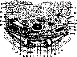
Figure 1. Cervical part of the thymus on a transverse section of the neck at the level of the upper edge of the sternum (child 15 months): 1-right and left lobes of the thymus; 2 and 3-mm. sterno-cleido-mast.; 4-edge of the sternum; 5-clavicle; 6-mm. sterno-hyoideus, st.-thyreoid., omo-hyoid.; 7-mm. scaleni ant. et post.; 8-body of vertebra; 9-m. longus colli; 10-a. anonyma; 11-a. carot. comm.; 12-a. subclav.; 13-v. anonyma; 14-trachea; 15-esophagus; 16-n. vagus dext. et sin.; 17-n. phrenic dext. et sin.; 18-n. recurrens dext. et sin.; 19-pi. brach; 20-lymph node. (After Testut)
inhalation it descends into the mediastinum and hides behind the sternum, during exhalation it rises toward the neck, sometimes approaching the lower pole of the thyroid gland. The anterior
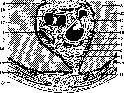
Figure 2. Thoracic part of the thymus, transverse section of the chest through II rib (child 15 m.): 1-right and left lobes of the thymus; 2-right and left lungs; 3-sternum with ossification points; 4-body of vertebra; 5-aorta (arch); 6-bifurcation of trachea; 7-v. cava sup.; 8-esophagus; 9-mediastinal pleura; 10 and 11-n. vag. dext. et sin.; 12-n. recurr. sin.; 13-vessels and n. phrenic, dext. et sin.; 14-a. mammar. int.; 15-m. pect. major; 16-mm. intercost.; 17-glands; 18-II rib. (After Testut)
surface of the cervical part of the thymus is covered by the descending laryngeal muscles, the middle and superficial cervical aponeurosis, and skin. In hypertrophy of the thymus gland, it is visible above the sternum. Its posterior surface in its middle part touches the trachea, with its right horn-the right innominate vein, the internal and anterior jugular veins, the right common carotid artery, the right vagus nerve, and the beginning of the right subclavian artery; with its left horn, usually more voluminous, rising higher on the neck than the right, the thymus touches the left innominate and inferior thyroid veins, the left subclavian and left common carotid arteries, the left vagus nerve and even (according to Cruchet) the left edge of the esophagus and the left recurrent nerve.-The thoracic part of the thymus is located below a horizontal line drawn through the sternoclavicular joints and descends to the III-IV intercostal space, occasionally to V (W. Felix). The anterior surface of the gland is flat, corresponding to the posterior surface of the sternum, with which it is in contact (sometimes with Bruch's membrane). The slightly concave posterior surface of the thymus in its lower part lies on the pericardium, in the middle-on the aorta and superior vena cava and in the upper part-on the large vessels arising from the aortic arch; in addition the thymus lies on the inferior cardiac nerve, at the
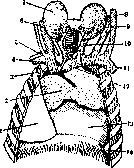
Figure 3. Topography of the thymus of a newborn: 1 and 14-right and left lung; 2-thymus; 3 and 12-v. anonyma; 4 and 11-v. subclavia; 5-a. subclav.; 6 and 7-v. jugul. int.; 8-gl. thyreoidea; 9-v. iugul. ext.; 10-v. thyre0cyMKaf:; (no"CS-" touches (sometimes with Bruch's membrane). The slightly concave posterior surface of the thymus in its lower part lies on the pericardium, in the middle-on the aorta and superior vena cava and in the upper part-on the large vessels arising from the aortic arch; in addition the thymus lies on the inferior cardiac nerve, at the
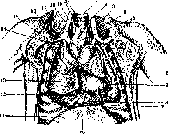
Figure 4. Position of the thymus: 1-trachea; 2 and 19-a. carotis comm.; 3 and 15-v. jugul.; 4 and 17-n. vagus; 5-gl. thymus; 6-left lung; 7-pericardium; 8-diaphragm; 9-VII rib; 10-sternum; 11-VIII rib; 12-plica adi-posa Luschkae; 13-right lung; 14-I rib; 15-a. subclavia; 16-clavicle; 20-ligaments supporting the thymus. (After Shevchenko.)
crossing of it with the aortic arch. The lateral surfaces of the thymus are flat, touching the lungs, the proper mediastinal pleura, n. phrenici and aa. pericardiaco-phrenicae. Fixation of the thymus. The thymus is covered by a fairly strong fibrous capsule, the outer surface of which is closely fused with the adjacent areolar tissue and intimately connected with the pericardium, the large vessels of the heart, and above-with the cervical aponeurosis and the carotid sheath. With the help of such a ligamentous apparatus, the thymus is suspended and to the trachea (fig. 4). The thymus is loosely fused with its capsule, which makes it possible to fairly easily enucleate it during operation.-Vessels and nerves. The thymus receives blood from the a. mammaria int., from the trunk itself or from a. mediastinal. anter., from the inferior thyroid artery, from the innominate artery and from aa. pericardiaco-phrenicae. The veins flow in the form of short thick trunks into the left innominate vein, the inferior thyroid vein and v. mammaria interna. Lymphatic outflows Severeanu divides into upper, anterior and posterior; they flow into the anterior and posterior mediastinal nodes, into truncus jugularis, tr. subclavius and ductus thoracicus, to which on the right corresponds tr. broncho-mediastinalis.-The nerves of the thymus are branches of n. sympathici and perhaps of the vagus (Testut et Jacob, H. Klose).-Topographic-anatomical conditions of the thymus are characterized by its close contact with the most important organs of the chest cavity. Hence arise in enlargement of the thymus purely mechanical changes in the narrow space of the anterior mediastinum; an inevitable consequence of this will be a disorder of blood circulation and compression phenomena from the constricted organs.-The parenchyma of the thymus is soft, yellowish or grayish-pink in color and consists of distinct lobules. The average weight of the gland (in grams) with age changes as follows (Hammar). Age Newborns 1-5 years 6-10 » 11-15 » 16-20
» 21-25
» 26-35
» 36-45
» 46-55
» 58-65
» 66-75
» Average weight 3. t. 13.26 22.98 26.10 37.62 25.58 24.73 19.78 16.27 12.85 16.08 6.0 Weight fluctuations 3. g. B-25 10-30 10-60 15-56 15-50 15-40 10-40 10-36 10-30 6-25 B-15 From these data it is evident that the weight of the thymus gland extremely fluctuates and not only with age, but also within the same age group; these fluctuations are primarily related to the nutritional status of the subject (thymus as a "barometer" of nutrition, Hammar), and since nutritional status can greatly change with various diseases, the weight of the thymus gland is subject to similar changes and moreover earlier compared to other organs. Finally, the presence of a large weight of the thymus gland does not yet say anything about the quantity and quality of its parenchyma; to resolve these questions histological research is necessary: "Glands of the same weight can differ in their content" (Sokolov). It is very probable that racial and geographical factors influence the weight of the thymus gland. Development. The thymus gland develops in the human embryo in the 1st month from the entoderm of the pharyngeal intestine (see.) in the form of two primordia on the ventral side of the III branchial pouches. Similar primordia of the IV pouch remain rudimentary. The primordia have the appearance of epithelial tubes, which very soon separate from the pharyngeal intestine and, continuing to grow, descend downward, with the primordia of both sides adjacent to each other. During histogenesis, their epithelial character is lost, and the gland takes on a lymphatic character. This occurs by the introduction of a large number of lymphocytes from the surrounding mesenchyme into the thickness of the epithelium, with the epithelium transforming into a reticulum, in the loops of which lymphoid elements are placed. The opinion of Stohr that lymphocytes also arise from the epithelium is rejected by the majority of scientists. Sometimes the thymus primordium remains connected to the pharyngeal intestine for a long time (ductus thymo-pharyngeus) and can give rise to accessory thymus glands. Comparative anatomy. The thymus gland exists in all vertebrates. In fish it develops from the dorsal side of all branchial pouches, and the gland formed by the fusion of primordia is located on the dorsal side of the gill apparatus. In amphibians (frogs) only the II branchial pouch participates in the formation of the thymus gland; the gland is located at the posterior angle of the lower jaw. In reptiles and birds development proceeds from the dorsal side of several branchial pouches (II, III, IV); the gland is located on the sides of the neck, stretching in birds into a long ribbon; in mammals also from II, III, IV pouches, but from the ventral side; the developed gland lies behind the sternum. In some species (lemurs) ductus thymo-pharyngeus persists throughout life. Structure. The thymus gland consists of cortical and medullary substance. If one dissects the lobules of one of the gland's lobes, one can be convinced that along its axis from top to bottom runs a medullary cord, the processes of which enter the lobules, forming their medullary substance; it is surrounded on the surface by a layer of cortical substance. In sections stained with hematoxylin and eosin, the blue cortical substance sharply differs from the pink medullary substance. The cortical substance is covered with a connective tissue capsule and consists of adenoid tissue, the reticulum of which has a cellular character with lymphocytes (thymocytes) laid in its loops. Lymphoblasts in the lobule do not form a single center of multiplication, as in lymph follicles, but are scattered throughout the cortical substance, appearing in the form of light spots. The medullary substance is generally built in the same way, but it contains fewer lymphocytes and more cells with light nuclei and vessels; the reticulum stands out more clearly and can take on a fibrous character. A distinctive feature of the medullary substance is the so-called Hassall's corpuscles, transient formations that already appear in the human embryo by the end of the third month. They have a round or elongated shape and considerable size, so they are well visible under weak magnification; in the center of the corpuscle usually lies one or several large cells, often in a state of degeneration, with dying nuclei and various kinds of inclusions; they are surrounded by other narrow cells, concentrically lying one on top of the other, so that as a whole Hassall's corpuscle has a layered structure. Sometimes the central cells disappear, and the corpuscle becomes infiltrated with leukocytes. Hassall's corpuscles are encountered consisting of one large cell of round shape with inclusions or a stratifying body. According to the accepted opinion, Hassall's corpuscles are transient formations: they arise, go through a cycle of development, and disappear. Views on their origin have not yet been unified. There are three main hypotheses: 1) Hassall's corpuscles arise from the epithelium remaining from the primary primordium of the thymus gland (W. Krause, His, Stieda, Maurer), 2) from the endothelium of dying blood vessels (Afanasiev, Nussbaum, Dustin), 3) from reticulum cells (Hammar); in favor of this latter view speak observations on the transplantation of the thymus gland. The significance of Hassall's corpuscles is also not clear. Recently, in connection with the recognition of the thymus gland as endocrine, Hassall's corpuscles are increasingly being assigned a secretory function, considering them as glands that die during secretion (of the holocrine type). In any case, no other cells to which a secretory function could be attributed have been found in the human thymus gland (in frogs they were described by Ver Eecke). It should be noted the repeated finding in the medullary substance of cross-striated fibers and bands (probably of muscular origin), the origin of which remains unexplained. Often in the thymus gland are encountered cysts-cavities lined with epithelium, inside of which is secretion; they arise from reticulum cells (Hammar). - Involution of the thymus gland begins with the onset of sexual maturity, which is reflected primarily in its weight (see above), since the size may remain unchanged. Involution consists in the gradual disappearance of the gland's parenchyma, mainly of the cortical layer, and its replacement with connective tissue. Subsequently the thymus gland becomes overgrown with fatty tissue, in which small islands of parenchyma remain.
V. Karpov, A. Sirotkin. Pathophysiology of the thymus gland. Judgment on the pathophysiology of the thymus gland is considerably complicated by the fact that cases of congenital aplasia of this organ in humans are extremely rare, and the clinical picture of cases of definite hypofunction of it is not exactly known. The experimental data are as follows. First of all, thymectomy can have an effect only in young, growing animals due to the involution of this organ from the period of sexual maturity; but also beyond this, apparently not for all without exception animals is the thymus gland an organ essential for life (in frogs thymectomy gives no results). There (apparently still in the overwhelming majority of cases), where the role of the thymus gland in this respect is great, it primarily appears as an organ of growth and nutrition. In very young (best of all-several days or weeks after birth) animals complete removal of the thymus gland is primarily reflected in growth, which
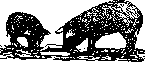
depends on the lesser length of the long tubular bones. But generally the entire skeleton becomes hypoplastic. The length, weight, and strength of the bones significantly fall short of normal (fig. 5 and 6). They become soft and flexible, prone to curvature and
fractures. The microscopic picture shows retarded bone development; the bone trabeculae are thinner and shorter, the marrow cavities are enlarged, there are fewer osteoblasts in the periosteum, and the epiphyseal zones are often widened. Chemically, the process amounts to insufficient deposition of lime in the cartilage, and since along with the hypoplasia of bones in length there is also thickening of the diaphyses in some places, first the bones become clumsy, and second the entire picture of changes in the bones begins somewhat to resemble rickets (Basch, Klose and Vogt). However, identification with true rickets should be considered not fully justified, and therefore premature. Depending on the mentioned changes, the gait also changes (which is especially noticeable in dogs): it becomes as if wider and more sweeping, but at the same time less free; the thighs and shins are turned outward; the front paws in turn curve forward somewhat; the animal when walking rests on the entire surface of its paw. At the same time, sluggishness of movement, muscular fatigue and weakness are striking, especially of the posterior half of the body: the animal moves reluctantly and prefers to sit. The latter depends not only on changes in the skeleton, but also on myasthenia, which finds its explanation not only in the loss of corresponding influence on muscle tone, but also in degenerative processes in the striated musculature. Nervous-psychic reactions are also sharply lowered, and sometimes such a significant retardation of mental development is observed that the authors find it possible to speak even of 'idiotia thyreopriva'. In such cases, in accordance with the symptoms during life, marked changes were found in the central nervous system: swelling of the brain, and in the cortex such phenomena as pyknosis of nuclei in ganglion cells, neuronophagia, proliferation of glia. The galvanic excitability of motor nerves is increased, and muscle tremors also occur, but as for the true, tetanic-like attacks sometimes observed in thymectomized animals, they must certainly be attributed not to the thymus gland as such, but to the additional epithelial bodies found there. Sensory functions and coordination of movements are also more or less disturbed later on. The general nutritional condition varies at different times after the operation: at first it is quite satisfactory, the appetite is even considerably greater than normal, but approximately a month after operation, a fairly significant deposition of fat is sometimes noted for a certain time (2-3 months). Later, however, in a number of cases cachexia develops. In other cases, on the contrary, the course of the postoperative period is more favorable. The influence of thymectomy on the bony skeleton is to some extent clarified by the disturbances in lime metabolism, namely-lime is excreted from the body in larger quantities in thymectomized animals compared to normal, and the center of gravity of the anomaly is apparently in the lowered ability of tissues to bind calcium, since the administration of calcium salts to such animals helps little. The cause of this phenomenon is presumably seen in the dissolution of lime salts by acid (as is thought, phosphoric), which accumulates in excess in the body and is normally neutralized by nucleoproteins. Hence-insufficient deposition of lime in growing bones, in cartilage, and osteoporosis in already more or less formed bones. Implantation of the thymus gland, as well as feeding with it, acts, as was to be expected, in the direction opposite to thymectomy, however not always. Classic in this direction are the experiments of Gudernatsch on tadpoles. Feeding the latter with thymus gland leads to a sharp increase in the size of their bodies ('giant tadpoles' compared to controls), but at the same time their metamorphosis is retarded, i.e. their transformation into frogs. The tadpoles besides appear as if swollen from the increased ability of tissue colloids to bind water (K. Scheer). As for the nature of the thymogenic substances stimulating growth, for some time they were identified with nucleoproteins, and then began to be associated with vitamins. Of other functions, the thymus gland is credited with an excitopoietic ability specifically with respect to lymphocytes (lymphocytosis from thymogenic extracts; fall in the number of lymphocytes in the blood following thymectomy; lymphocytosis in status thymico-lymphaticus; partly lymphocytosis in Basedow's disease). The hemodynamic effect (lowering of blood pressure from extract of thymus gland) apparently does not appear physiologically specific (is caused by choline, a product of lecithin decomposition passing into extracts). Similarly, the assumption of an alleged toning effect of the thymus gland on the parasympathetic nervous system should be considered unproven, with corresponding conclusions regarding the mechanism of the so-called thymic death. However, doubts go much further: not all are ready to classify the thymus gland as an endocrine organ, and even the above-mentioned experiments of Gudernatsch tend to be interpreted in the sense of non-specific action (assimilability of food). The important role of the thymus gland in the process of growth and nutrition of growing organisms, however, is beyond doubt, and the question is only whether in this case one can speak of hormones in the exact sense of the word or perhaps of trephones in the sense of Carrel. The clinical pathology of the thymus gland is still very inadequately developed. As mentioned, cases of congenital absence of the thymus gland are extremely rare, while tumors of this organ give neither symptoms of hyperfunction nor symptoms of loss. Cases of hyperplasia of the thymus gland have gained importance, with which the so-called 'asthma thymicum' and 'mors thymica' were thought to be connected. Nevertheless, it is necessary to keep in mind the following possible pathological deviations from this organ. First of all, along with the above-mentioned exceptional cases of congenital aplasia of the organ, congenital hyper- and hypoplasia of it are observed. The latter, for example, was described in cases of congenital myxedema. On the other hand, cases of the presence of small accessory thymus glands, located under the thyroid gland or in it itself, are not so rare. Of regressive processes, besides the gradual reduction of the parenchyma and calcification of Hassall's bodies with age, the greatest significance is attached to the sclerotic atrophy of the organ, developing in children in all those diseases that lead to exhaustion of the body (nutritional defects on the basis of artificial feeding, tuberculosis, syphilis, rickets, etc.), with the gland sharply decreasing in volume. Histologically, the process first amounts to the disappearance of the cells of the cortical layer, followed by progressive reduction of the medullary layer, as a result of which Hassall's bodies seem to approach each other. At the same time there is proliferation of both intra- and interlobular connective tissue, which gradually replaces the parenchyma. From physiological involution the process histologically differs in that in the first case there is a uniform reduction of the entire parenchyma, including here Hassall's bodies, with overgrowth of fatty tissue, whereas in sclerosis such uniformity is lacking, nor is there usually overgrowth of fatty tissue, at least in the first years of life. The amount of fat increases only in the cells of the reticulum, which already normally contain fat, and as for Hassall's bodies, they here appear even increased in volume.-In acute infections various degenerative phenomena are observed, predominantly in the cells of the cortical layer. Of circulatory disturbances it is necessary to mention the sharp venous engorgement of the organ in asphyxia of newborns, with hemorrhages also being possible. Of the vulgar forms of inflammation, abscesses found in the thymus gland in pyemias, in particular metastatic ones, e.g. in omphalitis, deserve special mention. Syphilitic granulomas in the thymus gland are extremely rare in adults, but in newborns in a certain percentage of cases in congenital syphilis a special kind of pathological formations, so-called Dubois' abscesses, are found, in the form of thin-walled, sharply delimited, the size of a cherry stone and filled with pus-like fluid cavities, in which syphilitic spirochetes are found. In miliary tuberculosis in the thymus gland tubercles are sometimes found, as well as caseous changes. In lymphogranulomatosis the gland is rarely involved in the process. Progressive pathological processes in the thymus gland (besides tumors) amount to two forms: the so-called thymus persistens and hyperplasia of the thymus. One speaks of thymus persistens in cases of delay of physiological involution of the thymus gland despite the corresponding age, when the size or, more correctly, the weight of the gland does not exceed normal limits, just as its histological structure, whereas the hyperplastic thymus gland can by weight exceed the normal two to three times, and as for the ratio of parts, Schridde emphasizes as the most common phenomenon an increase of the medullary layer at the expense of the cortical.
Hyperplastic thymus or thymus persistens is found in status thymico-lymphaticus, in Addison's disease (loss of the inhibitory influence of the chromaffin system), in eunuchoid states (similar loss of inhibitory influence of the sex glands), and in Basedow's disease (according to Capelle in 79% of cases), in which case the cause of hyperplasia remains unclear: both the 'mutual balancing' of the two glands—the thymus and the thyroid (v. Gierke), and the neutralization by the thymus of supposedly thyrotoxic substances (Hart), and 'functional parallelism' (Rossle), and the close nervous connection between the mentioned organs (Braeucker, Klose and Hellwig) are mentioned. From a histological standpoint, Schridde and Klose consider the enlargement of the medullary layer with reduction of the cortical layer as characteristic for Basedow's thymus. However, Hammar could not confirm this finding: the condition of the thymus in Basedow's disease varies in different cases, apparently depending on the heterogeneous pathogenesis of the latter. A number of authors (Bartel, Paltauf, Miloslavich) attribute a large thymus to suicides, but Hammar could not be convinced of the correctness of this indication. The so-called 'Thymussarkom' described by Wegert with 'metastatic' foci in muscles in myasthenia gravis pseudoparalytica, as was later clarified, represents nothing other than hyperplasia of the thymus as an expression of disturbance of endocrine equilibrium, and the 'metastases' are lymphocytic infiltrates. Thymus persistens, or enlargement of the thymus in lymphatic leukemia, was also noted by Virchow. Orth calls such an increase, caused by leukemic infiltration, lymphadenoma thymicum. - Among neoplasms in the thymus, tumors originating from the interlobular connective tissue and having the character of sarcoma are rarely encountered; more frequently the development of neoplasms from the parenchyma of the gland itself is observed, in which case the tumor either has the structure of a round-cell sarcoma (sometimes of the lymphosarcoma type) or consists of two kinds of cells corresponding to the normal cells of the thymus; rarely Hassall's corpuscles are found in the tumor. In view of the unresolved question about the nature of thymus cells, Simmonds proposed to call such thymus tumors THYMOMAS (thymoma).
G. Sakharov. Thymic death. The concept of thymic death (thymus exitus) is first encountered in F. Plater (1614). The basis for this was the observation of sudden death in children, autopsy of which indicated no pathological phenomena except for a large thymus. In such cases, difficulty in breathing was often observed before death. In connection with this, the term 'thymic asthma' (asthma thymicum) also arose. This doctrine was subjected to strong criticism by Friedleben (1858). He proved that the weight of the thymus depends on pathological processes, that in thymic death or thymic asthma a thymus with low weight is often found. Virchow (1862) returned to this doctrine and spoke in favor of the great importance of the mechanical factor, i.e., the pressure which a large thymus exerts on surrounding organs. However, the mechanical theory must still be considered poorly substantiated. There was also talk of hypertimization and distimization, interpreting the sudden exitus as a toxically conditioned 'cardiac death'. The idea of 'Vagustod' (preponderance of n. vagi over n. sympath.) encounters difficulty in the unproven assumption of a vagotonic effect of the thymus. Finally, with the development of the doctrine of lymphatic constitution, the concept of thymic death is transformed into thymic-lymphatic state [status thymico-lymphaticus according to Paltauf (1899)]. The individuals belonging to this condition are distinguished by lability of the cardio-vascular apparatus, biological instability, from which death occurs from insignificant causes. The morphological criteria of this constitutional anomaly are: a large thymus, an enlarged lymphatic apparatus at the root of the tongue, in the esophagus, intestine, spleen, and often also a narrow aorta. Later, hypoplasia of the adrenal glands and sex glands were also included among the morphological signs of thymic-lymphatic state. The hypoplasia of the chromaffin system was considered the cause of the instability of the cardio-vascular apparatus in thymic-lymphatic state (st. thymico-lymphaticus). Interest in this anomaly significantly increased with the appearance of Schridde's doctrine (1914) about the congenital nature of this condition. The signs of congenital thymic-lymphatic state should be considered: a large thymus and a well-developed lymphatic apparatus (follicles in the spleen). In this case, the thymus is distinguished not only by its large weight (20 or more grams), but, most importantly, microscopic examination reveals hyperplasia of the medullary substance in it. Later research showed that the cortical-medullary index (the ratio between the size of the cortical and medullary substance) of the thymus fluctuates within wide limits and that a large weight of the thymus occurs in well-nourished mature infants. Thymic-lymphatic state was used to explain all sorts of cases of death: from drowning, chloroform anesthesia, influenza, tetanus, the toxic form of diphtheria, electric current, poisoning (CO), combat gases, etc. However, in recent years it has been proven that the weight of the thymus fluctuates within wide limits, decreases in pathological processes, during starvation. A large thymus and a well-developed lymphatic apparatus are also found in healthy young individuals. Therefore, in healthy young individuals who suddenly died from the most diverse causes, a large thymus and a well-developed lymphatic apparatus are often found, but these findings cannot be given any significance in terms of explaining sudden death. Hyperplasia of the thymus is often found in Basedow's disease, which is connected with metabolic disturbances. However, the fatal outcome observed in such forms of Basedow's disease, for example during surgical interventions, chloroform or other types of anesthesia, should not be connected with the enlargement of the thymus, since sudden death was also observed in cases with low weight of the thymus. Thus, the concept 'thymic death' at present should be very limited in its application. In the pathological-anatomical department data of hospitals and maternity homes of the Moscow Health Department for 1923-27 (on 38,799 autopsies) there are 124 cases of 'status thymo-lymphaticus'; in a number of these cases, traumatic, infectious-toxic factors (operations, childbirth, anesthesia, acute infections of undetermined nature, etc.) are listed as directly preceding the onset of death. In connection with the above, it would be more appropriate to change the classification of these cases, distributing them into groups of traumatic shock, 'unidentified infections' and other more realistic designations. Undoubtedly, a significant number of so-called lightning-fast acute infections of hypotoxic and hyperergic type, as well as bright forms of idiosyncasy can simulate thymic death without being so in essence, Z. Morgenstern. Operations on the thymus. Resection of part of the thymus is performed in hyperplasia causing compression of the respiratory tract. First performed by Siegel (1896) on a 2½-year-old child, this operation, according to Rein's statistics, in 1920 reached the figure of 58, with fatal outcome in 14 cases; this result, considering the serious condition of most operated children, can be considered quite satisfactory. Authors speak at the same time of alleviation of symptoms of compression of blood vessels, esophagus and trachea from the thymus as a result of resection; however, it should be borne in mind that the danger of such compression was greatly overestimated until recently (see above about thymic death and asthma thymicum). The operation itself is performed as follows: a collar-shaped incision (according to Kocher) is applied, the cervical aponeurosis is cut lengthwise or crosswise, the mm. sterno-hyoidei and mm. sterno-cleido-mastoidei are retracted with hooks, after which the upper pole of the thymus is found on the anterior surface of the trachea. In a deeply situated thymus, it is necessary to resort to resection of the manubrium sterni. The capsule of the gland is grasped and it is opened, after which the part intended for removal is resected. At this time, there is usually quite significant bleeding. In the postoperative period, a significant increase in temperature usually occurs, which, according to Klose, depends on the enhanced absorption of the gland's secretion. - In the same way, the operation is performed for removal of part of the thymus in Basedow's disease, according to the proposal of Klose and others, and for removal of thymus tumors. However, the rationality of applying such an operation in Basedow's disease is very questionable. Moreover, in some cases, not resection but, on the contrary, transplantation of the thymus from animals was performed with good results. Apparently in different cases of Basedow's disease the condition of the thymus is different (true or false hypertrophy). In extirpation of the thymus in young animals, it is necessary in a number of cases to reckon with the possibility of pneumothorax, in order to avoid which a special apparatus or a special technique during the operation is used.
Related articles
Mentioned in
Cite this page
“Thymus.” Soviet Medical Encyclopedia. English translation of Bolshaya Meditsinskaya Entsiklopediya, 1st ed. (Moscow, 1928–1936), ed. N. A. Semashko. https://sovietmedicalencyclopedia.pages.dev/article/thymus/