Tracheotomy
Historical document, translated for reference. It reflects medical knowledge of the 1920s–30s and is not medical advice.
Summary
Tracheotomy is a life-saving emergency procedure performed to relieve upper airway obstruction caused by inflammation, tumors, foreign bodies, trauma, or edema. The article describes various techniques of tracheotomy, including upper, lower, and lateral approaches, as well as the types of tracheotomy tubes used and postoperative care.
Encyclopedia article (1928–1936)
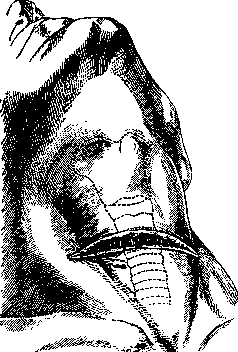
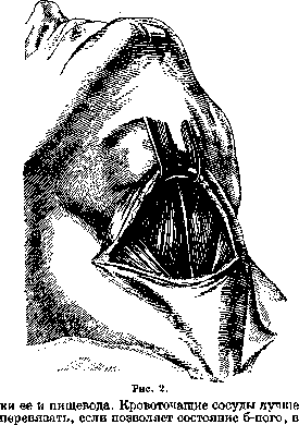
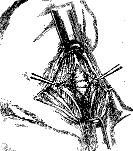
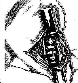
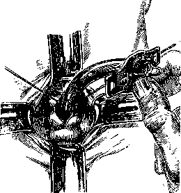
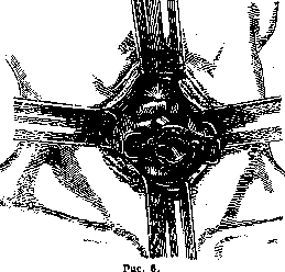
Tracheotomy, thrototomy, incision of the windpipe, is one of the most urgent life-saving operations; it is indicated for all more or less rapidly occurring difficulties in breathing in the upper respiratory tract, caused by inflammatory processes of the larynx, tumors, penetration of foreign bodies, trauma, edema, etc. Sometimes tracheotomy is performed as a preliminary act in operations on the mouth and pharynx, as well as in the larynx, with the aim of preventing blood from flowing into the respiratory tract (prophylactic, preliminary tracheotomy). Depending on where the narrowing of the lumen (stenosis) is located in the windpipe, either the trachea is incised—upper or lower tracheotomy—or the overlying parts of the respiratory tract are split: the cricoid cartilage alone or together with lig. crico-thyreoideum med., s. conicum (cricotracheotomy), or also the thyroid cartilage (laryngotomy); less frequently, individual parts of the windpipe are split—one thyroid cartilage or one lig. conicum. The upper part of the trachea from the lower edge of the cricoid cartilage to the isthmus of the thyroid gland is more frequently and more easily incised—upper tracheotomy; the splitting of the trachea below the isthmus of the thyroid gland is called lower tracheotomy; an intermediate tracheotomy was also proposed. Technique of upper tracheotomy. The operation is performed under general or local anesthesia, and in cases of severe asphyxia, also without anesthesia. The patient is placed on the back with the head thrown back, a roll is placed under the shoulders. The surgeon stands on the right side of the patient, fixes the larynx with the middle and index fingers with the left hand, finds the space between the thyroid and cricoid cartilages with the index finger, and makes a longitudinal incision along the midline of the neck from the middle of the thyroid cartilage downward or a transverse incision (Fig. 1) one transverse finger below the cricoid cartilage. The skin with subcutaneous tissue and aponeurosis are incised; arteries and veins are ligated and incised; then between the mm. sterno-hyoidei on both sides, the adhesion, which has the appearance of a white line, is separated; these muscles are spread apart with a blunt instrument (Figs. 2 and 3); they are held with blunt hooks; in the upper corner of the incision, the cricoid cartilage is found and pulled forward and upward with a sharp hook; along its lower edge, a transverse incision of the fascia is made, in the thickness of which is contained the isthmus of the thyroid gland; it is separated from the anterior wall of the trachea with a blunt instrument and pulled downward with a broad blunt hook; then, strictly following the midline, two or three rings of the trachea are incised, blunt hooks are immediately inserted into the incision, and a cannula is inserted into the expanded wound (Figs. 4, 5, and 6). When inserting the cannula into the trachea, one must ensure that its end passes into the lumen of the trachea and does not separate the mucosa or get between the mucosa and the cartilage; the tracheal incision must be large enough for the cannula to enter it without effort. When performing upper tracheotomy, one must carefully avoid injuring the cricoid cartilage, as this leads to the formation of
Figure 1.
persistent stenoses; when incising the trachea, do not go too deep so as not to injure the posterior wall; otherwise, leave it under a clamp. 728 Lower tracheotomy is a more difficult and dangerous operation than upper tracheotomy, because at this level the tracheon goes deeply backward and is surrounded by a dense network of venous vessels; the a. thyreoidea ima is also often found here. Even when the smallest branches are injured, profuse bleeding that is difficult to stop occurs. The skin incision is made strictly along the midline of the neck from the lower edge of the cricoid cartilage downward to the jugular fossa; after incising
Figure 3.
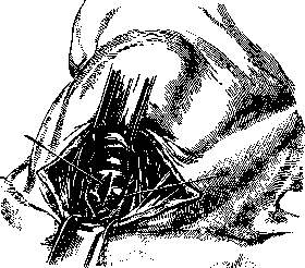
the skin, subcutaneous tissue and aponeurosis, one bluntly penetrates deeply between the mm. sterno-hyoidei, separates the loose connective tissue lying on the trachea, exposes the trachea, pulls it forward with a sharp hook, and incises it and inserts a cannula (Fig. 7). Lower tracheotomy is indicated where the upper part of the trachea is occupied by edema, tumor, scars, a foreign body, as well as in children with diphtheria.-Transverse tracheotomy. The skin incision is made transversely at the level of the cricoid cartilage, veins are ligated, muscles are separated along the white line, the trachea is incised with a small transverse incision along the
Figure 4.
lower edge of the ring, and a cannula is inserted into the opening. In cases where there is great hurry with the operation, a cricothyrotomy is performed: a simple or specially adapted knife (cricotome) is thrust into the upper part of lig. conici and, moving it down strictly along the midline, the cricoid cartilage and 2 or 3 rings of the trachea are incised; after stopping the bleeding, a cannula is inserted.
The tracheotomy cannula is a double tube, one fitting into the other, somewhat curved; the outer one is connected with a movable shield, which serves to fix the cannula on the neck; the inner one, easily removable and insertable, serves for cleaning. The diameter and length of the cannula are determined depending on age, configuration of the neck, and location of the obstruction in the trachea. Materials for cannulas include silver, platinum, nickel silver, aluminum, hard rubber, glass. Many modifications of tracheotomy tubes have been proposed, but in practice, silver tubes of Luer and hard rubber tubes of Trousseau have been retained. Tampooning tubes of Trendelenburg have a special design; they not only maintain free breathing but also do not allow liquids (blood, mucus) to pass deeply into the trachea due to the enclosing and inflating rubber balloon; Halm achieved the same by wrapping the tubes with compressed sponge. The cannula inserted into the trachea is left there, not removed, for 4-6 days; only the inner tube is removed for cleaning as needed; then during dressings, an attempt is made to remove the entire cannula and, by plugging the tracheotomy opening with a tampon, to see how passable the larynx has become; with good, free
Figure 7.
breathing, the cannula is no longer inserted; how much passability has been restored can be easily determined by using a cannula with a window (opening) on the upper convex wall and plugging the outer opening with a stopper. Removal of the cannula sometimes presents great difficulties (see Decannulation).
a. Pianov. Tracheotomy in operations on animals serves in physiology for establishing artificial respiration in all cases of physiological experiment, for some reason during the experiment natural respiration is turned off. T. is mandatory in experiments on curarized animals, in experiments requiring opening of the chest cavity or a high (below the medulla oblongata) transection of the spinal cord. T. is performed mainly on dogs, rabbits and cats as the most commonly used in experimental physiology and pathology of animals. The technique of T. coincides with the technique of the operation for applying a tracheotomy cannula and is the same for the listed species of animals. The operation is performed as follows. The animal is fixed on the operating table. The hair on the neck is shaved, and in rabbits it is removed with a depilatory. Under anesthesia, the skin is incised precisely along the midline from the lower edge of the thyroid cartilage downward for a distance of 4-6 cm. Then the subcutaneous tissue and the skin muscle (m. subcutaneus colli) are incised. After this, if the incision was made correctly along the midline, a narrow white strip of connective tissue is immediately found between the approximated edges of the sternohyoid muscles (mm. sterno-hyoidei). By pushing apart the edges of these muscles bluntly, the trachea is immediately exposed, which is also separated from the adjacent tissues bluntly. At the same time, care is taken that the n. recurrens definitely moves away from the trachea, because otherwise it may get into the ligature when tying the cannula. After freeing the trachea, a sufficiently strong ligature is passed under it and the trachea is incised transversely between the cartilaginous rings for two-thirds of its lumen. The tracheal end of a cannula selected for size is inserted into the incision and is firmly secured in the lumen of the trachea by tightening the ligature. If the operation was performed correctly, there are usually no complications. It should also be noted that the most convenient place for the tracheal cannula is the midpoint between the lower edge of the thyroid cartilage and the upper edge of the sternum. Tracheal cannulas used for physiological T. vary in shape but differ little in design principle. They are metal or ebonite tubes of various diameters corresponding to the most commonly encountered sizes of the tracheal lumen in the above-mentioned animals, and in factory production are usually sold as a set of five sizes of tracheal cannulas. Among the most commonly used, one can name the tracheal cannulas of Francois-Franck, Ludwig, and Verdin. In the simplest form, the tracheal cannula (e.g., of the firm Zimmermann, Leipzig) is a metal tube with olives at the ends and with a slit-shaped opening located in the lateral wall of the cannula. The slit can be closed by a movable shield to various degrees, which achieves regulation of the magnitude of pulmonary ventilation and facilitates the removal of exhaled CO2. The most complex tracheal cannulas are given a T- or Г-shaped form, in the latter case with rotating elbows, which significantly facilitates manipulation with them during the experiment. In addition, some types of tracheal cannulas have valves regulating the direction of air during inspiration and expiration and are used mainly in the study of air exhaled by the animal (e.g., the Verdin valve tracheal cannula). However, in case of need, one can easily do without special cannulas and successfully replace them with a T-shaped glass tee of suitable diameter, on the side outlet of which a rubber tube is fitted, narrowed to the required size by a screw clamp according to the desired magnitude of pulmonary ventilation. A very significant inconvenience when using tracheal cannulas of conventional design is the inability to maintain proper anesthesia, especially ether-chloroform, which is the most accessible and commonly used in the most diverse experiments, especially on dogs. In the simplest case, this inconvenience can be overcome by introducing into the lumen of the tracheal cannula through a side opening a wick made of gauze, moistened from time to time with ether or chloroform. However, this method is not very convenient and does not always achieve the goal. Therefore, it is better to use special apparatuses adapted for anesthetizing tracheotomized animals. The latter includes the simple and easy-to-use apparatus of Tigerstedt (fig. 8). The apparatus consists of a flask d, the lower end of which is connected with a tap e, passing into a cone, with a Y-shaped tee a, the straight leg of which is connected with an artificial respiration apparatus. Tube b also connects the latter with vessel d, which achieves equal pressure in the latter and in tee d, facilitating the outflow of ether or chloroform when tap e is open. The stopping of the outlet opening of tap e is done by a micrometric screw k, which regulates the required depth of anesthesia.
b. Indications. T. is indicated in cases of acute or chronic obstruction of the upper respiratory tract, when the obstruction cannot be removed by other means. The most common causes of such obstruction are: 1) foreign bodies in the larynx or trachea; 2) edema of the larynx or trachea (burns, injuries, diphtheria, etc.); 3) tumors of the larynx or trachea; 4) stenosis of the larynx or trachea (cicatricial, congenital, etc.); 5) paralysis of the laryngeal muscles. In addition, T. is performed in some operations on the neck and upper mediastinum to improve access to the surgical field and to facilitate the management of anesthesia. In military surgery, T. is used as a life-saving measure in cases of gunshot wounds of the neck with damage to the larynx or trachea.
c. Technique. T. can be performed under local or general anesthesia. The patient is placed in a supine position with the neck extended. The skin is disinfected and draped. A midline incision is made from the lower edge of the thyroid cartilage to the suprasternal notch. The subcutaneous tissue and platysma muscle are divided. The sternohyoid and sternothyroid muscles are separated in the midline. The trachea is exposed and a tracheal hook is inserted under it. The trachea is incised transversely between the second and third cartilaginous rings. A tracheal cannula is inserted and secured with tapes. The incision is closed around the cannula.
d. Complications. The most common complications of T. are hemorrhage, infection, subcutaneous emphysema, pneumothorax, and damage to the recurrent laryngeal nerve. Late complications may include stenosis of the trachea and granulation tissue formation. The mortality rate from T. is relatively low, but it depends on the cause of the obstruction and the patient's general condition.
Related articles
Mentioned in
Cite this page
“Tracheotomy.” Soviet Medical Encyclopedia. English translation of Bolshaya Meditsinskaya Entsiklopediya, 1st ed. (Moscow, 1928–1936), ed. N. A. Semashko. https://sovietmedicalencyclopedia.pages.dev/article/tracheotomy/