Foreign Bodies
Historical document, translated for reference. It reflects medical knowledge of the 1920s–30s and is not medical advice.
Summary
This article from the 1928-1936 Soviet Medical Encyclopedia describes various types of foreign bodies that can enter the human body, their locations, clinical manifestations, diagnostic approaches, and treatment methods, with special attention to historical medical practices and terminology.
Encyclopedia article (1928–1936)
FOREIGN BODIES, objects not natural to the organism but entering from outside into some part of the human body. If we do not speak of the penetration into the body of bullets and shell fragments, then in most cases foreign bodies enter accidentally. In mentally ill patients, foreign bodies are found, which have entered mainly into the stomach by swallowing. In outpatient practice, one often encounters fragments of needles left in the subcutaneous tissue, and sometimes deeper, in the muscles. The favorite locations are the hands, the outer surfaces of the shoulders, and the buttocks. In the hands, needles are most often found in the fingers, and then in the thenar and hypothenar areas (seamstresses, laundresses, domestic workers) (see separate table, figure 3). In the shoulder and buttocks, needles break and remain when some medicinal substance is injected subcutaneously, and since injections in the buttock area are intramuscular, the needle usually breaks at the base. Often such a needle fragment causes no clinical symptoms and can remain in the carrier's body for years without causing any unpleasant sensations. Cases of movement of a needle fragment are extremely rare and occur only when the needle is in a muscle, which is why it moves under the influence of the latter's contractions. Usually, however, the needle remains in the place where it first entered. Removal of needles often presents great difficulties. On the hand, if the needle is not felt completely clearly before attempting removal, it is necessary to take an X-ray, necessarily in two planes. Only then can one determine the location of the needle. The incision must be large enough, as it is very difficult to remove a needle from a small incision. Particularly great difficulties are presented by the removal of a needle from the buttock. X-ray usually helps little in these cases, and the abundance of fatty tissue sometimes creates insurmountable difficulties, and often, if the needle does not cause the patient any discomfort, it is better not to attempt to remove it. Besides needles, the role of foreign bodies in the skin and subcutaneous tissue can be played by wood particles ('splinters'), plant needles, etc. Furthermore, to foreign bodies detected within tissues are included dead parasites and their parts, threads of surgical ligatures, injected coagulated masses (paraffin); finally, epidermal scales and hairs that penetrate into tissue during ulcerative processes on the skin, salt deposits (lime, uric acid salts) and crystals (cholesterol). Patho-anatomically, the presence in tissue of all the aforementioned foreign bodies is accompanied by processes of encapsulation with the formation of so-called giant cells of foreign bodies. When foreign bodies contain pus-forming bacteria, suppuration develops. In the oral cavity, fish bones that become embedded in the tonsils, as well as in the mucous membrane of the pharynx behind the arches, have practical significance. A careful examination of the corresponding area easily detects the foreign body, which is removed either with a long, angled forceps or with a curved tonsil forceps. The situation with foreign bodies of the esophagus is significantly more complicated. According to data from Moscow morgues for 1923-27, foreign bodies of the esophagus were noted 28 times (in 33,609 autopsies). Most often, one has to deal either with accidentally swallowed meat or fish bones that have become stuck somewhere along the esophagus, or with swallowed artificial teeth and prostheses. Cases of extraction of a five-kopeck coin from the esophagus have been described. Patho-anatomically, the effect of foreign bodies on the esophageal wall can vary depending on the size and shape of the foreign body. With a smooth surface, the foreign body may not cause any changes in the esophageal mucous membrane, whereas foreign bodies with sharp ends or projections cause damage to the esophageal wall in the form of either a puncture or a pressure ulcer of varying depth and width; when such areas become infected, suppuration often develops, which represents a very serious complication, as it easily spreads to the mediastinum and causes purulent mediastinitis, which usually ends in the death of the patient. Some sharp foreign bodies (e.g., fish bone, pin, etc.), by perforating the esophageal wall, with their sharp ends penetrate into the trachea or bronchus, and sometimes into the aorta; perforation of the latter can cause acute fatal bleeding. Clinically, patients experience varying degrees of difficulty during the act of swallowing, depending on the size, shape, and location of the foreign body. X-ray easily determines bodies of larger size and those that do not allow rays to pass through. Otherwise, esophagoscopy should detect the foreign body (see separate table, figure 4). It is not recommended to try to push a stuck foreign body into the stomach with a probe to avoid injury to the esophageal wall, which can lead to the introduction of infection. If possible, every foreign body of the esophagus should be attempted to be removed by esophagoscopy. In case of failure, one has to operate; when the foreign body is located in the upper part of the esophagus, an external incision of the esophagus on the neck is made. With a lower position-gastrotomy with an attempt to remove the foreign body through the cardia. In clinical practice, one encounters foreign bodies that have accidentally entered the pleural cavity. Such bodies are usually rubber drains used in the treatment of purulent pleurisy. If they are not secured to the dressing or, as is practiced, the outer end is not pierced perpendicularly with an English pin, the drain can slip into the pleural cavity and, if left there, maintain a fistula. X-ray usually clarifies the cause of the unhealing fistula in such cases, and one only needs to remove the drain. Sometimes this can be done with a tonsil forceps through the fistulous tract without expanding it; if this is not possible, then the fistula needs to be expanded to the required size. Foreign bodies of the stomach impress with their diversity, and sometimes their quantity. Out of the total number of foreign bodies, 1% fall to the stomach. Incredible cases of foreign bodies that entered the stomach by swallowing have been described repeatedly. The most diverse metal objects have been extracted from the stomach. A case of removing 1,000 needles from the stomach of a mentally ill patient has been described. A special form of stomach foreign bodies are the so-called 'hair tumors' (see Bezoar), which are relatively rarely encountered. They form from swallowed hair in people who have the habit of biting the ends of their hair. Sometimes tumors identical to 'hair tumors' form from vegetable fibers. The swallowing of such fibers is associated with profession (upholsterers, spinners, seamstresses). The behavior of patients in the presence of foreign bodies is extremely diverse-ranging from false alarm to intentional denial of the existing foreign body. Therefore, first of all, one must be sure that there really is a foreign body, which is easily achieved with the help of X-rays. If the foreign body does not pass further into the intestine but remains in the stomach, then the further consequences for the patient depend on the size, mobility, and external shape of the foreign body, which may either cause no symptoms or give indefinite sensations in the stomach area that are not characteristic. Foreign bodies with sharp ends, such as nails, glass fragments, needles, dental prostheses, pose a danger of damage to the stomach walls up to perforation. The prolonged presence of a foreign body can contribute to the development of an ulcer with a number of subsequent complications. If one is sure that the foreign body, due to its size and shape, will not pass into the intestine, it is necessary to resort to its surgical removal by gastrotomy, which is usually a simple and easily accessible intervention if undertaken in time. According to the collective statistics of Belfler and Lieblein (Wolfler, Lieblein) out of 1,184 cases of foreign bodies in the stomach, a fatal outcome occurred in 190 cases from various causes (16%). According to the same statistics, in 34%, i.e., in 434 cases, the foreign body exited per vias naturales. Foreign bodies of the intestine can be, just as in the stomach, of the most diverse shape and different origin. A case has been described where in a hernia of the linea alba, X-ray examination showed the presence of a needle; in this same patient, needles were found in significant quantity in the stomach and intestine. Murphy's button can also become stuck in the intestine, causing symptoms of obstruction. A special type of intestinal foreign bodies are ascarids, which, when gathered into a ball, can give a severe picture of intestinal obstruction. Foreign bodies of the rectum are not uncommon; they enter the rectum either through the mouth, passing through the entire intestine, or from the side of the anus. Foreign bodies of the first group are most often encountered. The second group of foreign bodies consists of bodies introduced from the side of the anus. From the rectum, ends of enema tubes that accidentally slipped into the rectum have been extracted. Sometimes foreign bodies are intentionally introduced into the intestine to hide something. A known case when a patient introduced a hundred-bottle cap into his rectum. Usually, the foreign body can be easily felt with a finger or is even visible to the eye. Treatment consists of removing the foreign body after preliminary dilation of the sphincter, preferably under anesthesia. Foreign bodies of the abdominal cavity have special significance for the surgeon. This group consists of bodies that enter the abdominal cavity during surgery and are forgotten there.
The conditions under which a foreign body is left in the abdominal cavity sometimes come down to simple chance or the surgeon's forgetfulness, quite understandable to anyone who has performed complex operations in the abdominal cavity. In other cases, unexpected complications may arise that can confuse the operating surgeon. Mainly, gauze sponges and hemostatic forceps are left forgotten in the abdominal cavity. The latter, in case of a defective so-called 'spring' lock during the operation, unfasten and unnoticedly slip into the abdominal cavity. The fate of such foreign bodies varies. Some remain in the abdominal cavity for a long time, causing various clinical disorders, some, 'making their way' into the intestine, exit through the anus, while others, causing a bedsore of the intestinal wall, form around themselves a encapsulated peritonitis with a series of complex fistulas between the intestinal loops. Thus, Krause described a case where a hemostatic forceps accidentally left by him caused the formation of a series of fistulas between the loops of the small intestine and required an extremely heavy and painstaking operation to restore normal intestinal function and remove the forceps. To prevent such an accident, it is necessary to exercise special care during large laparotomies, paying attention to gauze sponges and forceps. The edges of the sponge are best grasped with clamps. The instruments should be counted before the operation and before suturing the abdominal cavity. It is also necessary to carefully examine the operated area before the end of the operation. The origin of foreign bodies of the bladder can be varied. A large group consists of foreign bodies that enter the bladder during unsuccessful medical manipulations. Casper mentions a gauze sponge left in the bladder during a high suprapubic cystotomy. In most cases, the matter concerns broken catheters (see separate table, figure 5) or bougies. The second group of foreign bodies consists of the most diverse objects that are introduced into the bladder during masturbation. Favorite objects for this purpose are women's hairpins. A case is described of removing a broken metal hanger from the bladder of a 17-year-old girl. Upon entering the bladder, a foreign body very soon causes the formation of cystitis and disturbance of urination. A careful examination of the bladder cavity with a cystoscope reveals the foreign body. If it is difficult to establish the size and shape of the object with a cystoscope, an X-ray should be taken, which will already bring final clarity, which is necessary for deciding on the methods and paths of removal. First of all, one must think about not injuring the mucous membrane of the bladder wall with the foreign body during removal. If removal via natural routes raises doubts, a high suprapubic cystotomy should be performed. However, it must be kept in mind that in women the urethra is short and easily stretches, and in persons who have practiced masturbation- especially. This circumstance often makes it possible to remove the foreign body naturally, without surgery. Finally, the last group consists of foreign bodies that enter the bladder from the abdominal cavity side (e.g., from the intestine, from abdominal cavity organs) by way of a previously formed fistula. Foreign bodies of the urethra are encountered much less frequently than foreign bodies of the bladder. Most often, pieces of catheters that break off during insertion into the bladder get into the urethra. In the female urethra, pins introduced for the purpose of masturbation have been found; the clinical picture comes down to disturbance of urination and painful sensations in the canal. Urethroscopy establishes the presence of a foreign body. Treatment consists of removing the object, if possible without incising the urethra. Foreign bodies of the vagina have no great practical significance. Forgotten tampons introduced for therapeutic purposes, pessaries introduced and forgotten have been found. In most cases, they do not give clinical phenomena and are an accidental finding. In some cases, foreign bodies remained in the vagina for several years before they were noticed. Finally, foreign bodies can sometimes also be found in the uterine cavity. In most cases, this is some object that got there during one or another medical procedure. During intrauterine cauterizations, pieces of cotton wool can easily enter the uterine cavity. Cases are known of finding fragments of instruments (fragments of a curette- see separate table, fig. 1) in the uterus. If the removal of the foreign body is associated with difficulty, it is necessary to recommend dilatation of the cervical canal and internal os and even hysterotomy.
a. Bakulev. Foreign bodies of the ear. Pebbles, beads, buttons, sunflower seeds, beans, pieces of paper, rags, grains of fruit, etc.-such are the foreign bodies of the ear, most commonly found in children (see separate table, fig. 6). In adults, forgotten pieces of cotton wool in the ear canal, pieces of ear of wheat (during field work), pieces of matches, etc., are most often found. Sometimes live insects, such as flies, bugs, fleas, beetles, get into the auditory canal, causing extremely distressing sensations. Foreign bodies can lie for a long time without causing any complications. Cases are described of the presence of foreign bodies in the ear for 40-45 years. The foreign body bothers the patient if it has sharp edges or, under the influence of body heat, swells, blocks, and irritates the walls of the ear canal. In rare cases, a foreign body can be the cause of reflex nervous phenomena (cough, convulsions, facial neuralgia, salivation, mental disorder). In most cases, however, foreign bodies bother the patient little or not at all. Therefore, foreign bodies by themselves do not represent a dangerous disease. The danger lies only in the unskillful removal of the foreign body, if the patient himself, or a nurse, or a doctor, attempting to remove the foreign body with the help of some sharp instrument (hairpin, forceps), wedges the foreign body even deeper, violates the integrity of the skin of the ear canal, and sometimes also the integrity of the eardrum, and introduces infection into the outer, middle ear, and sometimes deeper. With few exceptions, foreign bodies yield to removal by means of irrigation (a large syringe of 100-150 cm3, a powerful stream of water). If the foreign body is wedged, a blunt or sharp hook is introduced (which should be done by a specialist doctor under the control of a mirror) flat between the wall of the auditory canal and the foreign body, then the hook is turned so that its tip takes a downward direction, and the foreign body is easily removed in this way. If the foreign body is wedged in the bony part of the auditory canal and attempts to remove it through natural routes are unsuccessful, they proceed to the operation of bloody removal: an incision behind the ear, the edges of the incision are pulled back to the ear canal, the latter is incised, and the foreign body is removed. In rare cases, it is necessary to chisel away a part of the bony auditory canal with a chisel. With living foreign bodies (flies, etc.), it is best first to pour 5% Carbolglycerin or oil into the ear canal, and then only irrigate the auditory canal. When removing a foreign body from the ear canal in children, due to their restless state, anesthesia often has to be administered.
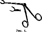
Foreign bodies of the nasal cavity. Method of entry: Foreign bodies are intentionally inserted into the nose by children, enter the nasal cavity during vomiting, through the choanae, in accidents-injuries (bullets, shell fragments). Symptoms are expressed 1) in obstruction of the corresponding nasal cavity and 2) in purulent, sometimes bloody discharge. With a long stay of a foreign body in the nose, calcium and magnesium salts are deposited around it, thanks to which a rhinolith is formed. The diagnosis is made on the basis of anterior rhinoscopy, sometimes on the basis of probing with a probe. Treatment consists in removing the foreign body, after which all inflammatory phenomena in the corresponding nasal cavity quickly subside. Before removal, it is better to smear or introduce a few drops of adrenaline (1:1,000) into the nose. The simplest method of removing a body from the nose is blowing the nose. If this method does not give results, the foreign body is removed under the control of a mirror: a hook is introduced behind the foreign body and the body is thus removed from the nose. Sometimes instead of a hook, Weingartner's forceps (fig. 1) is used. In exceptional cases-with very large foreign bodies-it is necessary to make an incision of the external nose. Foreign bodies of the larynx, trachea, and bronchi. The most diverse objects can enter the respiratory tract: parts of toys, pieces of food, bones, grains from berries (cherry, plum), beans, parts of grass stems, ear of wheat, metal objects (fig. 2 and 3), nails, money (see separate table, fig. 2); in the USSR, sunflower seeds most often enter the respiratory tract. Into the larynx, trachea, and bronchi, foreign bodies enter mostly from the oral cavity, rarely-from the outside (wounds). For a foreign body to enter the larynx or trachea, it must overcome the natural protective reflexes; these reflexes decrease during intoxication, in hysteria. The mechanism of entry is always the same: deep inspiration, fright, expressed in a cry, weeping. Most often, foreign bodies enter the respiratory tract in children. The explanation lies in the habit of children to put all objects in their mouths. A foreign body, overcoming the reflex cough and closure of the glottis, can get stuck in the larynx or penetrate deeper-into the trachea or bronchi. Hence the natural division into foreign bodies of the larynx and foreign bodies of the trachea and bronchi. According to data from Moscow morgues for 1923-27, foreign bodies of the respiratory tract were noted 75 times in 33,609
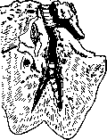
Figure 3. Part of a dental prosthesis in the bronchus of the right lower lobe. > Of
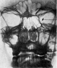
Fig. 1. Fragment of a curette left in the uterus during an abortion and extracted by hysterotomy. Figure 2. A ten-kopeck coin in the trachea of a child (its shadow during swallowing). Figure 3. A needle in the soft tissues of the palmar surface of the hand, extracted by operation (from the Surgical Clinic of the 1st Moscow State University). Рнс. 4. Esophagoscopic picture of a peach pit wedged at the entrance to the stomach (from Kirschner-Nordmann). Figure 5. The end of a broken glass catheter in a niche of the bladder (from Kirschner-Nordmann). Figure в. Foreign bodies extracted from the ear (from Feldman's collection). Figure 7. A metal fragment (o) in the left eye. (Figure I, 2 and 7-
From the X-ray Institute of the Clinical Faculties of the 2nd Moscow State University.) autopsies.-F. b. of the larynx. F. b. get stuck in the larynx either because of their volume or because of the unevenness of their edges. F. b. of the larynx mostly belong to the category of immobile, wedged foreign bodies. Symptoms: reflex cough, rapidly occurring hoarseness, often complete aphonia, shortness of breath. Cough should be considered as a protective reflex, with the help of which the patient tries to free himself from the F. b.; if the F. b. becomes wedged between the vocal cords and prevents their closure, then sudden aphonia occurs-this is almost a reliable diagnostic sign. If the F. b. is not so large and does not interfere with partial closure of the vocal cords, then hoarseness occurs. Finally, with a voluminous F. b., severe, sometimes inspiratory and expiratory shortness of breath (due to obstruction of the glottis) may occur, which in severe cases requires tracheotomy. The diagnosis is made based on 1) history, 2) the above-mentioned signs, 3) indirect (very difficult in children, sometimes impossible) and 4) direct laryngoscopy. Differential diagnosis: it can be confused 1) with diphtheria (history and temperature different), 2) with laryngeal papillomas (slow progression of aphonia), 3) with false croup {catarrhal phenomena are present in the nose and pharynx), 4) with laryngeal syphilis (history, other manifestations of the disease). In all cases, direct laryngoscopy decides the issue. The prognosis depends 1) on the size of the F. b. closing the larynx, 2) on the speed with which one turns to a specialist doctor, and 3) on the age of the child (the older the child, the more favorable the prognosis). Removal in children is best accomplished with the help of direct laryngoscopy, which requires no, neither local nor general, anesthesia. A laryngeal spatula or a wide bronchoscopic tube is inserted; the body is grasped with forceps, gently rotated movements are used to free it from the surrounding tissues and remove it. Due to the displacement of the foreign body, sudden asphyxia may occur, therefore instruments for tracheotomy should be ready. F. b. of the trachea and bronchi. F. bodies penetrate more often into the right bronchus than into the left. This is explained by 1) the larger diameter of the right bronchus, 2) the right bronchus as it were constitutes a continuation of the trachea and departs less vertically, the site of bifurcation is deviated "from the midline to the left (fig. 4). F. bodies of the trachea and bronchi can be mobile or immobile (volume, rough edges), they can cause immediate or distant symptoms. The first include: a sharp, reaching to vomiting, to suffocation attack of cough, with redness of the face, welling up of tears; sometimes cyanosis of the face; cough, recurring in attacks, during the day and Hoqbio (more at night) and- always accompanied by the release of sputum, sometimes bloody; by the force of the expiratory stream of air the F. b. is thrown upward (if it is mobile), strikes the spastically closing glottis, which gives a characteristic splashing sound, and falls back; this splashing can be established by auscultation, it is easily recognized also by palpation ];. zh. э. т. XI.
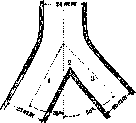
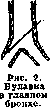
Figure 4. Anatomical justification for the most frequent entry of foreign bodies into the right bronchus: 1 and 3-right and left bronchi. The right one is significantly wider than the left, and the angle (2) formed by it with the left bronchus lies to the left of the midline. (fingers of the left hand on the jugulum sterni). To these symptoms is added a slight change in voice (hoarseness), shortness of breath, general restlessness and retraction of the skin under proc. xiphoideus. After some time, the mucous membrane of the trachea and bronchi gets used to the presence of the F. b., from which the cough becomes rarer, but its character-in attacks-does not change. Patho-anatomically, depending on the nature of its surface, the F. b. causes either insignificant damage to the mucous membrane, or more significant injury, or finally perforation of the wall (e.g., when a pin enters the trachea); a sharp F. b. can penetrate through the wall of the trachea or bronchus into the esophagus or aorta. A F. body, immovably fixed, e.g., in one of the main bronchi, gives rise in the corresponding place to a persistent change in the mucous membrane in the form of metaplasia of its epithelium into a flat multilayered one or the formation of a defrect-bed sore. Subsequently, due to the infection that is introduced by the F. body, an unresolved unilateral bronchitis (on the side where the F. body is located) develops, often accompanied by dilation of the bronchial lumens and usually passing into bronchopneumonia (Schluck-pneumonia), very dangerous in early childhood; sometimes chronic interstitial pneumonia with bronchiectasis and lung abscesses develop. If the F. body is immovable, its symptoms vary depending on its volume and the obstruction of the bronchus. If a first-order bronchus is obstructed, then the corresponding lung does not breathe, the amplitude of fluctuations of the chest wall is reduced, percussion gives a decrease in tonality (dullness), auscultation gives a decrease in vesicular breathing; unilateral compensatory emphysema (in the healthy lung) and atelectasis on the side of the diseased lung may develop. X-rayoscopy gives positive results if the F. body is metallic or if the shadow from the F. body (bone) does not coincide with the shadow of the ribs and large vessels. In addition, X-rayoscopy is mainly applicable in adults or in children after 4-5 years of age (the X-ray is taken during inspiration). Mortality from F. b., very high in the pre-bronchoscopic period (52% according to Eicken; 20-42% according to Markuzon), sharply decreased after the introduction of the bronchoscope into use (13.5% Eicken; 5.3% Kiilian; 1.7% C. Jackson; 7.5%-by N. A. Shneider). Mortality depends on age (it is greater the smaller the child), on the period of time that passes from the moment of entry of the F. body until bronchoscopy (the longer the period of time, the more often unresolved bronchopneumonias with fatal outcome occur) and on the nature of the F. body. The diagnosis is made based on the history and the above-mentioned symptoms, which together give reliable signs; if there are scattered symptoms, then the history is of decisive importance. The prognosis depends 1) on the size and nature of the F. body, 2) on the age of the patient and 3) on the general condition of the patient. A large F. body can cause sudden asphyxia (a nut obstructing the trachea). The prognosis is especially serious in a breast-fed child due to the narrowness of the respiratory tract and rapidly developing pneumonia. The prognosis is also serious in an emphysematous patient or in a patient suffering from tbc of the lungs. The outcome can be death from asphyxia during the strangulation of the F. body between the vocal cords, more often-protracted pneumonias or pleuropneumonias, sometimes with the formation of an abscess, which can also lead to death; in rare cases, spontaneous expectoration of the F. body may occur (a sunflower, a spike or a grass stem). Prevention should consist in the fact that all small parts of any toys that children put in their mouths should be securely fastened; in addition, it is necessary to wean children from the habit of putting everything in their mouths and not allow them to chew sunflower seeds. Treatment: tracheo-bronchoscopy and removal of the F. body from the respiratory tract. Anesthesia in children is general, in adults-local (10% cocaine solution).
A. Feldman. Foreign bodies in the eye occur according to observations by Russian authors in 18.3% (Kantsel) - 52.4% (Varshavsky) of all cases of eye injuries. Such a difference is undoubtedly explained by the fact that the authors dealt with workers from different industries. Foreign bodies can become lodged in penetrating injuries in the anterior chamber, iris, posterior chamber, lens, vitreous body, retina, and sclera. There can also be double perforation of the eye with penetration of a foreign body into the orbit. According to Varshavsky, out of 1,105 cases from various authors, in 17.9% foreign bodies were located in the anterior segment and in 82.1% in the posterior segment. They most often become lodged in the vitreous body (up to 70%). The lighter the foreign body by weight, the more quickly it is detained in the anterior segment of the eye, and the heavier it is, the more often it penetrates into the vitreous body. Sometimes foreign bodies are visible with side illumination or during ophthalmoscopy, but often they are not visible due to opacities of the lens, vitreous body, and hemorrhages. The presence of foreign bodies in the eye, besides the anamnesis and the appearance of the injury, which sometimes indicates the direction of the wound canal, is suggested by the discrepancy between the relatively mild external injury and severe intraocular changes. Sometimes observed bubbles of air inside the injured eye also have diagnostic significance. But more often, especially in non-fresh cases, it is necessary to resort to special methods to establish the presence of foreign bodies in the eye: sideroscope, large electromagnet, radiography, orthodiagnostic illumination. Unfortunately, the sideroscope, even in the most improved models, gives questionable results with small fragments. Large electromagnets render great service; when approached, in eyes containing foreign bodies susceptible to magnetic action, pain is felt in the vast majority of cases. In isolated cases, even the electromagnet does not give a painful reaction; radiography gives positive results (see separate table, Figure 7) for metallic foreign bodies, but with very small size, they cannot always be clearly distinguished on the film. In this case, it is best to take two films of the eyeball on the same plate with the head in a fixed temporal position: with the eye looking upward and downward (Kohler, Golovin). With orthodiagnostic illumination of the orbit using a well-centered X-ray tube, the eye is examined on a screen. The presence of infection plays the most important role in penetrating eye injuries; in this case, a picture of endophthalmitis develops in one form or another. The nature of the foreign body itself is also not indifferent to the outcome of the injury. The well-known fact is the phenomena of siderosis with iron-containing fragments, chalcosis and suppuration - with copper foreign bodies. Lead is better tolerated by the eyes, and aluminum, glass, stone, and wood are even better tolerated, if they are not infected. The size of the foreign body is also not indifferent to the eye. It has long been known that small fragments, under otherwise equal conditions, are better tolerated by the eye. Damage to the lens, according to the data of most authors, has an unfavorable effect on the outcome of the injury. The location of the foreign body is also very important; in this respect, the ciliary body is especially sensitive; as a consequence of reactive inflammation around the foreign body, retinal detachment can occur. The duration of the foreign body's stay in the eye undoubtedly has great significance for the outcome of the injury. Data from Russian (Varshavsky) and foreign (Lieb'er-mann) authors show that the earlier the fragment is removed, the less danger of infection developing and the greater the chances for a favorable outcome. Hence the importance of timely removal of the foreign body from the eye is clear. Iron and steel fragments are removed by most doctors with the help of large electromagnets by Haab, Volkmann, and Klingelfuess. Using various tips and sometimes changing the current strength with a rheostat (Klingelfuss), they regulate the magnetic attraction force to the desired effect. Fragments from the posterior half of the eye are moved to the anterior chamber. As experience has shown, the lens is usually not traumatized in this process. From the anterior chamber, foreign bodies are removed either through the fresh wound opening or through an incision at the edge of the cornea, and a manual Hirshberg magnet can also be used, introducing its flat tip into the anterior chamber. In cases of iris prolapse, iridectomy is performed. Some ophthalmologists, especially in America, remove foreign bodies from the posterior part of the eye with a small Hirshberg electromagnet, introducing its tip through an incision made in the sclera at the location of the foreign body. But most authors resort to a scleral incision only where it is not possible to move the foreign body to the anterior chamber with a large electromagnet. It is not possible to remove foreign bodies with electromagnets in all cases; the percentage of failures varies among different authors (Russian and foreign) from 5 to 11.4. Much greater difficulties are presented by extracting foreign bodies not susceptible to magnetic action, especially from the vitreous body. In this case, an attempt is made to extract the foreign body with forceps under ophthalmoscopic control, introduced through an incision made in the sclera; success in the operation is a matter of chance. Methods have been proposed to facilitate the operation: removal with the help of an X-ray screen on which shadows from the foreign body are visible, and forceps; diaskleral illumination of the eyeball with the help of a Sachs lamp to obtain a shadow from the foreign body and make a corresponding incision; finally, the telephone-forceps, when it grasps the foreign body, a current is closed and an audio signal is obtained, but all these methods require complex equipment and are accessible to few. But even after extracting the foreign body from the eye, the prognosis remains serious. In this case, 19% to 48% and even 55.6% of all eyes perish, and good vision is preserved in 23-40% (Varshavsky). The best prognosis is given in cases where foreign bodies became lodged in the anterior segment of the eye. The vast majority of ophthalmologists insist on the necessity of removing foreign bodies from the eye even in cases where they do not cause a reaction in the eye, since in most cases they eventually cause inflammatory phenomena. Treatment of inflammatory phenomena in different parts of the vascular tunic with foreign bodies in the eye is carried out according to general rules. For gaping wounds of the cornea, limbus, and sclera, it is recommended to perform conjunctival plastic surgery according to Kunt, and for large wounds, to place sutures on the sclera. Timely and skillful assistance with foreign bodies in the eye can do much to save the already injured eye. But in the fight against eye injuries in general and foreign bodies in the eye in particular, only prevention can have decisive significance. Improvement of production, its mechanization, introduction of devices for eye protection directly on the machines and machines, introduction of specially adapted protective glasses for various industries; strict supervision of the implementation of protective measures for the eyes on the part of both management and workers, with penalties imposed on the guilty from both sides; extensive cultural and sanitary-educational measures, periodic re-examination of workers, professional selection for a given industry - such are the foundations of the fight against eye trauma (Averbakh).
Related articles
Mentioned in
Cite this page
“Foreign Bodies.” Soviet Medical Encyclopedia. English translation of Bolshaya Meditsinskaya Entsiklopediya, 1st ed. (Moscow, 1928–1936), ed. N. A. Semashko. https://sovietmedicalencyclopedia.pages.dev/article/foreign-bodies/