Blepharoplasty
Historical document, translated for reference. It reflects medical knowledge of the 1920s–30s and is not medical advice.
Summary
This article from the 1928–1936 Soviet medical encyclopedia details the historical techniques of blepharoplasty, including sliding flaps, pedicle flaps, and the Italian method. It discusses the surgical challenges of restoring eyelid function and appearance following trauma or tumor removal.
Encyclopedia article (1928–1936)
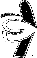
BLEPHAROPLASTY (from the Greek blepharon—eyelid and plassein—to depict, to create), a plastic surgery operation, the task of which is the restoration of the normal appearance and position of the eyelid. However, operations for certain types of eyelid ectropion—non-cicatricial (senile, paralytic, etc.), the correction of which does not require borrowing tissues from other areas—are usually described separately. Corresponding to the variety of causes and the degree of destruction of the eyelids, the methods of surgical intervention are also very diverse. Under ordinary conditions, a physician has to resort to blepharoplasty either for neoplasms or for the purpose of restoring tissues destroyed by a long-standing inflammatory process (anthrax). The most common method used previously was blepharoplasty by shifting flaps cut from the integuments directly adjacent to the defect. This method, proposed by Celsus, was especially cultivated in France (the French call it “par glissement”), which is why it is known by the name of the French method. Figures 1 and 2 present blepharoplasty of both eyelids (for malignant neoplasms) according to the Shimanovsky method. An essential feature is the long lateral angles, which makes it possible to pull together with sutures the edges of the defect resulting at the site of the moved flap. In plastic surgery of the lower eyelid, it is recommended, after freshening the edges of the eyelids, to suture the palpebral fissure tightly. In general, this method is easily applicable, mainly for skin and skin-muscle defects, at Figure 2.
in the destruction of the eyelid, however, blepharoplasty according to
Shimanovsky by lateral shifting of flaps [is used] ONLY for small defects. Blepharoplasty through the full thickness is used only for small defects.
The disadvantage of this method is that even with smooth healing, quite a few new, albeit linear, scars remain on the face; furthermore, in plastic surgery of the lower eyelid, the latter does not receive sufficiently strong support and often sags in the future. The impossibility of obtaining material on the spot (scars in the vicinity or large defects that cannot be closed by shifting) forced surgeons to turn to places more distant from the eyelid and take flaps of the desired shape and size from there, throwing them over the normal skin surrounding the defect or only moving them, while the connection with the mother soil is maintained by means of a more or less long pedicle. This method, proposed by Fricke, is more often called in Russian the transplantation of a flap on a pedicle. This method is widely used, in general, in plastic surgery for replacing old tissue losses (consequences of burns, ulcers, etc.) and fresh losses (during the removal of various
neoplasms). In the first case, it is necessary to carefully remove the scar tissue and completely stop the bleeding by means of pressure or temporary application of hemostatic forceps, with twisting of the vessels if necessary, but without ligatures, which, being a foreign body, do not favor the engraftment of the flaps. For the upper eyelid, the flap is usually cut from the skin of the temple, and for the lower—from the cheek, with the base of the pedicle having to be located on the line of the palpebral fissure, and the outer incision must, in plastic surgery of the lower eyelid, begin above the palpebral fissure, and for the upper—below it (see Figure 3). The transplanted flap immediately after its separation from the mother soil shrinks a little, which continues in the future; therefore, it is necessary to cut the flap with the calculation that its surface exceeds the defect by 1/3. When turning the flap by 90°, a significant thickening is obtained in the bend, which is why the pedicle must be made thinner here. Even with a completely smooth postoperative course and good engraftment of the transplanted flap, a somewhat unpleasant appearance is obtained from the pedicle, to eliminate which Professor Golovin proposed cutting the pedicle and moving it back to its former place by bloody means. With such an addition, the operation gains a lot cosmetically. To replace defects in the inner corner or inner parts of the eyelids, the flap has to be taken from the forehead or the lateral surface of the nose; in such cases, it is inevitably necessary to turn it by a minimum of 90°, which is undesirable. Morax proposed for such cases to cut the flap in the region of the frontal artery so that the vascular bundle (artery and vein) is not cut, but isolated, which makes it possible, by ensuring the best nutritional conditions for the flap, to turn it not only by 90°, but by 120° and even 180°. Most authors recommend, after cutting the flap, to suture first the wound obtained at the site of cutting, and only then proceed to fitting the flap.
The negative side of these methods of blepharoplasty is the inevitable new scar strips on the face, only sometimes covered by the hair of the head or beard. It is understandable, therefore, the desire to replace flaps from the face with those from other parts of the body, while fully ensuring, however, good nutritional conditions provided by the pedicle. As such, the Italian method was put forward—transplantation of a flap on a pedicle from the inner [Figure 3. Blepharoplasty with a flap on a pedicle. The dots indicate the dimensions of the flaps at the moment of cutting, and the lines—after their shrinkage] surface of the arm, for which the arm corresponding to the eye was fixed with the forearm on the head by means of a corset and bandages. Such a position had to be maintained for 10–14 days; this is so burdensome for the patient that sometimes it is necessary to separate the flap from the arm prematurely. To avoid this, Professor Elschnig in 1918 resorted to such a method: he cut a long (18 cm) flap starting above the middle of the clavicle and going down and inward to the xiphoid process (flap width 8 cm); under this flap, connected to the mother soil by its upper and lower ends, he placed a layer of gauze, and when the flap strengthened and good blood circulation appeared in it, its lower end (on the 21st day) was partially, and then after a few days completely, separated, turned almost 180°, and used for plastic surgery of the lower eyelid. But even in 1908, Morax reported on a successful blepharoplasty (according to Snydacker)—a flap 4 cm wide was cut by two parallel incisions starting at the mastoid process and going to the sternoclavicular joint. The lower end of the flap was given the desired shape, and the upper one remained in connection with the organism and served for nutrition. Thus, a flap with a very long pedicle was obtained, and after the engraftment of the lower end, it was possible to cut its beginning and obtain a flap again on
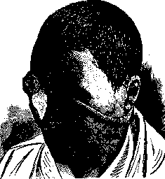
Figure 4. Plastic surgery of the two inner thirds
of both eyelids and the lateral wall of the nose using a flap on a round pedicle according to Filatov. The pedicle is used for further plastic surgery if necessary. This method is especially suitable for blepharoplasty in cases of extensive loss of skin and tissue, but its weak point was the frequent necrosis of the middle of the pedicle, which is why Professor Filatov proposed, without detaching the lower end, to suture the edges of the flap and thereby turn it into a round pedicle (see Figure 4). Simultaneously, the edges of the wound under the pedicle are sutured. This is the first preparatory step. A dressing is applied to the neck, and after complete healing (not less than two weeks), a newly formed circulatory system is obtained in the pedicle, which ensures the nutrition of the future flap, into which one can preliminarily implant mucous membrane, cartilage, etc. Since incisions on the neck leave noticeable scars, Filatov began to cut this pedicle in more distant areas covered by clothing and from there, by means of several transfers of one or the other end, bring it to the desired location. This method also does not produce new scars on the face, and, in case of failure, does not cause even greater disfigurement, which constitutes the main advantage of blepharoplasty. Unfortunately, this type of eyelid plastic surgery is not always applicable, since a transplanted free flap is in worse conditions in terms of nutrition than a flap with a pedicle, especially in the modification of Morax or Filatov. Thus, deep destruction of eyelid tissue up to the bone, with the preservation of only the ciliary margin and conjunctiva, which is observed after anthrax, or scars after local tuberculous processes with destruction of the orbital bone margin, are more suitable for the above-described methods, since with the necessary preparation and removal of scar tissue, a too deep defect and unfavorable conditions for engraftment are obtained. The method of plastic surgery using free flaps was proposed independently of each other by Le Fort and Wolf. Under the double name of the authors, it is known in the USSR, whereas in France it is known by the first name, and in Germany by the second. A skin flap is usually taken from the inner surface of the shoulder or thigh, where the skin is thin and in its character most closely approaches the skin of the face. At present, only autoplasty is used, i.e., tissues for transplantation are taken from the patient himself, since transplants from other people have extremely rarely succeeded. Transplantation from animals always ended in failure, and now such attempts have been completely abandoned. The site from which the flap is taken must be carefully prepared according to modern surgical rules, and it is recommended to shave this area on the eve of and on the day of the operation, not so much for the removal of hair as for the mechanical removal of the superficial layers of the epithelium. Some advise applying preparatory measures long before the operation, which have the task of strengthening the metabolism in the area of the future flap: massage, treatment with radiant energy—sunlight, quartz lamp, and, finally, X-rays. The same treatment is recommended for the site of the flap transplantation. It is necessary, however, to note that Ochapovsky invariably obtained good results even without this preliminary irritating treatment. If, when transplanting a flap on a pedicle, the state of the wound surface receiving it was of great importance (complete cessation of bleeding without the use of ligatures), then it plays an even greater role in free plastic surgery, since here the complete adhesion of the flap and the absence of cavities and gaps under it are of decisive importance for smooth engraftment. Upon separation from the donor area, the flap shrinks in its dimensions by 1/3. This circumstance must be taken into account when cutting, using a measure or the aforementioned pattern. Having outlined the boundaries of the flap with a knife, its edge is grasped with tweezers and, with the help of a scalpel, separated from the underlying subcutaneous tissue, trying to traumatize it as little as possible with the tweezers. The excised flap, if the site for transplantation is not yet quite ready, is best kept on a dry gauze compress, folding the flap so that the wound surfaces are in contact, which protects them from excessive drying. Kuhnt suggests, after separating the flap, to put it back on the donor site and leave it under a dry dressing for a day so that during this time it shrinks and assumes its final size. The fitting of the flap edges must be especially careful. If the bleeding has been well stopped, then the flap, placed and pressed to the wound surface with a gauze compress, adheres so well that there is no need to apply sutures along all edges, and a few are sufficient to prevent displacement when applying the dressing—a dry one, consisting of several layers of gauze directly on the flap, a layer of cotton wool, and a bandage. If, upon changing the dressing on the 3rd or 4th day, it turns out that the flap has turned blue and the epithelium is sloughing off, then one should switch to wet dressings of physiological saline: under the influence of moist heat, the flap often recovers. In an unfavorable course, it turns completely black and is rejected by the granulations developing under it. In general, even with a favorable course, the healing process lasts longer here than with a pedicle transplant and ends in the 5th or 6th week; but even then, the transplanted flap differs from the surrounding skin surface by its lighter color, which slowly (over a year or longer) evens out with the color of the neighboring skin. Transplantation together with adipose tissue is possible, but it is better, if necessary, to transplant fat to fill the depression later. Blepharoplasty with transplantation according to Thiersch-Eversbuch is used, mainly, in those cases where conditions do not allow resorting to one of the two above-mentioned methods, namely, in burns by chemical or physical agents and frostbite. For all large plastic surgeries, especially of the lower eyelid, temporary suturing of the freshened edges of the eyelids is recommended; likewise, to ensure complete immobility of the eyelids, it is preferable to apply a dressing to both eyes for the first 3-4 days. Through-and-through defects of the eyelids, if they are congenital, then always, and if acquired—often, have a triangular shape. Their operative elimination consists in freshening the edges of this triangular defect and splitting it into two layers—the skin-muscle layer and the conjunctiva-cartilage layer. Sutures are applied in two tiers, first to the conjunctiva together with the cartilage: they are tied from the side of the conjunctival sac, and then the skin-muscle sutures. The ciliary edges must be fitted especially carefully. If significant tension occurs, it is necessary to add two incisions according to the type of Professor Shimanovsky's operation: a horizontal one, which is a continuation of the palpebral fissure, and from the outer end of this incision—a second one, going at an acute angle downwards or upwards (see Figure 1), depending on which eyelid is being restored. If the defect in the upper eyelid is small, then one can cut a triangular flap from all its layers from the thickness of the lower one, without touching the ciliary margin, pull it through the palpebral fissure, and sew it into the freshened triangular defect of the upper one. Upon engraftment of the flap, its pedicle is cut, and the final plastic surgery of the eyelid margin—marginoplasty—is performed. The complete absence of the lower eyelid, in contrast to the upper one, does not pose a great threat to the eye and sometimes bothers the patient so little that he refuses operative help. The absence of cartilage in such cases makes blepharoplasty more difficult and complex. Budinger proposed using cartilage from the ear for plastic surgery: for this, in the region of the scapha, a vertically running incision of the skin is made on the posterior surface of the auricle, which is separated from the cartilage deep backwards, after which only the cartilage is dissected along the front edge of the wound (see Figure 5). After this, a vertical incision of the skin is made on the anterior surface, a little in front of the incision in the cartilage. The ends of the incision are connected by an arc, turned with its convexity backwards. Through this arcuate incision, after slight separation of the skin forward, an arcuate incision of the cartilage is made, and a concave cartilage plate is obtained, 10-12 mm long and 5-6 mm wide, covered on the concave side with skin. This plate is placed into the defect of the lower eyelid with the concave surface towards the eye; further, the edges of the skin cover are sutured with the conjunctiva, and the anterior surface of this skin-cartilage flap is covered by moving a flap cut from neighboring parts, or by a flap on a pedicle. With the conjunctiva preserved, Müller restored the lower eyelid by free transplantation of a skin-cartilage piece from the anterior helix of the ear, and Perimov and Krivonosov, by means of two parallel incisions through the same anterior helix, continued along the zygomatic arch, obtained the possibility of using a flap on a pedicle with a very good result when removing the lower eyelid entirely.
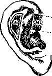
Figure 5. Blepharoplasty—skin-cartilage flap (according to Budinger); M—skin-cartilage flap according to Müller (shaded), converted by incisions along the dotted lines into a Perimov-Krivonosov flap on a pedicle.
for malignant neoplasms. This same method is also applicable for plastic surgery of the upper eyelid. Using a Filatov round pedicle flap, one can graft a mucous membrane, cartilage, etc., to the corresponding end, and then reconstruct the eyelid by applying a complex flap consisting of all the necessary layers. In cases of significant destruction of the entire lower part of the upper eyelid, one can utilize the lower eyelid for its reconstruction; for this purpose, the ciliary margin of the latter is severed, and then it is sutured layer-by-layer to the freshened edges of the remainder of the upper eyelid. After complete healing, a new palpebral fissure is formed by incision using a scalpel and a grooved probe, calculated so that the new upper eyelid is sufficient to cover the eyeball. In cases of loss of both eyelids, but with preservation of the transitional folds, Blepharoplasty according to Lowenstein is possible; the separated conjunctivae of the fornices are sutured with catgut sutures, leaving a small opening in the inner corner for the exit of tears. A skin flap (free or pedicled) is transplanted onto this wound surface; after its engraftment, only a small palpebral fissure is initially made, which can later be enlarged. When reconstructing the margin of the eyelids, it is necessary to cover it with mucous membrane from the lips or cheeks. The absence of eyelashes on such an eyelid necessitates resorting to the transplantation of a strip of skin from the scalp or tattooing.
Related articles
Mentioned in
Cite this page
“Blepharoplasty.” Soviet Medical Encyclopedia. English translation of Bolshaya Meditsinskaya Entsiklopediya, 1st ed. (Moscow, 1928–1936), ed. N. A. Semashko. https://sovietmedicalencyclopedia.pages.dev/article/blepharoplasty/