Foregut
Historical document, translated for reference. It reflects medical knowledge of the 1920s–30s and is not medical advice.
Summary
The foregut is the anterior section of the developing intestinal tube that appears first in the embryo. It forms within the head fold and connects to the yolk sac through the anterior intestinal gates, with the heart developing beneath it.
Encyclopedia article (1928–1936)
FOREGUT, the anterior section of the developing intestinal tube, which appears first in the embryo: it lies within the head fold—a crescent-shaped depression that forms before the neural tube and isolates the head end of the embryo (see figure). This fold turns inward the entoderm and forms the F. k. as a tube ending blindly in front; toward it later forms the oral cavity by depression of the ectoderm. The opening through which one can enter the F. k. from the side of the yolk sac is called the anterior intestinal gates. Under the foregut forms the rudiment of the heart; from its anterior section later develops the pharyngeal intestine (see).
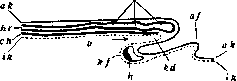
Schematic longitudinal section of a chick embryo in an early stage: ah. ik-outer, inner germ layer; eft-notochord; h-rudiment of heart; hb-brain vesicles; hd-cavity of foregut; kf-head fold; nr-neural tube; v-anterior intestinal gates; am-amnion. (According to Hertwig.)
The heart rudiment develops beneath the foregut; from its anterior section, the pharyngeal intestine subsequently develops (see).
Related articles
Mentioned in
Cite this page
“Foregut.” Soviet Medical Encyclopedia. English translation of Bolshaya Meditsinskaya Entsiklopediya, 1st ed. (Moscow, 1928–1936), ed. N. A. Semashko. https://sovietmedicalencyclopedia.pages.dev/article/foregut/