Esophagus
Historical document, translated for reference. It reflects medical knowledge of the 1920s–30s and is not medical advice.
Summary
This article from the 1928–1936 Soviet Great Medical Encyclopedia details the comparative anatomy, embryological development, and human anatomy and histology of the esophagus. It covers the organ's structural divisions, physiological constrictions, dimensions across different age groups, and anatomical relationships with surrounding thoracic and cervical structures.
Encyclopedia article (1928–1936)
Thermal and chemical injuries...
Esophagus (oesophagus, from the Greek oiso - I carry and phago - I eat; a term of Aristotle's; the Greeks often called the esophagus stomachos; later, in old anatomical texts, this term was incorrectly used to designate the stomach). Comparative anatomy. The esophagus is a division of the "foregut," which in lower vertebrates is short and often not yet differentiated from the stomach. In fish, it begins from a wide pharynx behind the last gill cleft; in reptiles, it passes mostly, though still without sharp boundaries, into a wide stomach; in crocodiles, the esophagus is narrow with strongly pronounced longitudinally arranged folds. In birds, in connection with the elongation of the neck, it is long and in grain-eating birds forms a crop (ingluvies) in the form of a protrusion of the ventral wall, located beneath the muscle that expands it (the crop is contracted by the activity of smooth musculature embedded in its wall); food is retained, swells, softens, and is partially digested in the crop; it is absent in insectivorous birds. In mammals, the esophagus is relatively narrow and long and serves merely as a conducting channel; it is wide in seals and carnivores; its abdominal section reaches considerable length in some marsupials and many rodents. Anatomy and histology of the esophagus in childhood, according to Gundobin, is funnels-shaped and widened at the transition into the stomach, with constrictions being weakly expressed.
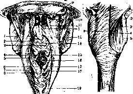
Figure 1.
Fig. 1. Anterior surface of the pharynx (posterior view): 1 - uvula; 2 - tonsilla palatina; 3 - tonsilla lingualis; 4 - vallecula epiglottica sinistra; 5 - plica aryepiglottica; 6 - tuberculum cuneiforme (Wrisbergi); 7 - recessus piriformis; 8 - incisura interarytaenoidea; 9 - condylus occipitalis dext.; 10 - recessus pharyngeus; 11 - plica salpingopharyngea; 12 - velum palatinum; 13 - papillae vallatae; 14 - foramen caecum linguae; 15 - plica pharyngoepiglottica; 16 - aditus laryngis; 17 - plica nervi laryngei; 18 - tuberculum corniculatum (Santorini); 19 - tunica mucosa oesophagi. Figure 2. Esophagus from behind and above: 1 - m. constrictor pharyngis; 2 - cornu superius cartilaginis thyreoideae; 3 - m. stylopharyngeus; 4 - m. pharyngopalatinus; 5 - cornu inferius cartilaginis thy-
reodeae. are weak; in the newborn, its length in situ is about 10 cm, by 2 years about 20 cm, in an adult on average about 25 cm on a cadaver; in the living person, however, the length fluctuates depending on the functional state and the position of the larynx and diaphragm, as well as the position of the cardia, which can shift by 6 cm or more. The esophagus is a tube flattened dorso-ventrally, with a characteristic star-shaped lumen. The boundary of its cervical part from the hypopharynx (pars laryngea pharyngis) at a middle position of the head corresponds to the body of the sixth cervical vertebra, its carotid tubercles, and the lower edge of the cricoid cartilage (in elderly people, due to the relaxation of muscles and the supporting apparatus, the larynx is located lower). This upper end of the esophagus is called the "esophageal mouth" (oesophagus-mund) in view of its ability to close during contractions of the lower annular bundles of the inferior constrictor muscle of the pharynx (Figures 1 and 2). By analogy, the lower end of the esophagus is called the "stomach mouth" (Magenmund). The place of origin of the esophagus from the pharynx, located at a distance of approximately 5 cm from the edge of the incisors, is situated in the median plane; the esophagus at this place is tightly connected with the posterior wall of the larynx and loosely with the prevertebral fascia clothing the longus capitis and longus colli muscles. In a living person with the head thrown back, the esophagus rises slightly and is wedged between the trachea and the spine; in a lying person, it is displaced dorsally; lateral displacements are also possible. The esophagus in the sagittal plane follows the curvature of the spine, to which it adheres until the bifurcation of the trachea, which is located in an adult at the level of the fourth thoracic vertebra; lower down, at the level of the fifth to sixth thoracic vertebrae, the esophagus is pushed away from the spine ventrally by the aorta, which it wraps around in the form of a very elongated spiral; in the living person, under the influence of muscle tone, the esophagus straightens out and is accessible to esophagoscopy along its entire extent.
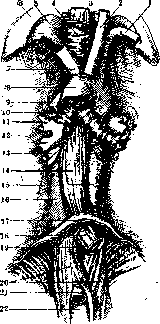
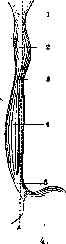
Figure 3.
Fig.
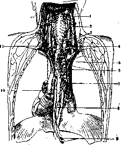
Figure 3. Esophagus with surrounding parts from the front: 1 - a. subclavia sin.; 2 - a. carotis communis sin.; 3, 14 and 15 - esophagus; 4 - trachea; 5 - cupula pleurae; 6 - costa I; 7 - a. anonyma; 8, 13 and 21 - aorta; 9 - v. azygos; 10 - bifurcatio tracheae; 11 - ramus bronchialis; 12 - vv. pulmonales dext.; 13 - rami bronchiales; 16 - columna vertebralis; 17 - diaphragm; 19 - hiatus oesophageus; 20 - pars lumbalis diaphragmatis; 22 - hiatus aorticus. Figure 4. Esophagus from the front with dilations and constrictions: AB - midline; 1 - upper constriction (14 mm); 2 - upper dilation (19 mm); 3 - middle constriction (14 mm); 4 - lower dilation (20 mm); 5 - lower constriction. Thus, the esophagus occupies a median position only in its initial section and at the level of the fourth and fifth thoracic vertebrae. The esophageal hiatus of the diaphragm is located ventral to the aortic hiatus, 2–3 cm from the cardia of the stomach; upon entering the stomach, the esophagus adheres to the left edge of the eleventh thoracic vertebra. This place is located about 3 cm below the esophageal hiatus of the diaphragm, at the level of attachment of the seventh costal cartilage to the sternum (Figures 3 and 5).
In the esophagus, three sections are distinguished: cervical, thoracic, and abdominal, the boundaries between which are determined by 1) a horizontal plane drawn immediately above the jugular notch of the manubrium of the sternum (incisura jugularis sterni) and corresponding to the gap between the second and third thoracic vertebrae, and 2) the place of passage through the diaphragm. The length of the esophagus, according to Jossel, in a living person with the head thrown back is determined approximately by measuring with a probe the distance from the gap between the spinous processes of the twelfth thoracic and seventh cervical vertebrae and from there across the shoulder to the mouth. The cervical part of the esophagus (pars cervicalis oesophagi), measuring about 5 cm in length, is slightly displaced from the median position to the left
beyond the left edge of the trachea; here the esophagus reaches the surgical intervention zone, and one must take into consideration the position of the common carotid artery. The thoracic part turns to the right, and from the bifurcation of the trachea - again to the left; thus, the esophagus is located in the median plane only in its initial section and in the middle of its extent, approximately up to the fifth thoracic vertebra. Its lumen presents a series of constrictions (angustiae oesophagi) and dilations (three constrictions are constant); the first constriction is located at the beginning of the cervical part (Figure 4); here, from the posterior wall of the hypopharynx, a venous plexus protrudes, forming the "lip" of the aforementioned "esophageal mouth"; the mucous membrane forms folds here, like in a tightened tobacco pouch. Due to the divergence of the longitudinal musculature of the esophagus into two lateral bundles, its posterior wall between them appears thinned and prone to protrusion (Zenker's diverticulum). The second constriction, middle or aortic, is located approximately in the middle of the esophagus where it is compressed by the aorta, lying to the left and behind, and the left bronchus, located in front; with chronic inflammation of the bronchial lymph nodes located here and subsequent adhesion of the esophagus to the aorta and bronchus, so-called "traction" diverticula of the esophagus arise; here is also the favorite site for the appearance of tumors. The third constriction, lower, is located at the place where the esophagus passes through the diaphragm; its crura also take part in its formation. The plane of the narrow esophageal hiatus of the diaphragm is located somewhat obliquely relative to its axis; the cardia of the stomach appears open. When the stomach is contracted, the abdominal part of the esophagus, which has a length of about 1.5 cm, stands out clearly; when the stomach is distended, it appears widened and shortened to the point of complete disappearance. Between the constrictions, the esophagus is significantly widened and can be stretched up to 35 mm. Its width in the cadaver of an adult, injected with formalin and filled with plaster, ranges from 7 to 22 mm. In the living person, longitudinally arranged folds are not visible. During probing of the esophagus in children younger than two months, according to Shkarin, the probe should be 7–8 mm in diameter,

in 6–9-month-olds - 9 mm, up to 2 years - 10 mm, in 6–12-year-olds - 12 mm (Gundobin).
Structure of the esophagus. From the inside out, the esophagus features tunica mucosa, submucosa, muscularis, and adventitia (Figure 6); serosa covers it for a minor extent. The epithelium of the esophageal mucosa is stratified squamous; in humans, not far from the transition of the esophagus into the stomach and in the upper part of its cervical region, small scattered islands of cylindrical gastric epithelium with its specific pits and glands are usually encountered: between the whitish smooth surface of the esophageal mucosa and the pinkish and uneven surface of the stomach, there is a sharp serrated border. The submucosa is quite thick and very loose, containing a thin layer of smooth, longitudinally disposed muscle fibers, the tone of which drops at the site of contact of the mucosa with sharp objects, thereby reducing the danger of wounding the esophagus by them, since its wall yields to their pressure. Numerous, predominantly mucous glands are located mainly in the submucosa and are usually visible to the naked eye upon preparation of the muscle layer; the aforementioned islands with glands of the gastric type lie deeper, internal to the muscularis mucosae. The muscle coat (tunica muscularis propria) consists of an outer longitudinal layer and an inner circular layer, in places even more powerful; the first passes below into the longitudinal bundles of the stomach muscles, above, splitting into 2 bundles, it attaches to the cricoid cartilage, without connecting with the inferior pharyngeal constrictor muscle; they are called cricoesophageal muscles (mm. crico-oesophagei); they play the role of an esophageal elevator (levator oesophagi); functioning as an esophageal sphincter (sphincter oesophagi) are the lower bundles Figure 6. Longitudinal section of the esophagus at the level of the esophageal hiatus: 1 - inferior pharyngeal constrictor muscle; 2 - longitudinal musculature; 3 - connective tissue between the esophagus and the diaphragm; 4 - diaphragm; 5 - cardiac notch. of the diaphragmatic musculature; the circular layer of esophageal muscles in the region of the upper
narrowing continues into the pharyngeal musculature, striated fibers of its extend beyond the middle third of the esophagus and into the circularly and obliquely running bundles of the stomach muscles. Both layers jointly perform the final, third phase of swallowing. The striated musculature of the esophagus functions automatically and with greater speed than the smooth. In the upper 1/5 of the esophagus there is striated musculature, in the next 2/5 - mixed, in the lower 2/5 - smooth, though in the circular layer mixed musculature begins almost from the very "oesophageal mouth". In dogs, sheep, and cattle, all esophageal musculature is striated. Strands of smooth muscle fibers of the esophagus continue onto most of the organs adjacent to it: into the longitudinal musculature of the left bronchus (sometimes also the trachea) - the bronchoesophageal muscle (m. broncho-oesophageus), to the left pleural sac - the pleuroesophageal muscle (m. pleuro-oesophageus), to the pericardium and aorta; they are mostly mixed with elastic fibers and frequently replaced by them. The muscular pillars of the diaphragm adjoin the esophagus laterally, but do not pass into its walls; the esophagus is connected to the diaphragm by dense connective tissue, which however does not prevent it from moving into the posterior mediastinal cavity (cavum mediastini post.) when it is cut away from the stomach. The outer coat (tun. externa, in the cervical part of the esophagus also called adventitia) is loose connective tissue enveloping the tun. muscularis and passing without sharp boundaries into the mediastinal cellular tissue and into the pharyngeal elastic membrane (membrana elastica pharyngis, Luschka). In the thoracic part of the esophagus for a short
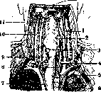
Figure 7. Lymphatic vessels and glands of the esophagus: 1 - common carotid artery (a. carotis communis); 2 - internal jugular vein (v. jugularis int.); 3 - deep cervical lymph nodes (lgl. cervicales prof.); 4 and 11 - supraclavicular lymph nodes (lgl. supraclaviculares); 5 - recurrent nerve (n. recurrens); 6 - tracheal lymph nodes (lgl. tracheales); 7 - right tracheobronchial lymph nodes (lgl. tracheo-bronchiales dex.); 8 - esophagus; 9 - cardiac lymph nodes (lgl. cardiacae); 10 - aorta.
extent it is clad by the pleura, which invaginates behind it (recessus mediastino-vertebralis); in the abdominal part it is covered by the peritoneum, except for the posterior surface. In newborns and in early childhood, occasionally also in adults, slightly above the passage of the esophagus through the diaphragm to the right of it there is a small cavity, which split off during its development from the omental bursa (bursa omentalis); in animals it can be more voluminous (bursa infracardiaca). Blood supply. Numerous fine arterial branches (esophageal arteries, aa. oesophageae) for the cervical part of the esophagus originate from the inferior thyroid artery (a. thyreo-idea inf.), for the thoracic part - from the aorta and bronchial arteries, for the abdominal part - from the left gastric artery (a. gastrica sin.) and inferior phrenic arteries (aa. phrenicae inf.). They form numerous anastomoses and networks in all layers of the esophagus down to the papillae of its mucous membrane. The veins correspond in their course to the arteries, draining into the azygos and hemiazygos veins (vv. azygos et hemiazygos) and gastric veins (vv. gastricae), through which they are in connection with the portal vein. This connection has special clinical and anatomical significance, because when blood flow in the portal vein is hindered, blood from the latter, passing through the esophageal veins, can stretch them, producing typical esophageal varices. Regional lymph nodes are located at various heights, partly directly adjoining the esophageal wall (bronchial and posterior mediastinal lymph nodes, lgl. bronchiales, mediastinales post.) and partly located at some distance from it, at the venous angle (the site of convergence of the internal and external jugular veins and subclavian vein) - the inferior deep cervical lymph nodes (lgl. cervicales prof. inf.). Lymph from the cervical and thoracic regions of the esophagus pours into these glands; from the latter also into the tracheobronchial and bronchial lymph nodes (in the angle between the esophagus and trachea) and the inferior posterior mediastinal lymph nodes (lgl. mediastina-les post. inf.); from the lower part of the thoracic region and from the abdominal region - into the cardiac lymph nodes (lgl. cardiacae) (Figure 7). In the mucous membrane of the esophagus are located lymphoid follicles and isolated lymphocytes. Innervation. External to the tunica muscularis are located larger trunks, forming a plexus embracing the esophagus - branches of the vagus nerve (the chordae oesophageae of older authors) and its recurrent branch, wherein the left vagus nerve, owing to the rotation of the stomach in the embryonic period, gradually shifts to the anterior surface of the esophagus, and the right nerve to the posterior surface (as it descends). To these trunks are admixed sympathetic fibers from the stellate ganglion (gangl. stellatum), from the aortic and laryngopharyngeal plexuses, and groups of cells embedded between the longitudinal and circular muscle layers (intramurally) (see Autonomic nervous system). Sensitivity of all kinds is insignificant; the sensitive fibers belong to the V thoracic segment. It should be noted: 1) that at the level of the lower edge of the cricoid cartilage, medially from the lateral lobes of the thyroid gland, the upper pair of parathyroid glands is usually adjacent to the upper section of the esophagus; 2) laterally, in close proximity to it, is located the sympathetic trunk, and at a distance of 1–2 cm the neurovascular bundle (carotid artery, jugular vein, and vagus nerve); 3) the aorta can (especially in the elderly) shift to the left of the spine, lying para- rather than prevertebrally; 4) to the left of the caudate lobe of the liver (lobus caudatus hepatis), on its posterior surface, is the esophageal impression (impressio oesophagea).
P. Karuzin. Methods of examining the esophagus. The examination of patients with diseases of the esophagus should always begin with a carefully taken case history: the time of onset of difficulty in swallowing, the appearance of pain, the nature of dysphagic phenomena, what kind of food the patient can still swallow, whether there is vomiting and what its character is, whether mucus is secreted in the intervals between meals, what position seems most convenient to the patient when swallowing, whether he has lost weight, whether he has become weaker, whether he copes with his workload, etc. After acquainting oneself with the general condition of the patient, one proceeds to the examination of the esophagus, starting with more accessible methods. Inspection. Dilated veins on the neck and chest give reason to think of a circulatory disorder in the chest cavity; a change in the contours of the neck makes one suspect the presence of a tumor or a diverticulum in the cervical part of the esophagus. Edema and redness indicate a phlegmonous process spreading, possibly, from the depths, from the walls of the esophagus. Percussion is applicable to a very limited extent and only on the cervical part of the esophagus. Diverticula, when overfilled with food, give a dull sound upon percussion, and, conversely, a tympanic sound when they are empty. Auscultation. By listening with the ear or a stethoscope to the left of the spine at the level of Dviii (Hamburger), one can distinguish a gurgling sound during the swallowing of water—the water passes together with air. In strictures of the esophagus, a similar sound is heard above the site of narrowing and may be absent below this site. Palpation is possible only in the cervical part of the esophagus, where it is possible to feel a dense tumor, dense glands, a compressible tumor at the site of a diverticulum in the cervical part, edema, fluctuation, or emphysema in inflammatory processes. Sometimes a finger can reach through the mouth to the initial part of the esophagus, especially in children, and feel, and sometimes even grasp, a foreign body stuck there. With sounding using a probe or some other instrument, it is possible to determine the location of the narrowing in the esophagus. For sounding, the so-called English rubber elastic probes with cylindrical or cone-shaped ends are best suited; they bend easily and are most easily sterilized after each use in a sublimate solution (1:1,000.0) for half an hour. Sounding is usually performed in the sitting position of the patient, who is asked to remain calm and make only one or two swallows at the moment of feeling the end of the probe on the posterior wall of the pharynx. A probe advanced deeper at the same time passes into the esophagus. In this way, in the vast majority of cases, it is possible to pass the probe into the esophagus. Frequently, patients cannot swallow or flatly refuse to do so; in such cases, one has to insert the left index finger into the mouth, press the tongue, and direct the probe into the pharynx with the tip of the same finger, advancing it forward with the right hand. The patient should sit, leaning forward slightly and lowering the chin to the chest; this head position increases the lumen of the pharynx. For sensitive patients, the fauces and the posterior wall of the pharynx are smeared with cocaine or novocaine. A mouth gag is inserted into children. When introducing a probe to nervous, anxious patients and children, it is recommended to put a metal finger cot on the index finger inserted into the mouth. For the most part, it is easy to pass the probe into the esophagus, but there are patients for whom sounding causes great torment. If the probe goes along a false path and instead of the esophagus gets into the larynx, a severe fit of coughing appears, the patient turns blue, tosses about, and jumps up from a chair or bed. It is necessary to immediately stop the sounding, let the patient calm down, and after some time, make another attempt to pass the probe. Under no circumstances should sounding be performed without a thorough general examination, especially of the cardiovascular system. Cases are known where the walls of the esophagus, thinned by the pressure of an aortic aneurysm, ruptured along with the aorta at the moment of introducing the probe. With dysphagic phenomena causing suspicion of a foreign body or a decaying tumor, the introduced probe can easily pierce the wall of the esophagus and get into neighboring tissues. Inflammatory processes around the esophagus—mediastinitis, pleurisy, etc.—affect the wall of the esophagus, which easily tears during sounding. If blood appears at the moment of sounding, the latter must be stopped immediately. Sounding makes it possible to determine the location of the narrowing in the esophagus. On the probe passed up to the stricture, the line of the front incisors is marked, it is withdrawn, and the length from the end of the probe to the marked line is measured, which indicates at what height the obstacle is located. The degree of narrowing is determined by passing thinner bougies or probes, which can be brought down to the finest numbers. The retention of the probe anywhere in the esophagus and the inability to advance it deeper towards the stomach indicate an existing obstacle in the form of a cicatricial stricture, a foreign body, a spasm, a diverticulum, a tumor of the esophagus, or some tumor compressing the walls of the esophagus from the outside. If the tumor is rapidly disintegrating, the diverticulum turns out to be empty, and so on, then the probe, which had previously encountered an obstacle in its path, passes easily without difficulty. Sounding, despite its simplicity, requires a maximum of attention and caution from the physician, and only under such conditions does this method of examination not lose its value, which recently laryngologists (Schlommer and others) have tended to deny. More precise data are obtained by examining the esophagus with an esophagoscope (see Esophagoscopy) and with X-rays. Fluoroscopic or radiographic examination of the esophagus is possible at the moment of passing a probe or swallowing a contrast mass; the latter gives a more visual picture of the passage of a barium lump or liquid gruel through the esophagus, makes it possible to see its peristaltic movements, the site of narrowing, the presence of a foreign body, and the nature of the latter. Observation of the passage of the contrast mass is best carried out in the so-called first oblique dorso-ventral direction (Holzknecht), i.e., when turning the patient with the right shoulder forward. In this position, a free space is visible between the heart and the spine where the esophagus is located, giving a shadow only when the contrast mass passes. Fluoroscopy is contraindicated in those cases where there is a fistula between the esophagus and the respiratory tract, since the contrast mass can get into the bronchi and cause aspiration pneumonia. (Regarding the examination with X-rays of pathological changes, see below.) Developmental defects of the esophagus are of no great practical importance, since children with such congenital defects are not viable and quickly perish. Most often, the division of the esophagus into two unconnected segments is observed: the upper one reaches approximately to the jugulum and ends in a blind pouch, from where a connective tissue-muscular cord stretches towards the blind gastric segment. Cases are known where the upper or lower segment of the esophagus communicated with the windpipe, located 1–2 cm above the bifurcation. Such underdevelopment, explained embryologically, is noted already by the end of the 3rd week, the moment of separation of the esophagus from the trachea. The remaining forms of stenoses and atresias are rarer and appear in a later embryonic period. Symptoms. At the very first attempt to swallow, the child turns blue, begins to cough and suffocates; these symptoms, as well as the impassability of the esophagus to a probe, make it possible to diagnose a congenital defect of the esophagus. These children usually die from aspiration pneumonia. Treatment. For the time being, it can only be a question of a gastric feeding fistula (see Gastrostomy), with the help of which the child could be fed. Until now, not a single such operated child has survived the operation (Lotheissen). Congenital strictures of the esophagus due to the presence of membranes are rare. Lotheissen collected 45 cases. The clinical picture is the same as in acquired cicatricial strictures of the esophagus. Examination with a probe, esophagoscopy, or fluoroscopy make it possible to make a diagnosis. After internal transection of the membrane or circular electrolysis, bougienage of the esophagus is begun. The results are good. Dysphagia lusoria is caused by the fact that the right carotid artery, as well as the subclavian artery, in very rare cases depart from the left of the aortic arch or its descending part and can compress the esophagus at the site of intersection. Injuries to the esophagus are external and internal. Isolated external injuries to the esophagus are observed extremely rarely, and then only in stab and gunshot wounds. For the most part, large vessels, the windpipe, the spine, the heart, the lungs, the pleura, and other organs located in the vicinity of the esophagus turn out to be damaged simultaneously. Incised wounds are found more often in suicides. Simultaneously with the esophagus, the windpipe is also severed, and both tubes, having contracted, go deep and infect the tissue of the mediastinum. Cases have also been described where the throat and the anterior wall of the esophagus turned out to be severed, while the posterior wall remained untouched and turned, as it were, into a bridge connecting both segments. Large vessels do not always turn out to be affected. Everything depends on the sharpness of the instrument, the scope, etc. In isolated gunshot wounds of the esophagus, a bullet can descend into it and be expelled through the mouth or by the natural route.
Sometimes a bullet, grazing the esophagus, becomes lodged nearby, over time destroys the walls of the esophagus, and leads to severe, sometimes fatal complications. The material from the recent war shows that cut wounds, despite the fact that initially they ran a very severe course and produced a grave impression, yielded a better outcome than stab and gunshot wounds, especially of the posterior wall of the esophagus. In cut wounds, the drainage of wound secretion is free, and healing generally proceeds without special complications, whereas stab wounds, and frequently also gunshot wounds, quickly glue together externally, while the opening in the esophagus in the depth gapes and serves as a source of severe infection. Symptoms boil down to the discharge through the wound of food or liquid taken by mouth. With narrow wound tracts, swelling, redness, and subcutaneous emphysema appear on the neck; patients complain of severe, shooting pain upon swallowing. Simultaneous damage to the trachea is accompanied by hoarseness, aphonia, bouts of suffocation, etc. From the gaping wound of the esophagus, the infection descends into the mediastinum, which is accompanied by a ominous deterioration of the general clinical picture of the disease. Treatment must be directed in the first place against life-threatening moments: severe hemorrhage must be stopped; in case of difficulty in breathing, tracheotomy is indicated. As for the esophagus, it is necessary first of all to take measures against the spread of infection from the wound. In fresh cases, an incision is made, the esophagus is exposed, and, if possible, its wound is sutured. According to war materials, the attempt to primarily suture the esophagus yields worse results than tamponade (Guleke). In those cases where the esophagus turns out to be severed, and the lower segment has retracted deep down, primary suture does not achieve the goal. It is better to sew both segments into the skin wound, pack widely around, and for feeding the patient, establish a gastric fistula. If the patient is admitted with phenomena of spreading inflammation, the site of the wound on the esophagus should be exposed by a wide incision—collar mediastinotomy (Hacker)—and the lower corner of the wound tightly packed. The patient is allowed nothing by mouth for 3–4 days, and up to 2–3 liters of normal saline or 5% glucose are administered daily subcutaneously or intrarectally. A complication is the formation of esophageal fistulas, observed in connection with periesophageal phlegmons. Fistulas can be of such sizes that food masses are constantly discharged through them to the outside, which affects the nutrition and strength of the patient. In the presence of a fistula between the esophagus and the trachea, patients cough and choke when taking food. Treatment can only be surgical and consists in exposing the esophagus above and below the fistula, freshening the edges, and applying sutures. Damage to the esophagus from the inside is possible during the passage of a probe, an esophagoscope, upon the introduction of a foreign body, during a trial excision of a piece of mucosa, etc., as well as in the form of a spontaneous rupture of its altered wall. Ruptures of the normal esophagus have also been described, associated with contusions upon being hit under an automobile, between car buffers, falling from a bridge, from an airplane, etc. The pathogenesis of spontaneous esophageal ruptures has still been little studied, but there are literary indications that such ruptures are observed at the moment of strong vomiting movements when the upper section of the esophagus is narrowed by a scar or compressed by a goiter or a tumor. Spasm of the upper section of the esophagus (Beneke, Mohr), chronic esophagitis (Ach, Mackenzie) can also serve as the cause of spontaneous rupture during a strong bout of vomiting. If, during unsuccessful probing, esophagoscopy, etc., only the mucosa is found to be damaged (erosion), patients complain of pain upon swallowing, sometimes radiating to the back; this pain soon disappears. Such superficial injuries end for the most part in recovery and in rare cases with the formation of a cicatricial stricture. With deeper injuries, sometimes penetrating through the wall of the esophagus into adjacent tissues, the course of the disease is likewise, as in spontaneous ruptures, for the most part severe. The moment of perforation or rupture is accompanied by sudden pains in the esophagus, depriving one of the ability to swallow liquid even in small amounts. Sometimes bleeding through the mouth appears depending on the caliber of the wounded vessel. Soon, subcutaneous emphysema develops, spreading from the posterior mediastinum upward in the direction of the supraclavicular region, and from there throughout the whole body. Pneumothorax is frequently observed at the same time. The pulse becomes small, barely perceptible, the general condition sharply deteriorates, patients turn blue, suffocate, lose consciousness, and perish. In perforation of the abdominal section of the esophagus, severe peritonitis develops. However, cases are known where perforation proceeded almost without any symptoms and was discovered only at autopsy. In chronic cases, when perforation is a secondary phenomenon on a previously altered wall of the esophagus, the picture of the disease is significantly weaker. Thus, for example, upon breaking through into the trachea, esophageal cancer leads to the formation of a tracheoesophageal fistula. In an inflammatory process in the wall of the esophagus, the moment of perforation even brings as it were temporary relief. Treatment can only be surgical (Venglovsky) and must be directed toward the earliest possible exposure of the rupture site transpleurally or through the posterior mediastinum (collar, dorsal mediastinotomy), toward wide tamponade and the creation of an alimentary fistula on the stomach. In perforations of malignant neoplasms, there can hardly be any question of any intervention. Thermal and chemical injuries of the esophagus. Burns by hot substances do not penetrate into deeper layers, but are limited mainly to the mucosa alone, the surface epithelium of which hyperemizes shortly after this and then turns into a white thin film with fine wrinkled folds. With bed rest and the ingestion of cold liquid food inside, patients recover in a few days. Prophylaxis: avoid too hot food, do not eat hurriedly. Chemical injuries of the esophagus by acids or caustic alkalis, drunk for the purpose of poisoning or by mistake, are frequently encountered [see separate table (p. 223–224), fig. 3 and 4]. The quantity and concentration of the drunk solution is important for the further course and outcome of the poisoning. The surface layer of the mucosa, burned by acid or alkali, is immediately necrotic, and depending on the concentration of the solution, the action of the latter extends to deeper layers of the walls of the esophagus. Changes in the walls of the esophagus upon swallowing caustic substances consist of the following. Sulfuric acid: the mucosa is uniformly grayish-white, in long folds; little mobile, brittle and hard. The submucosa is infiltrated, vessels are engorged with disintegrated blood corpuscles. Subsequently, ulcers with a diphtheritic plaque develop and a scar forms. Hydrochloric acid: the same changes as with sulfuric acid, but with a short exposure more superficial, and in the upper sections uneven formation of scabs, between which the tissue is edematous, blood-infiltrated. Nitric acid: yellow or yellowish-brown scabs; with a more concentrated solution, deeper destructions. Chromic acid: brownish-red scabs with a transition to green color. The walls of the esophagus are dry and dense. Microscopically, crescentic deposits in the submucosa are characteristic (according to Rössle). Oxalic acid: the upper layers of the mucosa are whitish-gray, at the bottom brownish-black. The mucosa is dense, but not brittle, as if folded into long folds. At the bottom, frequent scab-like self-digestion, since oxalic acid together with stomach pepsin digests proteins. With more prolonged exposure, calcium oxalate crystals are found in the esophagus. Carbolic acid: the esophagus inside is grayish-white in color, as if covered with a layer of lime: the walls are dark in color, dry and brittle. Lysol: the swollen mucosa is slippery, as if soaped, brownish-red to grayish-white in color. Citric acid (tartaric and acetic acids): the mucosa is whitish-gray in color; more superficial layers are affected. Alkalis: severe changes, liquefaction necrosis. Scabs are initially white, soft, subsequently dense, dry. Sublimate, zinc chloride, copper sulfate, silver nitrate give coagulation necrosis, more superficial than with the mentioned acids; with copper sulfate, the scabs are bluish-green, while with silver nitrate they are grayish-white, and sometimes black. The esophagus is less hard than with acids.
Swallowing weak solutions commonly used in households destroys only the superficial layers of the epithelium, which soon regenerates. With deeper damage, the dead mucosa sloughs off in parts in the form of a tube, and by the 7th to 9th day, a bleeding granulation surface is visible beneath it. The latter subsequently scars gradually, which sometimes leads to the formation of valves or folds due to the tension of the unchanged part of the mucosa, but most often cicatricial strictures develop in this place. Depending on the depth of the damage, the scarring process can also involve the periesophageal tissue and lead to the formation of dense calloused scars, either shorter (2–3 cm in length), so-called annular, or longer (10–15 cm), so-called tubular stenoses. In rare cases, only a single continuous longitudinal scar may be found at the site of the burned esophagus. The cicatricially altered walls of the esophagus are densely penetrated by thick fibrous tissue and thickened throughout their extent. When the esophagus is burned in several places, multiple scars form ( Hacker), but most often deeper damage to the walls of the esophagus and the formation of scars are noted at the sites of physiological narrowing—at the esophageal sphincter, the bifurcation, and the cardia, from which the scarring process can spread upward and downward. The destruction from the swallowed poison can in some cases be so deep that scarring does not occur. The inflammatory process, spreading deeper into the walls of the esophagus, leads to the formation of phlegmons in the periesophageal tissue, from which patients die. Symptoms. Initial symptoms of esophageal burns reduce to severe pain in the mouth, pharynx, esophagus, and stomach. If the initial part of the larynx is also burned, patients begin to suffocate. Vomiting occurs with bloody mucus and fragments of the burned mucous membrane. In mild cases, all these symptoms are not so brightly expressed, gradually weaken, and patients begin to feel somewhat better. In 3–4 weeks, signs of dysphagia appear in connection with the scarring process. In severe cases, the sensation of burning and pain does not disappear, bleeding from the esophagus often appears, excruciating thirst, temperature rises, and with severe septic phenomena in connection with periesophageal phlegmon or peritonitis, patients die. Mortality from acid poisoning is 50%, and in the remaining 50% various degrees of esophageal stenoses remain. Upon swallowing alkalis, mortality is 25%, in 25% minor stenoses develop, and in 50% severe strictures (Hacker). Treatment. When delivering the patient immediately after poisoning, it is recommended to make an attempt and immediately wash out the stomach with a neutralizing solution. Morphine is prescribed against pain, subcutaneous infusions of physiological solution, a 5% glucose solution, or drop enemas against thirst. In case of cardiac decline, camphor, caffeine, digalen, and so forth are prescribed subcutaneously. In later stages, when the swollen mucosa closes the lumen of the esophagus or when, in connection with deep anatomical and pathological changes (ulceration of the mucosa, communication of the esophagus with a periesophageal abscess), the patient is deprived of the ability to swallow, a gastric fistula should be applied without delay, and in cases where the pylorus is also burned (see Stomach), a jejunostomy is performed. Preventive measures against the formation of narrowing scars of the esophagus after burns consist of early bougienage of such patients (Roux, Salzer). In milder cases, a bougie can be inserted as early as the next day, in more severe ones—after a few days, as soon as the patient recovers a little. According to Salzer, out of 34 patients treated with early bougienage, only one died, and that from an accidental disease. Bleeding from the esophagus is observed, according to Pitert (Schmerz), most often from the vessels of the esophagus. Furthermore, bleeding is possible from injuries through the esophagus of vessels lying next to it, upon the rupture of a vessel into the lumen of the esophagus, and upon the growth of an esophageal tumor into the descending part of the aorta, the pulmonary, subclavian, inferior thyroid artery, the azygos vein, or another large vessel. In the absence of perforation or rupture of the esophagus, bleeding is possible from varicose veins in the walls of the esophagus of a congenital (Marchand, Friedrich, and others) or acquired character; acquired dilations occur much more frequently than congenital ones and sometimes reach a considerable magnitude. The cause of varicose veins in the lower section of the esophagus is stagnant phenomena in the portal system, observed in cirrhosis, syphilis of the liver, and thrombosis of the portal vein. In the upper section, dilated veins are observed in malignant goiter. Bleeding can recur, appear without any symptoms, and any of them can become so abundant as to lead to death. Most often, a slight tickling in the throat appears, a peculiar taste in the mouth, and then vomiting of scarlet or foamy, and sometimes dark blood if it previously flowed into the stomach. With significant bleeding, dizziness, pallor, restlessness, a sense of fear, thirst, and other signs of increasing anemia appear. It is extremely important to precisely determine what the source of esophageal bleeding is. Cirrhosis of the liver, malignant goiter, and so forth will indicate the possible source of bleeding. In complicated cases, a sound is introduced into the patient's stomach after having them drink a few sips of water. A bloody spot on the sound will indicate the location of the bleeding source in the esophagus. Diagnostic errors have given occasion for laparotomies. Treatment. In case of severe bleeding, a probe-like rubber instrument is introduced, which can be inflated with air in the manner of a colpeurynter and thereby compress the bleeding vessel. Through a probe passed somewhat below the bifurcation, the esophagus can be washed with hot water, which sometimes succeeds in stopping the bleeding. Recurrent bleeding requires the use of all those measures that are used for any lingering bleeding—introduction of gelatin, serum under the skin, blood transfusion, and so on (see Bleeding). With injuries to very large vessels, patients die immediately. Inflammatory diseases of the esophagus are acute and chronic. Acute ones include catarrhal inflammations of the esophagus, which occur more often than commonly thought. In scarlet fever, diphtheria, dysentery, and other infectious diseases, in thermal and chemical damages, catarrh of the esophageal mucosa and even its diphtheritic inflammation are observed not infrequently. In such patients, complaints come down to pain upon swallowing, which passes in about 3 days. In more severer cases, pain upon swallowing is more intense, palpation of the esophagus on the neck, turning and tilting of the head are also painful. Vomiting movements and the discharge of mucus are excruciating. Upon esophagoscopy, a reddened, swollen mucosa is visible, with erosions noted in places. Treatment comes down first of all to complete abstinence from swallowing even liquids, but since patients suffer from thirst, they are allowed to swallow pieces of ice or cold milk in small sips. In severe infectious diseases, in uremia and severe enteritis in children, fibrinous films appear on the esophageal mucosa (esophagitis fibrinosa, pseudomembranosa, or crouposa), which subsequently fall off, leaving bleeding ulcers (esophagitis necroticans). In diphtheria, the process rarely (only in 1.1%) spreads below the pharynx (Reiche). In scarlet fever, on the contrary, in almost 89% the inflammatory process passes from the pharynx into the esophagus (Oppikofer). In smallpox, the entire catarrhally altered mucosa is dotted with smallpox pustules, especially in the upper sections of the esophagus. Thrush (soor), observed in severe diseases in children and adults, spreads from the mouth down into the esophagus, which indicates a sharp weakening of the organism. Such films can clog the lumen of the esophagus (Fraenkel) or grow into the deeper layers of its walls. If thrush is localized only in the esophagus, its diagnosis can present great difficulties, and only microscopic examination of the extracted film will allow an accurate diagnosis of the disease. Films sometimes depart spontaneously during vomiting, as well as after mechanical cleaning with tufts moistened in a 3–5% borax solution. Of other forms of acute inflammations of the esophagus, the following are known: 1) Esophagitis follicularis, or cystica—suppuration of the glands in the upper section of the esophageal mucosa, which then ulcerate and turn into follicular ulcers; part of the glands can become cystically distended. 2) Esophagitis dissecans superficialis, or exfoliativa—sloughing off upon an elevated temperature of a mucosal tube 15–20 cm in length; observed rarely; the separated tube consists of delicately fibrous tissue infiltrated with round-cell elements and the surface epithelium of the esophagus; the symptoms of the disease subside upon the departure of the membranous tube. 3) Esophagitis dissecans profunda—rejection of the necrotic mucosa, e.g., after burns with acids, lysol, and the like. 4) Esophagitis phlegmonosa—purulent inflammation of the submucosa of the esophagus refers to very severe diseases, occurring rarely, observed in the presence of infectious diseases—angina, lymphadenitis, etc., as well as a complication after abrasions and injuries to the mucosa. Sometimes the cause of inflammation remains unclear.
In phlegmonous esophagitis, the inflammatory process spreads only to the submucosa. Phlegmonous esophagitis is divided, according to Zenker, into general diffuse and local. The latter are observed more frequently, often rupture into the esophagus, after which recovery ensues. More dangerous and formidable in their course are diffuse phlegmons of the esophagus, which are encountered significantly less often. The inflammatory process extends to almost all layers of the esophageal walls, so that upon autopsy the latter appears as a cord saturated with pus. The course of the disease is extremely severe. Severe pain occurs upon attempting to take even a small swallow of water. Chills, elevated temperature, and pain in the sternal region upon turning the head exhaust the patient, who dies within 2-3 days amidst severe septic phenomena. Anamnestic data, the presence of a purulent infection in the vicinity, and the evacuation of pus through the mouth provide the occasion for esophagoscopy, with the aid of which it is possible to see an as-yet-unruptured abscess protruding into the lumen of the esophagus in the form of a small cushion; at the site of a ruptured abscess, an opening is visible from which pus sometimes still oozes. Treatment is symptomatic and directed at eliminating pain. An existing abscess may be opened through the esophagoscope. A trapped foreign body that served as the cause of the inflammation must be removed without fail (Hacker). Chronic inflammations of the esophagus are observed in the form of chronic catarrhs associated with irritation of the mucosa by alcoholic beverages, food masses constantly retained above a stricture, a tumor, etc. Compensated heart defects with congestive phenomena are accompanied by chronic catarrh of the esophagus. Upon esophagoscopy, the mucosa appears turbid, edematous, with tortuous veins, and is covered with mucus. Aside from eliminating the primary causes of the disease, local treatment may be applied (only in advanced cases): the mucosa is swabbed with a 1-3% tannin solution or a 1-3% silver nitrate solution, with anesthesia provided by a 3-10% cocaine solution or a 2-3% eucaine, novocaine, etc., solution. Tuberculosis of the esophagus occurs as a complication of pulmonary tuberculosis or as a result of the spread of infection from peribronchial or tracheal glands, and is on the whole extremely rare. The anatomical structure of the esophageal mucosa and the rapid passage of tuberculous sputum are not very conducive to this disease. Abrasions, caustic alkali burns of the esophageal mucosa, cancerous narrowing, and thrush facilitate the fixation of the tuberculous infection in these places. Most often, tuberculosis of the esophagus is found in the region of the bifurcation (up to 50%), somewhat less frequently in the upper third, and least frequently in the lower third, and is described in the form of two types: 1) superficial or deeper ulcers of insignificant size, oval shape, with persistent, uneven edges and a floor covered with pale granulations; 2) sclerosing infiltrates of a delimited or diffuse character that thicken the wall of the esophagus and narrow its lumen (sometimes simulating a cancerous tumor). Symptoms depend on the form of the process; the presence of ulcers is accompanied by a burning sensation during the swallowing of food and by pain that often disappears in the intervals between acts of swallowing. In the sclerotic form, phenomena of dysphagia predominate. The general condition of the patients is severe, since the primary process (in the lungs, glands) begins to proceed more acutely due to the inability to swallow and the rapid decline in general nutrition caused thereby. Diagnosis sometimes presents significant difficulties, especially in differentiation from syphilis and cancer, which is achieved with the aid of biopsy. Treatment: strictures are dilated by bougienage, ulcers are cauterized with silver nitrate or lactic acid; for severe pain, cocaine, novocaine, anesthesin, etc., are prescribed. The prognosis is generally poor and depends on the underlying affliction. Syphilis of the esophagus is rare, predominantly occurring in the tertiary period and mostly in the upper section, whence the process spreads from the pharynx. Gummatous nodules, appearing initially in the submucosa, break down, ulcerate, and then scar, subsequently narrowing the lumen of the esophagus. In the presence of ulcers, complaints come down to pain, and upon scarring, to dysphagic phenomena. Esophagoscopy allows the observation of gummatous areas, ulcers, or star-shaped scars. In differential diagnosis with tuberculosis and cancer of the esophagus, it should be kept in mind that cancer and tuberculosis can also occur in syphilitics. Treatment is specific. Cicatricial strictures are bougied. Actinomycosis...
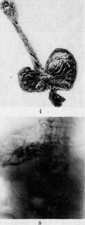
Fig. 1. Zenker diverter of the esophagus. Fig. 2. Traction diverticulum of the esophagus. Figure 3. Stricture of the esophagus after a burn with acetic essence. Figure 4. Burn of the esophagus and stomach with caustic alkali. Figs. 5 and 6. Cancerous narrowing of the esophagus. Fig. 7. Cancerous narrowing of the esophagus in the lower part. Figure 8. Pneumoconiosis in a Donbass miner with 25 years of service.
To the article Esophagus. Pneumoconiosis of the esophagus is extremely rare. The presence of actinomycosis in one of the adjacent organs and the onset of pain during swallowing give grounds to think of the transition of this disease to the esophagus, which can be confirmed or refuted by biopsy. In actinomycosis of the esophagus, patients die quickly. Treatment: see Actinomycosis. Peptic ulcer of the esophagus is roundish in shape with dense calloused (edges and is observed most frequently in its lower section. It is associated to a certain extent with cardiac insufficiency, pyloric stenosis, and other factors contributing to the constant regurgitation of gastric juice into the esophagus. Patients complain of pain radiating to the back, bleeding, regurgitation, and dysphagia; for fear of pain, they starve, lose weight, and weaken. At the same time, cases are known where ulcers did not manifest themselves during life and were discovered accidentally at autopsy. During X-ray examination, a niche can be detected. Probing and esophagoscopy are unsafe. Treatment of a peptic ulcer of the esophagus is therapeutic, as with a gastric ulcer (see Stomach, treatment of gastric ulcer); in cases unresponsive to therapeutic treatment, an attempt is made to cauterize the ulcer with silver nitrate or zinc chloride, applying a temporary feeding fistula to the stomach or jejunum. Acute hemorrhagic erosions of the esophagus are also observed, similar to those in the stomach. Softening (oesophagomalacia) of the esophagus is observed during life rarely and depends on the regurgitation of gastric juice into its lower section, which has a digesting effect. The wall of the esophagus turns out to be so thinned that it allows fluid and air to pass into the surrounding tissues and cavities. Strictures of the esophagus. Strictures of the esophagus are observed due to the impストgement of foreign bodies, due to compression of the esophagus by a tumor or formation located outside the walls of the esophagus (by an aneurysm, diverticulum, etc.), due to spastic contractions of the walls of the esophagus, frequently in cicatricial strictures after burns with chemical substances [see separate table (article 223 - 224), figure 3], but most often due to cancer tumors (up to 80-90% of all stenoses of the esophagus) (see below). Cicatricial strictures of the esophagus, considered more or less benign strictures, usually develop after burns with chemical substances, mostly in places of physiological narrowing. Hypertrophy of the walls of the esophagus above the stricture deserves attention in connection with the constant striving to push the food mass through the narrowed place. Gradually, the muscular membrane of the walls of the esophagus stretches, becomes weak and flabby, swallowed masses are delayed, stagnate, irritate the mucosa, which often leads to inflammation and the formation of ulcers capable of rupturing into adjacent tissues. Symptoms of esophageal stricture begin to manifest themselves shortly after the burn and develop in parallel with the scarring process. The first most frequent sign is considered to be difficulty in swallowing, as a result of which patients begin to eat slowly, chew food thoroughly and wash it down with water, eat soft or even liquid food, making all sorts of movements of the head, as if trying by this to push the swallowed lump through the narrowed place. The spasm that often joins completely closes that small opening at the site of the narrowing through which a part of the liquid food still passed. Patients indicate the site of the narrowing and complain of pain that does not always coincide with the site of the actual narrowing. With a stricture in the upper section of the esophagus, regurgitation is observed immediately after the first gulp, and with deeper located strictures - after 5-6 gulps. Larger quantities of food are swallowed by the patient in those cases where the walls of the esophagus above the site of the narrowing are significantly stretched. After some time, belching appears, vomiting with the discharge of swallowed food. The feeling of nausea is absent in this case, only the discharge of a significant amount of mucus is noted. In connection with all this, patients gradually lose weight, grow thin, weaken, and lose their working capacity. In the diagnosis of an esophageal stricture, the anamnesis is of great importance. Probing, X-ray examination, and in some cases esophagoscopy, make it possible to recognize the location and nature of the narrowing. With a probe of medium thickness, the site of the stricture can be determined. To orient oneself in the degree and course of the narrowing, thin English bougies are used, which are passed very slowly and cautiously to avoid creating false passages. Less suitable for this purpose is a thin metal probe, which should be passed in front of the screen in the fluoroscopic room. During X-ray examination, it can be seen how the shadow, somewhat expanded above the site of the narrowing with even, regular edges, narrows conically or in the form of a funnel towards the stricture. Complete retention of the contrast mass above the stricture does not always indicate complete obstruction of the esophagus (Seiffert). Only after the disappearance of the accompanying spasm is it possible to accurately determine the degree of esophageal narrowing. When examined with an esophagoscope, it is possible to see white stripes and scars on the inflamed mucosa above the site of the narrowing. Respiratory and pulsatory movements are preserved with superficial scars and absent with deeper ones (see Esophagoscopy). For the most part, it is possible to determine the location and degree of the stricture radiologically. In differentiating from cancer of the esophagus, uneven, irregular, and seemingly eroded edges are considered characteristic for cancers, and, conversely, even, smooth edges for benign strictures. Doubtful, confusing cases can be resolved by biopsy through the esophagoscope. In functional spastic strictures of the esophagus, the X-ray picture may differ little from the picture in cicatricial stricture. Antispasmodic agents (atropine, papaverine) often eliminate the spasm and make it possible to make an accurate diagnosis. Rarely encountered cicatricial strictures of tuberculous, syphilitic (I. I. Grekov, Solovyov, and others), diphtheritic character, scars after peptic ulcers of the esophagus, narrowing of the esophagus from compression by a tumor, aneurysm, etc. can also be distinguished from cicatricial stricture of the esophagus after chemical burns. Anamnesis, anatomical position of the narrowing, its nature, changes in adjacent organs, and finally the entire clinical course of the disease make it possible to make a correct diagnosis. The prognosis in strictures depends on the nature of the anatomical changes and the time of intervention. In children, the walls of the esophagus are more pliable, and in the process of growth, it is easier and better to stretch them. Treatment of cicatricial strictures of the esophagus boils down to expanding the site of the narrowing, and in cases where this fails, to the formation of a new esophageal tube. In expanding the formed cicatricial stricture, bougies filled with lead or mercury, Trousseau probes, and others are used. Dense English bougies or probes through which patients can be fed simultaneously are quite suitable. When passing probes with rounded or conical pointed ends, it should be borne in mind that the walls of the esophagus stretched above the site of the narrowing create favorable conditions for perforation. Initial attempts to pass the probe should be painless and short. A few drops of adrenaline (1:1000.0) or 10-12 drops of 1% morphine eliminate the spasm and facilitate bougienage. Anesthesia with cocaine and others is considered contraindicated by Haslinger, since it is easier to perforate the wall of the esophagus in this case. If the probe can be passed, it is carefully withdrawn after 5-10 minutes. The sessions of leaving the probe are gradually lengthened to 1/4-1/2-3/4 of an hour and more. With a smooth course, patients can be probed twice a day. If the temperature rises or pain appears in the side, bougienage is suspended until the general condition improves. If it is not possible to pass even the thinnest probe, they try to probe with a string thread or a hollow bougie brought to the stricture, through which they try to push one or several string threads into the narrowed place (Hacker). The passed string threads swell after 10-15 minutes and somewhat expand the lumen of the stricture, after which it is already possible to pass a thin probe. Passing thin probes using an esophagoscope is a complex procedure. Cauterization of films or bridges at the site of the stricture with chemical substances or a galvanocautery is feasible under the control of an esophagoscope. Cicatricial strictures of the esophagus have also been tried to be treated with electrolysis (Hacker, Lotheissen, Sletov, Zeldovich, and others). According to Frenkel, Michaelis, Stratievsky, Bramson, and others, the cicatricial area of the esophagus is easier to bougie after injection of fibrolysin. The general condition of the patient (emaciation, exhaustion, etc.) requires great attention. Against pain sensations at the site of the stricture, bromine, belladonna, and conventional narcotic substances are prescribed. By such measures, it is far from always possible to expand cicatricial strictures of the esophagus, which gradually become narrower and can reach such a degree that the patient may subsequently be threatened with death from starvation. In such cases, one or another surgical intervention is indicated, aimed at raising the strength and nutrition of the patient, as well as restoring the patency of the esophagus. Internal or external section of the esophagus, excision of the scar as a method of treatment of esophageal stricture, is used at present extremely rarely.
External section of the esophagus is still feasible in cases of stenosis in the cervical portion of the esophagus or somewhat deeper. Excision of the scar, which is permissible in the cervical portion, has been performed 6 times (5 cases by Schmieden and 1 case by V. L. Pokotilo) with one fatal outcome. If the stenosis is located in the lowest division of the esophagus, one can establish a communication between the stomach and the dilated part of the esophagus above the stricture (Heyrovsky) or an anastomosis between the brought-up small intestine and the same part of the esophagus (E. R. Hesse). Safer is the creation of a gastric fistula through which the patient begins to be fed, thereby increasing their strength, and then an attempt is made to pass a thin probe (ureteral catheter) or a thread, with the help of which bougies are then gradually advanced through the stricture, starting with the thinnest numbers. To the thread brought out on one side through the gastric fistula and on the other through the mouth, a passing probe is tied, and to one of the ends of the probe another thread, which upon extraction of the probe remains again in the esophagus. In this way, patients are bougied endlessly, "ohne Ende" (according to Hacker). The most difficult part of this manipulation is passing the thread or thin bougie and catching it from the stomach. Swallowing small lead pellets on a thread (Socin), passing a thin ureteral catheter under fluoroscopic guidance, and various other techniques make it possible, with a certain persistence and patience on the part of both the physician and the patient, to eventually achieve the passage of the thread through the stricture. Bier failed to pass the thread in only one case, and that only because the female patient prematurely interrupted treatment. According to Lotheissen, out of 257 esophageal stenoses, complete impenetrability of the stricture occurred in only one case. If it is not possible to pass a thread or ureteral catheter from above, one attempts by means of retrograde esophagoscopy (Rovsing) to pass a catheter from below through the gastric fistula and cardia upwards into the oral cavity. In long tubular strictures of the esophagus that completely fail to pass even liquids, the question arises of complete exclusion of the esophagus and the construction of a new esophageal tube via antethoracic esophagoplasty. This operation—the formation of an antethoracic esophagus—proposed by Bircher (H. Bircher, 1894), has been developed at the present time in all its technical details and is frequently applied with success. The main methods of antethoracic esophagoplasty are the methods of forming the esophagus: 1) from skin—the constructed skin tube is connected at its lower end with the stomach and at its upper with the severed esophagus brought out to the exterior (Bircher, Esser, Braytsev) (Figure 8); 2) from intestine—a small intestine loop...
Figure 8. Plastic surgery of the esophagus and intestinal tube. ...so that its nutrition is not impaired, i.e., the mesentery must be ligated and severed in such a way that a certain number of branches from the jejunal arteries and veins are preserved; such a segment of the small intestine is brought out under the skin onto the anterior chest wall and subsequently connected at its upper end with the brought-out esophagus and at its lower end with the stomach (Roux-Herczen) (Fig. 9a, II); 3) the esophageal tube is constructed partly from the small intestine (Lexer, Wullstein) or large intestine (Kelling, Vulliet), partly from skin (Fig. 9a, I; Fig. 9b, III, IV; Fig. 9c, V); the formed skin-intestinal tube is connected at its ends with the esophagus and stomach; 4) the material for forming the esophagus is the wall of the stomach (Hirsch, Jianu-Halpern) or the entire stomach—the latter is severed at the cardia and brought out subcutaneously (Kirschner); the stomach is severed at the pylorus and, turned over, brought out subcutaneously (Fig. 9c, VI). All these methods are multi-stage and require great patience and endurance on the part of the patient and physician, but in successful outcomes they restore the patient's working capacity. A number of cases are known in the literature where patients lived after such operations for 17 years (Stieda), 13 years (Hintz), 9 years (Voelcker), 3 years (V. R. Braytsev), etc. Still, these operations cannot be considered safe yet, but the most difficult thing is that in a number of cases it is not possible to bring them to completion for various reasons. In 1931, V. G. Veksner collected from the world literature 214 cases of antethoracic esophagoplasty performed for benign esophageal stenoses by various methods, with a mortality rate of 18.7%; only 115 cases (53%) were brought to completion, and in 49 cases the operation remained unfinished. Of the above material on antethoracic esophagoplasties, Russian surgeons account for 74 cases with 16 deaths and 24 unfinished operations. Esophageal diverticula. An esophageal diverticulum is generally considered to be a localized pouch-like blind protrusion of its walls, lined with normal mucous membrane. A protrusion of the esophagus associated with an inflammatory or cicatricial process and not lined with mucosa is considered a false diverticulum. Diverticula can be pulsion and traction (see Diverticulum). Diverticula are most often encountered in the initial part of the esophagus, the so-called Zenker's diverticula [see separate table (pp. 223–224), Figure 1]; they are also called pharyngo-esophageal, boundary diverticula (Grenzdivertikel) and are for the most part pulsion diverticula. In the remaining part of the esophagus, pulsion diverticula are rare, while traction diverticula are more frequently observed, which may subsequently, as they develop, become pulsion, or rather traction-pulsion diverticula. Traction diverticula [see separate table (223–224), Figure 2] are encountered more often than pulsion diverticula. Out of 472 diverticula, 188 were pulsion and 284 traction (A. V. Sudakevich). Zenker's esophageal diverticula. The site of development of such diverticula is the border of the transition of the lower part of the pharynx into the esophagus, directly above the esophageal sphincter. In this place, there is the so-called Laimer's triangular space, where the walls of the esophagus are less developed and more susceptible to pressure from the inside, especially during traumatic injuries of this section, as well as in other diseases (lodgment of a foreign body, spasm, tumors, goiter, etc.) hindering the passage of food. Zenker's diverticula are located along the posterior wall of the esophagus somewhat to the side and left. The size of the diverticula ranges from a pea to an adult human fist or even a child's head. They communicate with the esophagus via a narrower or broader slit-like circular passage depending on the size of the diverticulum. As it grows, the diverticulum seems to push out from its initial position between the spine and the esophagus and appears on the side of the neck, increasing during those moments when it becomes overfilled with food or liquid. In connection with the retention and stagnation of food masses in the diverticulum, a chronic inflammation of its mucosa is observed, which ulcerates in places. The inflammatory process spreads into deeper tissues, as a result of which adhesions form around the walls of the diverticulum in the neck and in the upper thoracic cavity. Zenker's esophageal diverticula are encountered more often in men (77.5%), and predominantly at an older age, over 50 years. Symptoms. The initial signs of the appearance of a diverticulum are so insignificant that patients cannot precisely indicate since when they began to notice difficulty in swallowing. A significant portion of patients recall that already long ago, sometimes 20–30 years ago, they began to notice more abundant salivation, a feeling of irritation in the throat, expectoration of a significant amount of sputum, despite dryness and a constant cough, which often ends in vomiting. The sensation in the throat of a foreign body in the first...
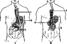
Figure 9a. Esophagus plastic surgery. I - 1st method: 1 - skin tube; 2 - loop of small intestine; 3 - end-to-end suture of the small intestine. II - 2nd method: 1 - skin tube; 2 - end-to-side anastomosis of the small intestine; 3 - isolated loop of small intestine.
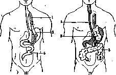
Figure 9b. Esophagus plastic surgery. III - 3rd method: 1 - skin tube sutured to the esophagus; 2 - sutured lower end of the newly formed esophagus from the small intestine into the stomach; 3 - side-to-side anastomosis of the small intestine. IV - 4th method: 1 - skin tube; 2 - formation of the esophagus from the transverse colon; 3 - descending colon.
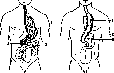
Figure 9c. Esophagus plastic surgery. V - from the transverse colon (colon transversum) with preservation of the middle colic artery (a. colica med.) (3); 2 - end-to-end anastomosis of the transverse colon. VI - esophagus plastic from the stomach: 1 - stomach brought out antethoracically; 2 - tightly closed stump of the stomach; 3 - anastomosis of the stomach stump with the small intestine. cannot be considered safe yet, but the hardest part is that in a number of cases it is not possible to bring them to completion for various reasons. In 1931, V. G. Veksner collected from the world literature 214 cases of antethoracic esophagoplasty performed for benign esophageal stenoses by various methods, with a mortality rate of 18.7%; only 115 cases (53%) were brought to completion, and in 49 cases the operation remained unfinished. Of the above material on antethoracic esophagoplasties, Russian surgeons account for 74 cases with 16 deaths and 24 unfinished operations. Esophageal diverticula. An esophageal diverticulum is generally considered to be a localized pouch-like blind protrusion of its walls, lined with normal mucous membrane. A protrusion of the esophagus associated with an inflammatory or cicatricial process and not lined with mucosa is considered a false diverticulum. Diverticulum.
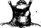
Figure 10. Appearance of the neck in a large esophageal diverticulum.
The initial period of the disease is replaced in later stages by a sensation of an obstruction, forcing the patient to swallow well-chewed food slowly and cautiously. All these sensations are caused by the fact that in the early stages of the disease the diverticulum, still of insignificant size, fills during one of the first swallows, remains overfilled during the entire meal and long after it, without disturbing the act of swallowing. In later stages, when the diverticulum has already reached a significant size, it quickly overfills during a meal and thereafter compresses the esophagus, thus preventing further swallowing of food. Many of such patients begin to choke, make vomiting movements, and regurgitate the food masses retained in the diverticulum. The diverticulum is thereby emptied, and the patient gains the ability to swallow freely again. However, nausea, vomiting, or regurgitation do not always appear. Sometimes the esophagus, compressed by the filled diverticulum, does not let through even a single swallow. Patients rush about, assume various poses, turn and tilt their head in different directions, and squeeze their neck, striving in this way to empty the diverticulum. Pressing on the site of the diverticulum also succeeds in emptying the latter. Part of the food stuck in the diverticulum is emptied, and the remainder stagnates and decomposes, as a result of which patients develop a foul breath and a number of dyspeptic phenomena associated with the swallowing of decomposed masses. Upon examination of such patients, one can see a tumor filling the supraclavicular fossa on one side of the neck (Fig. 10). If air and fluid are present in the diverticulum, specific sounds and gurgling are noted by the patient and those around him even before attacks of dysphagia develop. The inflammatory process of the walls of the diverticulum, spreading to adjacent tissues, is accompanied by pains radiating into the esophagus, neck, back of the head, chest, retrosternal region, shoulder blade, etc. Adjacent organs—the trachea, cervical and brachial plexus, recurrent nerve, and cervical vessels—may be subjected to constant pressure. Patients complain of constant thirst, hunger, lose weight, gradually weaken, and die from phenomena of exhaustion. According to Lyudin, 16-17% of patients die from pneumonia, pulmonary gangrene, and similar diseases associated with severe septic processes spreading from a perforated diverticulum.
Diagnosis. The periodic appearance of a tumor on the neck during a meal, its disappearance at the moment of compression, and the above-described specific gurgling noises give a reason to suspect the presence of a diverticulum. Upon percussion of an empty, air-filled diverticulum, a tympanic sound is heard, and conversely, an overfilled diverticulum gives a dull sound. In cases where the diverticulum drops into the upper section of the chest cavity, tapping may produce a high tympanitis, suggesting a cavity (Loeffler). It is difficult, almost impossible, to determine small diverticula by sounding, but on the other hand, with large protrusions of the esophagus, the probe almost always falls initially into the diverticulum, stops at a height of approximately 20 cm, and goes no further. At this moment, it is possible to probe the end of the probe through the skin on the corresponding side of the neck. The probe that initially got stuck in the diverticulum can nevertheless be passed into the stomach with repeated movements. In the same way, it is sometimes possible to pass another probe into the stomach next to the probe located in the diverticulum, which is a valuable diagnostic sign in the presence of a diverticulum. It is easiest to get into the diverticulum by sounding with special Stark probes, curved at the end like Mercier catheters. Upon esophagoscopy, a transverse slit is visible, opening upon a deep breath and appearing in the form of a rosette or funnel, where the tube of the instrument passes. The mucosa visible in the empty diverticulum is pale, covered with thick, viscous mucus, in some places stretched, in others in folds, with separate inflamed and even ulcerated areas. When transilluminated with X-rays, one can see how the contrast mass falls directly into the sac, fills it, and appears as a round-oval shadow with smooth edges [see separate table (p. 2:23-2:24), figure 1]. The uneven edges noted in some cases indicate adhesions of the walls of the diverticulum with surrounding tissues.
Treatment. Preventive measures should be aimed at eliminating the stricture contributing to the protrusion of the walls of the overlying section of the esophagus. Not always can patients indicate the moment of trauma that caused the stricture, especially since the initial symptoms of the disease are very poorly expressed. Patients seek help mostly with phenomena of dysphagia, i.e., already when the diverticulum reaches significant dimensions. The removal of stagnant masses is possible by periodic washing of the diverticulum, which significantly eases the patient's condition, but with each meal the diverticulum sac fills again, compresses the esophagus, and again makes swallowing difficult. Meanwhile, the diverticulum stretches even more, is filled even easier by swallowed food, and squeezes the esophagus even stronger. Conservative measures cannot break this peculiar circulus vitiosus, and therefore one should not waste much time on all these therapeutic techniques and it is better to subject the patient to surgical treatment. With a drop in nutrition and expressed weakness of the patient, it is recommended to apply a gastric fistula, through which the patient should be fed and thereby raise his strength somewhat, and only then remove the diverticulum surgically. Excision of the entire diverticulum...
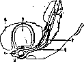
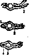
Figure 11. Excision of the esophageal diverticulum: 1 - diverticulum; 2 - line of section of the diverticulum; 3 - probe; 4 - esophagus; 5 - thyroid gland; 6 - trachea. Figure 12. Scheme of the esophageal suture during diverticulum surgery: 1 - muscular layer; 2 - mucous membrane; 3 - probe; 4 - lumen of the esophagus.
...
was proposed by Kluge as early as the beginning of the 19th century, and since then this method has been the most radical and leading to a complete cure. Subsequently, a number of other methods aimed at the same goal were proposed. Surgical treatment of diverticula is currently performed using several methods. 1. Girard's method is aimed at not opening the lumen of the esophagus. The isolated diverticulum is invaginated into the esophagus, and the outer walls of the latter are sutured with 2-3 stitches. The method is applicable for small diverticula, since invaginated large protrusions can close the lumen of the esophagus. 2. The displacement method (Verlagerung)—the isolated pouch is moved under the skin and fixed to the muscles of the pharynx (Schmidt). The method of Lieble and Bogoras is approximately the same, in which they sutured the isolated pouch subcutaneously, moving it upwards. Goldmann separates the isolated and displaced pouch after 9 days with a thermocautery. 3. Excision of the pouch at the very neck and careful application of a two-tier suture to the esophageal wound is the most reliable method (Figs. 11 and 12). Out of 241 cases of surgical treatment of a diverticulum, 10% of patients died, and for the period 1912-1924, out of 156 operated patients, 14 people died, i.e., 8.9% (Hacker, Lotheissen). Deeply located esophageal diverticula. Diverticula in the deeper parts of the esophagus between the sphincter and the cardia are much less common. Sometimes a traction diverticulum is formed first, which then turns into a traction-pulsion diverticulum. In any case, the pathogenesis of these diverticula is apparently not the same. These diverticula occur at a later age, over 40 years, more often in men, and are of various sizes—from a pea to the fist of an adult man—and of various shapes—round, pear-shaped. Diverticula above the diaphragm are called epiphrenic, in contrast to epibronchial ones, located at the site of intersection with the left bronchus (Rosenthal). Symptoms appear only when the diverticulum has already reached significant proportions; in these cases, the course of the disease resembles the clinical picture of border (Zenker's) diverticula. In addition to all of the above, patients with large diverticula in the thoracic cavity also complain of pain behind the sternum, palpitations, and lack of air, which disappear immediately after vomiting. Only large, sac-like diverticula can press on the esophagus and make swallowing difficult. Diagnosis. Repeated probing determines the height of the location of the diverticulum—25-30 cm for epibronchial and 40-42 cm for epiphrenic. Starck's probe can easily enter the diverticulum, determine its depth, and more accurately determine on which side, right or left, the diverticulum is located. During esophagoscopy, it can be difficult to find the opening connecting the esophagus with the diverticulum; significant diverticula in the lower section can be mistaken for diffuse expansion of the esophagus. X-rays almost always make it possible to diagnose the presence of a diverticulum, determine its shape, size, and location. Treatment is indicated only for large diverticula and should be reduced to washing with weak antiseptic solutions, after which the patient can be fed through a probe. Surgical treatment of deeply located diverticula presents significantly greater danger than that of Zenker's diverticula. Lotheissen, Sauerbruch, and others recommend invaginating the isolated diverticulum into the esophagus or, if its location is very low, connecting it with an anastomosis to the stomach wall stretched through the diaphragm. Traction diverticula are associated with inflammatory changes in the para-esophageal glands. The latter, having shrunk, constantly pull the wall of the esophagus outward and serve as the main cause of the occurrence of traction diverticula (Rokitansky, Ziegler, and others). The walls of such diverticula usually have the same three layers as the walls of the esophagus, only at the apex the mucosa is replaced by a scar. The diverticula are located on the anterior or lateral wall, mostly at the level of the bifurcation. The communication with the esophagus is wide, oblong-oval, from 6 to 8 cm in diameter. Traction diverticula are observed mostly at the age of over 30 years. Out of 200 traction diverticula, 127 were found in persons older than 30 years, and 8 in children younger than 10 years (Starck). Symptoms are absent in the overwhelming majority of cases (Oekonomides). The existing pain may depend on the inflammatory process in the wall of the diverticulum or in the gland located outside. Only in those cases when traction diverticula cause pain or interfere with the swallowing of food, patients are examined with a probe, an esophagoscope (Starck), or X-rays (Helm et al.) [see separate table (pp. 223-224), figure 2]. During probing, one should be careful, as cases of rupture of a traction diverticulum have been described when trying to pass a probe (Economides, Marx, et al.). Treatment. If a foreign body gets stuck or food masses are delayed in the diverticulum, the stuck object should be extracted or the delayed food removed with a jet of liquid. Repeated washing can eliminate the cause of chronic inflammation of the walls of the diverticulum. If the diverticulum breaks through into neighboring organs—an extremely dangerous complication—the question of surgical intervention may arise. Neuroses and atony of the esophagus. Esophageal neuroses are observed mostly in neurasthenics and hysterical subjects. According to F. Kraus, the entire clinical picture—the sudden appearance and disappearance of symptoms, the absence of organic changes, the connection with all kinds of mental trauma, etc.—all this indicates the functional nature of the disease, which manifests itself either in the form of a spasm or in the form of a disorder of the sensitivity of the esophagus. Esophagismus is observed in all its parts, but most often in the initial part, at the sphincter, and at the end, at the cardia. Spasm of the thoracic part of the esophagus is extremely rare. Spasm of the esophageal sphincter is much less common than spasm of the cardia (see Cardiospasm) (Guisez), and can cause the development of a pharyngocervical diverticulum. Esophageal spasms can appear suddenly—so-called acute spasms, observed mostly in the upper part at the sphincter; they can repeat with various light intervals—so-called chronic spasms (primarily in the lower part of the esophagus). The appearance of such esophageal spasms obviously has a connection with infectious-toxic processes, with diseases of the central nervous system, with the abuse of alcohol and nicotine, and with the general state of the nervous system, with a special predisposition to spasms. In some patients, esophageal spasms are noted in diseases of the stomach—with a callous ulcer, cancer on the lesser curvature. Treatment should be directed primarily against the underlying condition; in addition, it should also be symptomatic (atropine, cocaine lubrication, and in severe cases, bougienage). Regarding the treatment of spasm of the lower part of the esophagus, see Cardiospasm. Sensitivity disorder of the esophagus is often observed in the form of paresthesia or hyperesthesia, a feeling of pressure, globus in general neuroses, and also often in connection with increased stomach acidity. Atony of the esophagus (hypotonia) is observed, according to some authors (Holzknecht and others), very often, although clinically it manifests itself very little. Palugyay also believes that such changes in the esophagus are very common, which he was able to ascertain when examining with X-rays in a position with the pelvis elevated. M. Lüdin holds a different opinion, who, based on the clinical material of the Basel clinic, argues that patients with esophageal atony are rare. Complaints boil down to difficulty in swallowing, pressure behind the sternum, and a feeling of a lump of food getting stuck in the esophagus, while the probe passes unhindered; often both the probe and the esophagoscope are passed even surprisingly easily due to the expansion of the esophagus [see separate table (pp. 239-240), fig. 6]. This disease is best diagnosed by X-ray examination. The esophageal phase of the act of swallowing is lengthened, the shadow of the esophagus itself is significantly wider than normal. Treatment should be aimed at strengthening the entire organism and at drugs that have a tonic effect on the walls of the esophagus—strychnine, pilocarpine (Engels). Tumors of the esophagus. Among benign ones, fibromas, papillomas, lipomas, myomas [see separate table (pp. 239-240), figure 5], angiomas, and cysts are found. All these tumors are immobile, usually sit in the submucosal layer, less often on the mucosa of the esophagus, and narrow its lumen very little. At the same time, cases have been described where fibromyomas were arranged in a ring and narrowed the lumen (Anichkov), as well as cases where tumors hung on a thin stalk in the esophagus in the form of a polyp. Of all the listed tumors, which are rarely observed and generally have no clinical significance, fibromas and fibromyomas of a soft, elastic consistency, ranging in size from a pea to a walnut, are found more often than others. Growing, these fibromas can hang down in the form of polyps. Pure mucosal polyps originating from the anterior wall at the level of the cricoid cartilage, bifurcation, or cardia have also been described. Reaching a significant size, such polyps or fibromas close the lumen of the esophagus and significantly impede swallowing. Cough attacks and vomiting appear, and at this moment they can fall out into the mouth during a vomiting movement and even outward through the mouth.
With polyps in the cardial region, patients complain of pain in the epigastrium. Angiomas, characterized by pronounced bleeding, make themselves known early. The diagnosis of all these tumors is not easy, especially since it is necessary to differentiate them from the more frequently occurring cancer of the esophagus. With the help of probing, esophagoscopy, and X-rays, it is possible to establish the presence and nature of the tumor. In doubtful cases, the question is resolved by biopsy. Discovered tumors can be removed with a galvanocaustic snare through an esophagoscope, or by means of pharyngotomy or esophagotomy. Polypoid tumors protruding into the mouth are grasped with forceps, pulled taut, and severed after ligation of the pedicle. Malignant tumors. Sarcoma of the esophagus is observed very rarely, occurs at an advanced age, and mostly in men. Sarcomas of the esophagus can be primary or secondary, growing from neighboring organs. Primary sarcomas soon give metastases to the lungs, pleura, pericardium, and other organs. Circumscribed polypoid sarcomas are more benign, while diffuse ones are more malignant and rapidly metastasize (Starck). Microscopically, sarcomas of the esophagus are either round-celled or spindle-celled; lymphosarcomas are also encountered. The symptoms of the disease appear early. Patients complain of pain and an obstacle in swallowing; blood frequently appears in the vomitus, and sometimes pieces of the tumor, especially when the latter ulcerates. The course of the disease is more severe than in cancer of the esophagus, and patients soon die with a rapid decline in nutrition and symptoms of severe cachexia. Diagnosing sarcoma of the esophagus in the initial stages of the disease is very difficult. Constant boring pains radiating to the back, a more rapid course, and early appearance of metastases speak rather for sarcoma than for cancer, which proceeds significantly slower. More precise data are obtained by esophagoscopy, which allows examination of the tumor and taking a piece for examination. Surgical treatment is possible only in the very early stages of the disease, if the tumor is located in the cervical part of the esophagus. The prognosis is poor. Patients with sarcoma of the esophagus die on average 5-6 months after the onset of the disease (Hacker). Cancer of the esophagus. Of all diseases of the esophagus in general, cancer is encountered more often than others (Hacker, Gieses, Venglovsky, and others). As for the ratio of esophageal cancer to all cancer patients, according to Aschoff, out of 4,574 cancer patients, the esophagus was affected in 226 cases. Out of 17,794 autopsies, there were 1,694 cancer patients, of which esophageal cancer accounted for 294 cases (Sauerbruch). In Switzerland, esophageal cancer occupies second place among organs affected by cancer. In our country in the USSR, esophageal cancer is not uncommon. According to relatively old autopsy data in Leningrad, esophageal cancer was found in 15-20% (Kruzenshtern-Albrecht). According to Moscow prosectoriums for 1923-1927, esophageal cancer now occupies a place in cancer mortality after stomach and bronchial cancer, making up 12.3% of all cancers. According to Clairmont, up to 25,000 people die of esophageal cancer in Europe annually. Esophageal cancer occurs at a more mature age, after 40 years, predominantly between 50 and 60 years. Cases of esophageal cancer in young people are known: 24 years (Venglovsky) and 19 years (Heymann). Men contract esophageal cancer more often than women: 88% of men according to Heymann, 91.5% according to Gieses, 85.8% according to Venglovsky, and 78.3% according to Nisnevich. Cancerous neoplasms of the esophagus are more frequently located in the deeper sections. Out of 2,829 cases of esophageal cancer (collected statistics of Lotheissen), the tumor was found in the upper third in 579, in the middle in 888, and in the lower in 1,362 cases. A cancerous tumor is observed in the esophagus mostly as primary; it is significantly more rarely secondary, i.e., caused by sprouting from adjacent affected organs: the cardial part of the stomach, bronchi, pharynx, thyroid gland. The tumor may be localized in some area of the esophageal wall, but it can apparently also appear in several places at once. According to their consistency, esophageal cancers are dense (scirrhus) and soft (medullary). Scirrhuses appear as protrusions into the interior of the esophagus; the mucosa covering them is pale gray, immobile, and fused with the submucosa. The tumor, growing further into the deeper layers of the esophagus, makes its wall dense and brittle. At the site of the tumor, the lumen of the esophagus is narrowed, above the site of narrowing the esophagus is somewhat stretched, and the muscular layer in this part is hypertrophied. Subsequently, the tumor ulcerates, which increases the lumen of the esophagus at this place. Medullary cancers affect the walls of the esophagus over a significantly greater distance than scirrhuses and ulcerate early, as a result of which the edges appear as if undermined, often drooping in the form of flaps. The early ulceration of the tumor explains the fact that these patients have relatively fewer complaints of dysphagic phenomena. Microscopically, esophageal cancers are mostly squamous cell carcinomas with a tendency to keratinization (scirrhus). Round-cell and polymorphic cancers of the esophagus are also encountered. The tumor may turn out to be simultaneously an adenocarcinoma and a squamous cell carcinoma. The old proposition of Billroth regarding the low tendency of esophageal cancers to metastasize needs only the correction that distant metastases (beyond the mediastinum) are indeed not frequent. According to Petri, metastases in esophageal cancer occur in 59%, according to Redlich in 75%, and according to Starlinger, in 66% of those who died from esophageal cancer, no metastases were found. Metastases are more often observed in cancers of the lower third of the esophagus (Venglovsky). Metastases of esophageal cancer, spreading along the lymphatic vessels, appear first of all in the peri-esophageal, tracheal, and bronchial glands, and when spreading along the blood vessels, in the lungs and liver. Cancerous neoplasms of the esophagus can grow through its walls, spread to adjacent tissues and organs, forming large and dense tumors pressing on the trachea, sympathetic, vagus or recurrent nerves, pleura, aorta, pericardium, vertebrae, etc. Out of 604 esophageal cancers autopsied in 1920-21 in German pathologo-anatomical institutes, perforation into the lungs was noted in 3.8%, followed by perforation into the aorta, right pleura, bronchi, and pericardium. From a pathologo-anatomical point of view, esophageal cancers are classified as follows: 1) by localization: cancers of the upper section—up to the bifurcation, middle—from the bifurcation to the hiatus, and lower—from the hiatus to the cardia (the height in centimeters from the upper front teeth can also be indicated); 2) by form: a) insular or nodular, b) circular, and c) multiple; 3) by character: a) cancers in the initial stage, b) wart-like, villous, and c) ulcerating, disintegrating cancers. Symptoms. An important and early symptom of esophageal cancer is dysphagia, which, gradually increasing, can reach the point that patients cannot pass even a gulp of liquid. Depending on the localization and growth of the tumor, regurgitation and vomiting appear. When the tumor is located in the upper third, the patient regurgitates immediately after 1-2 gulps. With a deeper position of the tumor and dilation of the esophageal walls above this place, the patient can make significantly more gulps, but after some time vomits everything back. The patients' indication of the place of the obstruction in the esophagus does not always correspond to the actual position of the tumor, since in esophageal cancers, spasm of the walls is noted significantly higher than the location of the tumor. Viscous transparent mucus mixed with blood and pus is secreted from the mouth in later stages. By this time, a heavy odor from the mouth appears. The improvement that appears at times depends on the better patency of the esophagus in connection with the ulceration and disintegration of the tumor. The pains observed in some patients are not very intense. Only a few of them complain of pain appearing independently of the act of swallowing, radiating to the back, spine, sternum, epigastrium, and intensifying at nights. The growth of the tumor through the walls of the esophagus and compression of adjacent organs are accompanied by a number of characteristic, diagnostically valuable signs. Growth into the sympathetic nerve is revealed by Horner's syndrome, compression of the recurrent nerve is accompanied by hoarseness and loss of voice, severe attacks of coughing every time during eating, and coughing up previously taken food masses indicate a fistulous tract formed as a result of the tumor growing into the trachea or bronchus. Communication with the bronchial tree usually leads to gangrene of the lung and putrid pleurisy. The general condition of the patient with esophageal cancer becomes severe, nutrition drops, strength disappears, temperature fluctuations associated with the disintegration and suppuration of the tumor join in, which exhausts the patient even more and rapidly leads to a fatal outcome. Patients with esophageal cancer live from 1 to 2 years and die with severe phenomena of sharp exhaustion from thirst, hunger, and cancer cachexia. Diagnosis. Age, sex, absence of other factors capable of serving as the cause of dysphagic phenomena (foreign bodies, cicatricial strictures, etc.), noticeable emaciation, loss of weight, pallor, decline in strength, increasing difficulty in swallowing—all this gives reason to think of esophageal cancer. The presence of metastases, growth of the tumor into adjacent organs leave no doubt as to the correctness of the initial assumption. Often patients arrive late, when recognition does not present any special difficulty.
Of those who apply at an earlier period, far from all can accurately indicate the onset of their disease and describe all the symptoms of the illness. In such cases, it is necessary to check the patency of the esophagus and the degree of stricture by using sounds of various thicknesses. The sound may pass unhindered in the initial stages of the illness and during the breakdown of the tumor. In the latter case, there are traces of blood and sometimes pieces of the tumor at the tip of the sound and in its opening. Abundant discharge of bloody-purulent fluid from the esophagus and an aortic aneurysm are contraindications to sounding. During fluoroscopy, it can be seen how the contrast mass descends along the esophagus, reaches the site of the tumor, and here passes along the wall with uneven, as if eroded edges. The picture obtained during radiography of cancer of the esophagus is very diverse and depends on the shape and character of the tumor [see separate table (pp. 223-224), figures 5, 6, and 7]. The data of X-ray examination are decisive, but in the very initial stages of the disease, when the tumor is still very small and the picture of the disease is not sufficiently clear, it is not possible to determine the exact nature of the disease by X-ray. In these cases, esophagoscopy makes it possible to examine the suspicious area, check the respiratory movements of this section of the esophagus, examine the mucosa, its color, the presence of folds, infiltration, ulceration; in doubtful cases, a piece from the suspicious area can be taken under visual control for examination. Despite the fact that cancer of the esophagus occurs comparatively very often, it should still be differentiated from a number of other diseases—benign strictures, tuberculosis, syphilis, actinomycosis. Treatment. The only method of treatment for cancer of the esophagus is still, as with cancers of other organs, the timely removal of the tumor by surgical means. The technique of operations on the esophagus is sufficiently developed, but at the same time the number of operated patients compared to the number of sick patients is negligible. All this is explained by the late seeking of surgical help by patients, which can be explained, on the one hand, by the peculiarity of the clinical course of cancer of the esophagus, and on the other hand, by the deeply skeptical attitude of a wide circle of doctors toward the successful feasibility of such operations on the esophagus. A timely diagnosis and early operation make it possible in some cases to remove the cancerous tumor from the esophagus. The location of the tumor is of great importance in operations on the esophagus. A distinction is made between, so to speak, operations for cancer of the cervical, thoracic, and abdominal parts of the esophagus. Resection of the cervical part was developed mainly by Hacker and consists of the following: by an incision along the inner edge of the left sternocleidomastoid muscle, from the angle of the lower jaw to the jugulum, the esophagus is reached; along the way, the omohyoid muscle is cut and the inferior thyroid artery is ligated. On the exposed esophagus, the tumor is felt, which is gradually isolated from the surrounding tissues so that the esophagus can be resected in a healthy part. If the recurrent nerve is not yet captured by the tumor, it is desirable to preserve it. In case of adhesions to the larynx, it is recommended to remove it simultaneously. The operation is complicated, but healing proceeds better. Out of 13 cases of simultaneous removal of the esophagus and the affected larynx, only one patient died, who was already suffering from pneumonia before the operation (cited according to Hacker). If the defect of the esophagus after removal of the tumor is less than 4 cm, joining the ends is still possible. If it is not possible to connect both ends of the esophagus with a direct suture, a tube should be constructed from skin according to Hacker and used to replace the defect in the esophagus (see above). Of the 81 cases of resection of the cervical part of the esophagus for cancer collected by Hacker, 23 patients died—23.39% mortality. Schmiz reports 69 resections of the cervical part of the esophagus with 19 deaths (27.5%). Geselevich collected 45 resections of the same part of the esophagus with a mortality rate within up to 3 months of 43.5%. Tumors located in the upper section of the chest, 2-3 cm below the jugulum, can still be approached via the cervical route (Sauerbruch). Below this place and up to the hiatus, the esophagus is approached either retropleurally or transpleurally. The retropleural approach to the esophagus was developed by Nasilov, who believed that when the tumor is located in the upper sections of the esophagus, it is better to excise the III-VI ribs on the left side, and for deeper-seated tumors, the lower ribs on the right. Upon excision of the corresponding ribs, the pleura is carefully pushed aside and the corresponding part of the esophagus is exposed. By further studies of Rudnev, Starkov, Savvin, Rehn, Tuffier (L. Rehn, Tuffier) and many others, the technique of the retropleural approach to the esophagus from behind has been sufficiently developed, but nowadays the fear of wounding the pleura has disappeared and many surgeons (Rehn, Lexer), preferring to go retropleurally, nevertheless always have an apparatus ready for operation under elevated pressure. Of all operations on the esophagus performed by the retropleural route, Lilienthal's patient (cancerous tumor) and Enderlen and Nehrkorn's patients (removal of a foreign body) survived. Transpleural resection of the esophagus is feasible, firstly, in the region of the bifurcation and, secondly, between the latter and the diaphragm. During the operation above the bifurcation, a skin-muscle flap is formed on the right by an incision along the clavicle down along the line of the internal mammary artery to the lower edge of the IV rib. The II and III ribs are resected along their cartilaginous part and 3 cm of the bony part. Upon separation of the sternoclavicular joint, the clavicle is retracted upward, after which it is easy to resect the first rib. The chest cavity is opened with a vertical incision and the lung is pushed to the side and downwards. Deep down, the mediastinal pleura is visible, which is carefully cut between two forceps and bluntly separated up and down, after which the esophagus located behind the trachea is felt in the depth (Sauerbruch). Wendel, Torek (Wendel, Torek) and others approach this part of the esophagus transpleurally, but from behind, which gives them significantly more room. The literature cites a number of cases of operations for cancer of the thoracic section of the esophagus with a successful outcome (Torek) (the female patient lived 10 years after the operation) (Lilienthal, Hellström, Zaaijer, Eggers). Nevertheless, the mortality rate in these operations is very high and, according to Schmiz, is almost 98.75%. The transpleural route to the lower section of the esophagus passes in the VI intercostal space, which, upon excision of one or two ribs, provides wide access to the chest cavity and to this part of the esophagus. Küttner, Kümmell, Zaaijer, Meyer, Payr, and many others introduced a whole series of modifications: sewing with special buttons, operating in two stages, etc., but the main obstacle in the technical performance of operations for cancer of the esophagus is the unreliability of the sutures connecting both ends of the transected esophagus. In successful cases (Torek, Hellström, Eggers, etc.), sutures were not applied at all, and the ends of the esophagus were brought outward and connected by a rubber tube. Back in 1896, Lewy (W. Lewy) proposed completely removing the esophagus by invagination and pulling it down into the stomach. Asch, Kelling, Rehn, Enderlen, and Götz tested this method experimentally and on the operating table. None of the patients survived. In 1927, Golrbandt reported on one patient in whom he removed the entire esophagus. The patient lived for 1.5 years after the operation. Patient K. P. Sapozhkov lived for 53 days after the removal of the esophagus by the invagination method and died from another disease. Cancers of the abdominal part of the esophagus are operated on by the thoracic and transabdominal routes.
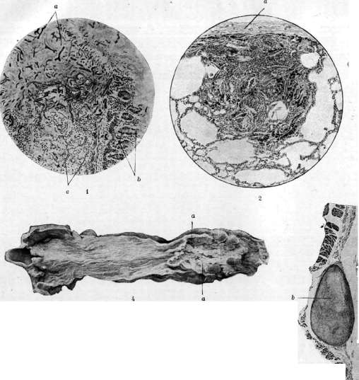
Fig. 1. Mold fungi (a) growing into a gastric ulcer; b—glands of the gastric mucosa; c—infiltration of the submucosal layer. Figure 2. Pneumoconiosis; nodule in the lung: accumulations of coal and silica needles are visible; a—pleura. Figure 3. Dilatation of the veins of the esophagus (a); b and c—mucous and muscular membranes of the esophagus. Figure 4. Cancer of the esophagus with ulceration in the central parts (a), tuberous edges of the tumor are visible. Figure 5. Myoma (b) of the esophagus; a—epithelium (magnified through a magnifying glass). Figure 6. Dilatation of the esophagus: a—stomach; b—sharply dilated esophagus; c—cardia; d—pylorus.
To the abdominal section of the esophagus one approaches by opening the chest cavity in the seventh intercostal space and cutting the diaphragm down to the esophageal hiatus. In this way it is possible to isolate the lower section of the esophagus and the upper part of the stomach, which is displaced into the thoracic cavity and, after excision of the tumor, is connected to the lower end of the esophagus. Sauerbruch divided this operation into three stages and operated on such a patient via a thoracoabdominal approach. Comparatively better results are obtained in resection of the cardiac section by the abdominal route. The abdominal section of the esophagus can be bluntly mobilized and lengthened, which makes it possible, after removal of the tumor, to suture the end of the esophagus to the stomach. Unfortunately, even with these cancers, patients present late, that is, at a stage when the tumor has progressed so far and reached such dimensions that it is essentially impossible to determine whether one is dealing with cancer of the abdominal part of the esophagus or with cancer of the cardiac part of the stomach. To date, 48 patients with cancer of the cardia have been operated on, of whom 14 persons survived, although it must be kept in mind that among these cardiac cancers a significant proportion belonged to the stomach. Surgical treatment should include the formation of an artificial food fistula on the stomach, which makes it possible to nourish such patients and thereby alleviate their grave condition. Often after such an operation, patients improve, even gain weight, and, in connection with the disappearance of the spasm, often begin to swallow. If the fistula is created not too late, patients live on average from 6 to 12 months (Lotheissen). Conservative treatment, or rather palliative treatment, aims to restore to some extent the patency of the esophagus through therapeutic measures. Antispasmodic agents (atropine, morphine, adrenaline) are prescribed internally, since the swallowing disorder in cancers of the esophagus is further aggravated by the accompanying spasm. Bed rest, complete quiet, and nutrition per rectum for 1 to 2 days also frequently eliminate the spasm, and patients begin to swallow more freely. The section of the esophagus narrowed by the cancerous tumor is bougiejated with appropriate probes, starting with the thinner ones, and they are left in the esophagus each time for a certain period. Proposals to introduce laminaria (Senator), a bladder inflated with air (Reichmann), or intubators (Simmonds, Leyden) are not widely used at present, especially since the expanded walls of the esophagus above the tumor are thinned out and can easily be ruptured. Perforations during probing occur significantly more often than is commonly thought (Kraus). Conservative methods of treatment for cancerous tumors of the esophagus also include treatment with radiant energy, mainly radium (Beck, Hotz, and others). Cases of recovery of such patients lasting up to 21/3 years are known. The patients began to swallow and improved to such an extent that they began to work. Treatment of esophageal tumors with radium is not devoid of a number of hazards in the form of rapid tumor breakdown, perforation of the esophagus, and finally the formation of a stricture at the site of the tumor.
Related articles
Cite this page
“Esophagus.” Soviet Medical Encyclopedia. English translation of Bolshaya Meditsinskaya Entsiklopediya, 1st ed. (Moscow, 1928–1936), ed. N. A. Semashko. https://sovietmedicalencyclopedia.pages.dev/article/esophagus/