Stomach
Historical document, translated for reference. It reflects medical knowledge of the 1920s–30s and is not medical advice.
Summary
The stomach is an expanded section of the digestive tract with specialized glands that serve as an important digestive organ. This article describes the anatomy, development, and variations of the stomach across different vertebrate species.
Encyclopedia article (1928–1936)
STOMACH. (gaster, ventriculus), an expanded section of the intestine, which, due to the presence of special glands, serves as an organ of particular importance for digestion. The clearly differentiated "stomachs" of many invertebrates, especially arthropods and higher mollusks, are of course not directly comparable to the stomachs of vertebrates, which are characterized by their lining of single-layered cylindrical epithelium with special tubular glands that differ in structure and the nature of their secretions. However, even in vertebrates, the stomach is not always clearly demarcated from the esophagus, and in some lower vertebrates it is absent altogether. In most fish and all terrestrial vertebrates, a clearly distinguishable stomach is present, but the external boundary of the stomach, even in mammals, may not coincide with the physiological boundary determined by the nature of the epithelium and glands (figs. 1 and 2). Sometimes elongated in animals with elongated bodies, the stomach is more often curved in the shape of the letter U, forming a blind pouch-bottom, so that it allows one to distinguish the concave "lesser curvature" and the convex "greater curvature." It is always demarcated from the small intestine by a circular pyloric fold. Even in some fish and amphibians, two types of glands are distinguished in the stomach—glands of the fundus and pyloric glands. In crocodiles, a very significant musculature develops in the walls of the middle part of the sac-like stomach. In birds, differentiation goes further and leads to the isolation of two separate departments—the anterior, slightly dilated glandular stomach, corresponding to the cardiac region, and the pea-shaped muscular stomach, developed from the pyloric region, the glands of which secrete a hard, horn-like lining that serves for grinding food.
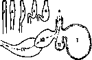
Fig. 1. 1-5—simplest forms of the stomach (according to Kingsley): 1 - stomach of the belone (Belone), a fish from the order Physostomi; 2 - stomach of the tailed amphibian-proteus (Proteus); 3 - stomach of the common grass snake (Natrix); 4 - stomach of the goby (Gobius), a fish from the order Gasterosteiformes; 5 - stomach of the shark; 6 - diagram of the stomach of a ruminant (the dotted line shows the path of food); insufficiently chewed food first enters the first compartment (I - the rumen), from which it is propelled by peristaltic movements into the second compartment (II - the reticulum); from the reticulum, the food is regurgitated, chewed again, swallowed again, and directed straight to the third compartment (III - the omasum), and then to the fourth compartment (IV - the abomasum), where it is digested.
In the middle part of the sac-like stomach, a very significant musculature develops. In birds, differentiation goes further and leads to the isolation of two separate departments—the anterior, slightly dilated glandular stomach, corresponding to the cardiac region, and the pea-shaped muscular stomach, developed from the pyloric region, the glands of which secrete a hard, horn-like lining that serves for grinding food. The stomach of mammals is characterized by significant development and differentiation of glands, of which the most constant are still the fundic glands and pyloric glands; less constant are the cardiac glands, which are reduced in carnivores and primates. A considerable part of the stomach is often lined with the same multilayered flat epithelium as the esophagus. The shape of the stomach can be complicated by the formation of sac-like protrusions in its various parts, which is especially observed in herbivorous forms (in some rodents, in even-toed ungulates). The most complex is the stomach of ruminants; however, of its four compartments—the large rumen with numerous strong papillae, the reticulum with a mesh-like inner surface, the omasum with leaf-like longitudinal folds, and the elongated abomasum with lower longitudinal folds—only the last represents a true stomach with fundic and pyloric glands. The first three compartments are lined with multilayered flat epithelium. The rumen and reticulum should be considered as parts of the esophagus in which food is only moistened and ferments. The omasum has obviously secondarily separated from the stomach, as can be seen from the structure of the camel's stomach.
I. Shmalgausen. The development of the stomach begins immediately after the formation of the anterior intestine, in the human embryo—at the 1st month (4 mm). The head section of the intestine forms the pharynx with gill slits and an anlage of the lungs; at some distance from it, a spindle-shaped expansion of the stomach forms; the anterior intestine is connected at this place with the walls of the body cavity by the anterior and posterior mesentery (mesogastrium). On the 5th week, the stomach acquires the characteristic shape of a retort with the greater curvature turned backward, but it is still symmetrical with respect to the median plane. Further changes consist in the bending of the stomach and its rotation along the axis, with the greater curvature turning to the left (fig. 3) (see Peritoneum, fig. there). The glands of the stomach begin to develop on the 10th week (Toldt) in the form of depressions of the epithelial layer (openings), from which then narrow pouches (gland bodies) grow inward. Lining cells differentiate only at the end of the 4th month. At the end of intrauterine life, the number of tubes opening into one opening reaches 7; after birth, it decreases to 2-3. Phylogenetically, in the development of the various glandular fields of the stomach (fig. 4), a certain sequence is noted. Thus, chief and parietal cells differentiate only in mammals; even later, pyloric and cardiac glands appear.
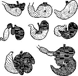
Fig. 2. Various forms of the stomach in mammals. Horizontal lines indicate the multilayered epithelium of the esophagus and esophageal part of the stomach; diagonal lines indicate the area of cardiac glands; dots indicate the area of fundic glands; crosses indicate the area of pyloric glands: 1 - stomach of a rat; 2 - dog; 3 - human; 4 - ruminant; 5 - horse; 6 - hamster; 7 - peccary (American wild boar); 8 - sloth (South American edentate, feeding on kidneys, young shoots, and fruits). (Fig. 1-6 according to Hesse, 7-8 according to Shimkevich.)
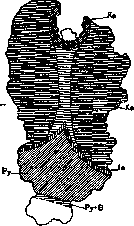
V. Karpov. Anatomical and topographical data. As in other vertebrates, the human stomach is an expanded part of the digestive tract, located between the end of the esophagus and the beginning of the duodenum. Its upper (inlet) opening, through which food enters, is called the cardiac, i.e., heart opening (from Greek cardia - heart), because it is closer to the heart than the outlet; the lower (outlet) opening, through which food passes from the stomach into the duodenum, is called the pyloric, i.e., sphincter (from Greek pylorus - gatekeeper). The stomach has two surfaces—1) anterior-superior and 2) inferior-posterior, and two edges: anterior (greater curvature) and posterior (lesser curvature). Like any hollow organ consisting mainly of muscular tissue, the stomach can undergo temporary changes in its volume and shape depending on the amount of its contents, on the state in which this contents is (solid, liquid, gaseous), and on the physiological requirements of the given moment of digestion. The glandular fields of the human stomach: a - cardiac glands; b - glands of the body; c - intermediate zone; d - pyloric glands; e - pyloric glands with parietal cells; Ru - lesser curvature.
Fig. 3. Forms of the stomach during its development: 1 - form of the stomach in an embryo of 5 mm length; 2 - in an embryo of 8 mm length; 3 - in 10 mm length; 4 - in 19 mm length. (According to Broman and Lewis.)

diac, i.e., heart (from Greek cardia - heart), as it is closer to the heart than the outlet; the lower (outlet) opening, through which food passes from the stomach into the duodenum, is called the pyloric, i.e., sphincter (from Greek pylorus - gatekeeper). The stomach has two surfaces—1) anterior-superior and 2) inferior-posterior, and two edges: anterior (greater curvature) and posterior (lesser curvature). Like any hollow organ consisting mainly of muscular tissue, the stomach can undergo temporary changes in its volume and shape depending on the amount of its contents, on the state in which this contents is (solid, liquid, gaseous), and on the physiological requirements of the given moment of digestion. The glandular fields of the human stomach: a - cardiac glands; b - glands of the body; c - intermediate zone; d - pyloric glands; e - pyloric glands with parietal cells; Ru - lesser curvature.
therefore the diversity of stomach forms is observed, which is found in autopsies and encountered in descriptions of the shape of the stomach by various authors. The nomenclature of the individual parts of the stomach also differs among different authors. In this article, the nomenclature of Lewis (Lewis; 1912) is given. Lewis gives the following scheme of the stomach and its anatomical subdivisions (fig. 5). The plane passing through the incisura angularis (a point on the lesser curvature where the concavity running along the pars cardiaca turns into the convexity running along the pars pylorica) forms on one side the left boundary of the opposite expansion on the greater curvature—on the other (vesti
Fig. 5. Diagram of the stomach and its anatomical subdivisions (according to Lewis).
bular part of the stomach; on one side it forms the left boundary of the opposite expansion on the greater curvature—on the other (vestibular part of the stomach; on one side it forms the left boundary of the opposite expansion on the greater curvature—on the other
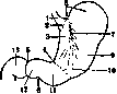
1-cardiac antrum; 2-cardia; 3-gastric canal; 4-angular incisure; 5-pyloric sulcus (pylorus); pyloric bulb), divides the stomach into two parts: ns; s_fUndus; s-left (cardiac) and pus; ; o-the boundary between corpus and vestibu-The right (pyloric). boundary between corpus and vestibulum pyloricum; *a-antnim/iuo-ca usually represents denale'(По Lewis *> a small bulge. Sometimes at this place there is a thickening consisting of muscle fibers, apparently belonging to the deep oblique layer. With full expansion of the St., this boundary disappears. The cardiac part occupies approximately 2/3 of the St., and the pyloric-1/3. The cardiac part consists of the corpus and fundus. The fundus is that part of the St. which protrudes above the horizontal plane passing through the St. at the level of the cardia. The pyloric part in turn is also divided into 2 parts: the right, or pyloric canal (canalis pyloricus, antrum pyloricum) and the left, or vestibule (vestibulum pyloricum). The plane dividing the pyloric part into the 2 mentioned parts passes at a right angle to the long axis of this part through the intermediate sulcus (sulcus intermedius) located on the greater curvature, approximately 2.5 cm from the pyloric sphincter. The gastric canal (canalis gas-tricus, road of the stomach, pu ) /~") or Magenstrasse Wal-\^_^-У L~^r-J deyer's) is called a narrow f>/~~\ o/-\ section of the cavity of the St., stretching \Z\_J <J^~^ in the form of a groove ^Г/Ч ^/^) along the lesser curvature from fir y f\/s-J cardia to incisura angu-УГу, ^S laris (fig. 30, p. 115). flyj {У/Э During the process of diges-----^ C^ tion (fig. 6) the St. is divided \-^^ \jf", "by a muscular constriction \-^^ \jf", "Ha into a large expanded left lobe and a narrow, co Fig. 6. change in the right, shaped like a tube ЛГ^оЖ right. The muscular con- digestion. (По ТЯЖКа usually прОХОДИТstriction usually passes cannon'y.) through the body of the St., and therefore often does not coincide with the anatomical boundary line between the body and the pyloric part. As the process of digestion moves forward, the aforementioned constrictive ring moves to the left, i.e. an increasingly larger part of the previously expanded body of the St. now takes on a tubular form and in this way gradually increases the tubular (pyloric) part. It is clear from this that the physiol. cardiac part can correspond to the anatomical only at a certain stage of gastric digestion. When moderately distended, the St. has a pear-shaped curved form. Its upper two thirds are located b. or m. longitudinally, while the lower third is more transverse. When the St. is relaxed, it tends to assume a more vertical position. Knowledge of the shape and position of the St. in a living person became more accurate after it became practice in the study of the St. to prescribe food in advance, to к-рой were added X-ray opaque salts of bismuth. The shadow from the St. was fixed on a photographic plate. On the basis of this method of study, Hertz gives a picture of the shape and position of a moderately distended stomach in a living person when standing (fig. 7). The lower boundary in this case is located slightly below the navel. By voluntary contraction of the muscles of the anterior abdominal wall, this boundary can be raised by 5-13 cm. In the lying position, the lower boundary of the St. is raised upward. This displacement of the lower boundary of the St. upward is not associated with a noticeable elevation of the level of the pylorus. Addison places on the corpse the lowest point of the lower boundary of the St. (caudal pole) 2 cm below the pylorus. Groedel determines this distance in a living person in the vertical position at 8 cm. The higher position of the lower boundary of the St. on the corpse is explained first of all by the fact that the corpse is usually examined in the horizontal position, and secondly by the fact that the diaphragm on the corpse is in the position of exhalation, and therefore the St. should occupy a correspondingly higher level. Gastroscopic anatomy with its color spatial pictures complements the shadow pictures of living anatomy and the data obtained on corpses. Gastroscop-ich. anatomy immediately considers the St. in the space of 3 dimensions. According to Sternberg, the axis of the St., i.e. that line which connects the midpoints of moderately expanded " '" "g "food masses, with der- cross щ "Fig. 7. In vivo shape and position of the St., " "shaving salts of bismuth: 1-esophagus; 2-bottom of the St. filled with the pelvis; 3-angular incisure; 4-pylorus; 5-navel. (По Hertz -u.) individual sections of the St., represents a helical line; the St. is thus a spiral tube with a right-hand thread (thread to the right). Gastroscopically-cardia and pylorus lie surprisingly close to each other along the air line. The boundary between the body of the St. and its pyloric part always exists gastroscopically, and at this place the passage of the gastroscope always encounters resistance. It is at this place that a fold running from the angular incisure is noted. Moynihan, who repeatedly observed spasmodic muscular contractions of the walls of the St. during operations, says that he saw such contractions on the body of the St. and on the pylorus, but never saw them on the fundus. Dimensions of the stomach. The stomach greatly increases (Scammon) in the last months of intrauterine life and in the first three months after birth. On the corpse, the average (anatomical) capacity of the stomach at birth is determined at 30 cm3, while the average physiol. capacity, determined during life by weighing before and after eating, is 7 cm3. During the first day, the average physiol. capacity doubles, on the 3rd day it quadruples, on the 4th day it increases 7 times, on the 10th day-more than 11 times (about 81 cm3). After the 4th day, the anat. and physiol. capacities approach each other. At the end of the first month, the capacity of the stomach is from 90 cm3 to 100 cm3. Then the increase is slower and by the end of the first year it reaches 250-300 cm3, during the 2nd year-from 300 cm3 to 500 cm3, during the 3rd year-from 400 cm3 to 600 cm3. In the period from 4 to 7 years, there is a slowdown in the increase in the capacity of the St., and after 7 years acceleration again sets in, and at 10-12 years the capacity of the St. reaches 1,300-1,500 cm3. In an adult, a moderately distended St. has a length (from the apex of the fundus to the lowest level of the greater curvature) of approximately 20-22 cm. The distance between the inlet and outlet openings varies from 7 cm to 15 cm. The transverse diameter at the widest part is 8-9 cm. The capacity of the St. in an adult 5
located on top of the small intestines, and finally by the transverse colon. The anterior wall of the gastric bed is formed 10 11
12 1.3 14 Fig. 8. The St. in its bed. The liver is removed, 1-right adrenal gland; 2-right kidney; 3-first part of the duodenum; i-second part of the duodenum; 5-fragments of the inferior vena cava; 6-portal vein; 7-hepatic artery; 8-common bile duct; 9-pylorus; 10-transverse colon; 11-right gastroepiploic artery, omentum removed exactly at the level of this artery. (Ho Dayis'y.) on average is determined at about 3 liters, but it varies greatly depending on the amount of food usually taken, and especially drinking, and on the degree of tonicity of the musculature of the St. itself as well as the abdominal walls. Topographic relations of the St. The part of the abdominal cavity in which the stomach is located is called the "gastric bed" (fig. 8). This bed has a posterior wall and a ceiling b. or m. vault-shaped, a lower wall (floor), somewhat sloping forward, and an anterior-Fig. 9. View of the liver and НЮЮ wall b. OR pa St. from above and behind по- m_ straight. Part of the removal of the diaphragm: "пггппкя обпячокя-j_stomach; 2-left lobe of the ceiling oopadUBd liver; г-esophagus; 4-le- of the visceral surface of the вая triangular ligament; liver; 5-spleen; в-transverse f ттппи ттршши" dissected diaphragm; Б0И Д°ли of the liver, 7-right lobe of the liver; the remaining part of the PO-v-inferior vena cava and the liver and posterior walls-печоночные вены; 9-anterior "пйпячоияны ттия- are formed by the dia-nal ligament; 10-right triangular phragm (fig. 9). ligament; 11-aorta; The floor rep- 12 - falciform ligamentresents "пйпя nrniTiv ня кп-liver (По Farabeufy.) СОООЙ полк} , on KO torom is located the lower-posterior surface of the St. The posterior part of the floor is formed by the apex of the left kidney (with its adrenal gland) and the gastric surface of the spleen (fig. 10 and 11); more forward-the broad upper surface of the pancreas; still more forward-the transverse mesocolon, extending from the anterior edge of the pancreas and
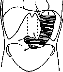
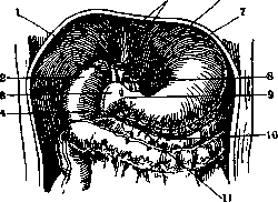

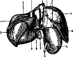
Figure 10. Relationship of the Stomach to surrounding organs. Posterior view. The figure is made from a model constructed by the reconstruction method: 1- stomach; 2- spleen; 3- part of the liver surface not covered by peritoneum; 4- liver; 5- right kidney; 6- ureter; 7- pylorus; 8- Spiegelian lobe of the liver; 9- omental tuber of the liver (tuber omentale); 10- left lateral (triangular) ligament of the liver; 11- esophagus; 12- inferior vena cava; 13- right adrenal gland; 14- coronary ligament of the liver. (After Cunningham.)
on the area of the anterior abdominal wall, bounded on the left by the left costal arch and on the right by the liver (fig. 12). The gastric bed is completely filled by the Stomach when it is distended. When the Stomach is contracted, it occupies only
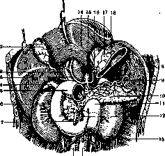
19th Figure 11. The liver is pulled upward. The Stomach is largely excised to show the organs to which its posterior surface is adjacent: 1-gallbladder; 2-portal vein; 3-hepatic artery; 4- right adrenal gland; 5-initial part of the duodenum; 6-right kidney; 7-superior mesenteric vein; 8-spleen; 9-splenic artery; 10-pancreas; 11- duodenojejunal flexure; 12-superior mesenteric artery; 13-inferior mesenteric artery; 14-liver; 15-aorta; 16-left gastric artery; 17-stomach; 18-splenic vein; 19-inferior vena cava; 20-aorta. (After Testut.)
the lower part of its bed; the rest of the bed is filled at this time by the transverse colon, which as the Stomach contracts gradually turns upward and finally lies in relation to the Stomach in front and directly under the diaphragm,- a fact which must be remembered during clinical
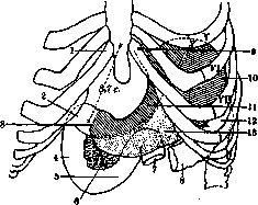
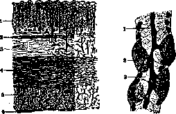
examination of this area. The cardiac opening of the Stomach is located at the level of the XI thoracic vertebra in front of the aorta. It is situated approximately 10 cm behind the anterior chest wall and 2.5 cm from the left edge of the sternum- behind the VII left costal cartilage. When the Stomach is not filled, the pylorus is located approximately 2.5 cm to the right of the midline at the level of the I lumbar th vertebra. This level quite accurately corresponds to the midpoint of the line connecting the incisura suprasternalis (jugularis) with the upper edge of the symphysis ossium pubis. When the Stomach is distended, the displacement of the pylorus to the right of the midline can reach 7.5 cm. Structure of the Stomach. In the wall of the St., as in other parts of the digestive tract, three layers are distinguished: 1) the mucous membrane (tunica mucosa), 2) the muscular (t. muscularis) and 3) the serous (t. serosa) (fig. 13). The mucous membrane has a thickness from 0.5 mm (in the cardia region) to 2 mm (pylorus), pale pink color, reddening during digestion. In the collapsed Stomach, it forms longitudinal folds (plicae mucosae), which smooth out when it is filled. On the surface of the mucous membrane, polygonal fields (1-4 mm2) protrude, bounded by shallow grooves (areolae gastricae) and caused by the accumulation of glands (status mamillaris, etat mamelonne, see); in the area of the fields, numerous openings (9:-18 per 1 mm) are visible, leading into the mouths of the glands (foveolae gastricae). The mucous membrane includes: the surface epithelium, glands, own tissue, muscular layer and the submucous membrane, which is sometimes isolated as a separate layer. The epithelium represents a special form of mucous epithelium, forming a secretory epithelial surface (see Glands); it consists of cylindrical cells with a nucleus in the middle and mucigen grains in the upper part, where the centrosome and Golgi apparatus are also located (fig. 14). The upper end of the cells is usually open on preparations, for which reason they were long considered goblet cells; it is now proven that the free surface is always closed by a thin membrane and the secretion is released through it in small portions; it gives mucous reactions. Due to the presence of fat droplets, this epithelium is sometimes credited with the function of absorption. The Glands of the Stomach (glandulae gastricae) belong to the type of branched tubular
Figure 13.
Fig 14.
Figure 13. Section of the stomach wall at low magnification. On the right, injected vessels. 1-mucous membrane; 2-muscularis mucosae; 3- submucous membrane; 4 and 5-muscular membrane; 6-serous membrane. (After Lavdovsky.) Figure 14. Surface epithelium of the Stomach: 1-lumen of the glandular tube; 2-covering cells with secretory capillaries; 3-main cells. (After Zimmerman.) and are located extremely densely. Their beginning serves as the foveolae gastr., which, deepening, form a funnel-shaped mouth of the glands, lined with surface epithelium. After it follows a thin short section- the neck, from which 2-3 glandular tubes go, more or less winding and expanded at the ends. The total number of glands in an adult is about 25 million, in a newborn about 2 million (Toldt). In the human Stomach, 4 types of glands are found: a) Glands of the fundus (gl. gastr. propriae, Magensaftdrusen), the most common type, occupying the larger part of the body and fundus of the Stomach. They have a length of 1.5-3 mm, with 11/4-3/4 of which falls on the mouth; the tubes are straight or slightly winding, 30-50 μ in width, the blind ends are slightly expanded. Besides the membr. propria and flat branched cells, they contain glandular cells of two types (fig. 15); some are called the main cells of the fundus (R. Heidenhain) or adenomorphic cells (see), others-covering cells (Belegzellen, R. Heidenhain) or delomorphic cells (of distinct shape). There are more main cells, and they border the lumen; they have cubic or pyramidal shape, the nucleus is closer to the basal end, in which a rod-like striation is noticeable. In life, fairly large, strongly light-refracting grains of pepsinogen are visible in the cell; after fixation, the cell body becomes light and stains with hematoxylin. Covering cells are fewer in number, they are located further from the lumen, covered by the main cells and protruding membr. propria; there are most of them in the neck and least in the fundus of the gland. In size they are larger than the main cells, have round, oval or polygonal shape, the nucleus in the middle; in life they contain fine granularity and on fixed preparations they appear darker than the main cells; they stain with acidic dyes. Silvering according to Golgi reveals inside the covering cells secretory capillaries, which in the form of a basket surround the nucleus and communicate with the lumen by a short neck. The main cells secrete pepsin, the covering cells-HCl. The latter however is not secreted as such, but in some compound, which becomes active only with the participation of the covering epithelium. b) Pyloric glands, occupying the area near the pylorus for a distance of 4-5 cm (sometimes up to 14), are characterized by longer mouths and a shorter, strongly winding body (fig. 16). The lumen is lined with one type of light cylindrical cells, with nuclei closer to the base and fine granularity in life. Previously they were considered mucous (Magenschleimdrüsen, Koelliker), but they do not give mucous reactions; then they began to be identified with the main cells of the fundus glands (R. Heidenhain), from which however they differ in appearance and character of the grains, and finally-recognized as elements sui generis (Oppel). Pyloric glands without a sharp boundary pass into the Brunner's glands of the duodenum; there is an opinion that they secrete pepsin. As a variety of pyloric glands, the so-called accessory cells (Nebenzellen of German authors) are described, which secrete a serous or seromucous secretion. They are located in the area of the neck of the fundus and body glands; they increase especially in number toward the pyloric region, where they displace the main cells, forming together with the covering cells the so-called intermediate zone, c) Cardial glands, serving as a continuation of the esophageal glands, occupy in humans a very narrow area (5-6 mm) at the entrance to the Stomach and are similar in structure and character of cells to the pyloric ones. They do not secrete mucus and sometimes contain covering cells, d) Lieberkühn's glands, quite similar to the glands of the small intestine, very often
Figure 16. Pyloric
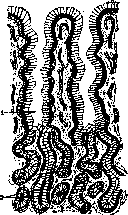
glands: 1-openings of the glands; 2-glandular tubules. (From Lavdovsky.) They occur in islands in the area between the fundus and the pyloric part, rarely in the cardiac zone. The diversity in the structure of different parts of the stomach mucosa can under pathological conditions become even greater; thus, in chronic gastritis, pyloric glands are found in islands in the body of the stomach, Lieberkühn's glands are sometimes found in the cardiac area, etc. These phenomena are based not only on the factor of heterotopia, but represent a product of pathological regeneration, i.e., various glands arise in place of the covering epithelium.-The proper tissue of the mucous membrane (lamina propria) consists of delicate connective tissue of a reticular nature and is always infiltrated with lymphocytes, which in places can form follicles. Under the base of the glands there is a plate of dense connective tissue (stratum Zeisseli). Following it is the proper muscular layer of the mucous membrane (muscularis mucosae), in which, in addition to the outer longitudinal and inner circular layers of smooth muscles, a layer of oblique fibers adjacent to the Zeissel membrane is also described. The submucous membrane consists of loose connective tissue and sometimes contains fat cells. The muscular coat of the stomach consists of three layers of smooth muscle fibers: outer, middle, and inner (fig. 17). The outer layer consists of longitudinal fibers, which are a continuation of the longitudinal fibers of the esophagus, which in the cardia area begin to diverge radially over the surface of the stomach. These fibers are best expressed along the lesser and greater curvatures; on the anterior and posterior surfaces they appear as a very thin scattered layer, which toward the pylorus again transforms into a uniform, well-expressed layer; above the pyloric sphincter the longitudinal fibers split into 2 layers-superficial and deep; the superficial layer continues into the corresponding layer of the duodenum, and the deep one is directed to the pyloric sphincter, where it ends in the form of diverging bundles, many of which reach the submucosa. The middle layer of the muscular coat of the stomach consists of circular fibers, which are located throughout the entire length of the stomach, except for its fundus. Toward the pylorus this layer becomes thicker. The pyloric sphincter is formed by this layer. The inner layer of the muscular coat consists of circular and oblique fibers. The circular
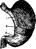
Figure 17. Preparation of the three muscle layers of the stomach wall: 1-inner layer-oblique fibers (fibrae obliquae); 2-middle layer-circular fibers (fibrae cir-culares); 3-outer layer-longitudinal fibers (fibrae longitudi-nales). (After Cunning-ham.)
fibers of this layer completely surround the fundus of the stomach, just as the circular fibers of the middle layer surround the rest of the stomach. Starting as a series of rings at the top of the fundus, they extend in the form of a layer of rings located at right angles to the axis of the stomach to the cardia. Below the cardia this layer continues as oblique fibers, which diverge radially from the left side of the esophageal opening downward and to the right on both surfaces of the stomach. Some of them almost reach the antrum pylori. The oblique fibers end by sharply turning toward the greater curvature and passing into the circular fibers of the middle layer. The oblique fibers are best observed if the mucous membrane is removed. The upper part of the oblique muscles of the inner layer of the human stomach serves to form a special kind of groove (see above "stomach channel"). It is believed that along this path saliva and liquid parts of food can pass from the esophagus directly into the pyloric part, bypassing the cardiac part. In ruminants, the oblique fibers form a complete ring and cross in the second stomach. By their contraction they close the entrance to the first and second stomach and conduct regurgitated and chewed a second time food directly to the 3rd and 4th stomach. The oblique fibers act as sphincters, and peristalsis is apparently not inherent in them. During vomiting, usually originating from the pylorus, the oblique fibers pull the pyloric part toward the cardia, shortening the path between them. A special prepyloric sphincter as a separate anatomical formation apparently does not exist. The right anatomical boundary of the cardiac part of the stomach is the right-sided area of distribution of the oblique muscles; their closure forms the physiological prepyloric sphincter. As the cardiac sac empties, larger and larger parts of the oblique fibers, and apparently also the circular ones, are drawn into contraction and form an increasingly elongated tubular section extending to the left. The filling of the pyloric part throughout the entire time of digestion remains constant due to such a mechanism, and the pressure in different parts of the stomach as it empties of food can be maintained at the same level. Forssell believes that the fibers of the oblique layer

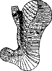
Figure 18. Left-longitudinal muscle fibers of the stomach; right-transverse muscle fibers of the stomach. (After Forssell*».)
are located in certain bundles (fig. 18) (sling 1st, 2nd, 3rd), which by their contractions limit certain areas.-Tunica serosa. The outer layer of the stomach is formed by the peritoneum, which surrounds it on all sides, except for two areas extending along the lesser and greater curvature of the stomach and intended for large blood vessels. The arteries of the stomach originate from three main branches of the a. coeliacae: 1) from a. gastrica sinistra (also a. coronaria ventriculi super. sinistra), 2) from a. hepatica and 3) from a. lienalis (fig. 19). The branches intended for the stomach,
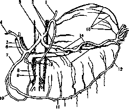
Figure 19. System of the celiac artery-a. coelia-sa (3): 1-a. oesophagea; 2-a. gastr. sin., seu a. coron. ventric. sup. sin.; 4-a. cystica; 5- v. portae; 6-a. hepatica; 7-a. gastro-duode-nalis; 8-v. mesent. sup.; 9-a. mesent. sup.; 10-a. pancreatico-duoden.; 11-a. gastro-epipl. dextra; 12-a. gastro-epiploica sin.; 13-aa. ga-stricae breves; 14-a. lienalis; 15-v. renal.; 26-a. gastrica dex., seu a. coron. ventric. sup. dex. (After Cunngham'y.)
anastomosing with each other, form two main arterial arches located along the greater and lesser curvature between the converging peritoneal sheets covering the anterior and posterior surfaces of the stomach. The arterial trunks connect with the stomach wall very weakly, which is extremely important in view of the large changes in the size of the stomach: when the stomach contracts, the arteries twist, when it stretches, the arteries straighten. The thin arterial branches penetrate through the muscular layer to the submucosal layer, where they divide into even thinner branches that freely anastomose with each other. The branches branching off from this vascular plexus are distributed in the mucous membrane and in the muscular layers.-Veins, just like arteries, form a plexus in the submucosal layer, from where blood through the left gastric omentum (v. gastro-epiploica sin.) and short gastric veins goes to the splenic vein (v. lienalis), through the right gastric omentum to the superior mesenteric vein (v. me-senterica superior) and through the coronary vein and the stomach exit vein directly into the portal vein (v. portae). These veins, like other branches of the portal system, have a well-expressed muscular layer and contain numerous valves (valvulae).-Lymph. vessels of the stomach begin in its mucous membrane around the gastric glands; then in the submucosal layer they form a plexus, from where the trunks emerge, directed together with the blood vessels to the greater and lesser curvature and receiving along the way branches from the subperitoneal lymphatic plexuses (fig. 20).
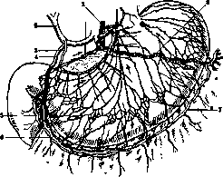
Figure 20. Lymphatic vessels and lymph, glands of the stomach: 1-left gastric artery; 2- hepatic artery; 3-right gastric artery; 4- gastroduodenal artery; 5-artery of the duodenum and pancreas; 6-right artery of the stomach and greater omentum; 7-left artery of the stomach and greater omentum; 8-splenic artery. (After Taylor'y.)
The lymphatic ducts, running together with the blood vessels, are connected: 1) with the upper lymphatic gastric glands, located along the lesser curvature, 2) with the lower glands, located along the greater curvature, and 3) with the splenic glands, located between the layers of the gastro-splenic omentum, right near the place of its attachment to the spleen. Although there are free communications between the lymphatic vessels of the stomach wall, nevertheless in certain areas of the stomach the flow of lymph goes in known, strictly defined directions. If from the upper point of the bottom of the stomach a line is drawn in the direction of the pylorus, passing at the level of the junction of the middle and lower thirds of the gastric wall, this line will serve as the boundary between the upper lymph flows, directed to the lesser curvature, and the lower ones, directed to the greater curvature. A vertical line going from the cardia downward to the greater curvature will mark with a considerable degree of probability the place of divergence of the right and left gastric-omental ducts. In cancer of the stomach (Lengemann), the glands located along the lesser curvature are affected in 50% of cases, the glands along the greater curvature in 37% of cases, and those located under the pylorus in 60%. Innervation of the stomach. In detail - see Autonomic nervous system. At the same time, it must be indicated that there is an opinion that the cardiac half of the stomach is supplied with both motor and inhibitory fibers through the n. vagus, while the pyloric half of the stomach is supplied with motor fibers through the n. vagus (Fig. 21), and with inhibitory fibers through the sympathicus by means of the plexus Solaris. Starling says: "The predominant motor effect of the n. vagi in the area of the fundus (cardial part) will be an increase in tone, and in the pyloric part - an increase in peristaltic waves".
a. deshin. II. Physiology. Secretory functions. The stomach in a functional relation is divided into 2 parts: the fundal and the pyloric. The first part has besides the chief cells also parietal cells and produces in addition to enzymes also free HCl (about 0.5%), which is among other things an important factor in disinfecting food masses. The pyloric part has only chief cells and secretes a pyloric juice of an alkaline reaction, viscous and transparent. Among the enzymes, pepsin (Schwann; 1836) must be mentioned, which breaks down protein substances to peptones in an acidic environment, with the optimum of action at about 0.15-0.2% HCl. When the juice is mixed with food, an acidity close to the optimal is obtained. The gastric juice very easily digests connective tissue. Hammarsten (1872) proved the enzymatic nature of the clotting of milk by gastric juice and named this enzyme chymosin. However, I. P. Pavlov expressed the view that the milk-clotting and protein-dissolving actions may belong to the same enzyme, since in all cases both actions change in exactly the same way. Volhard pointed out
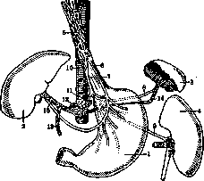
Figure 21. Diagram of the relationship of the branches of the vagus nerves to the stomach: n. vagus dext. spreads along the posterior surface of the stomach and gives connecting twigs to the celiac plexus, to the splenic and hepatic plexuses; n. vagus sin. spreads along the anterior surface of the stomach and gives connecting twigs to the right vagus nerve and the hepatic plexus. 1-stomach; 2-liver; 3-spleen; 4-left kidney; 5-esophageal plexus; 6-left vagus nerve; 7-right vagus nerve; 8-branch of n. vagi to the splenic plexus; 9-branch of n. vagi to the renal plexus; 10-aorta; 11-left gastric artery; 12-celiac plexus; 13-a. gastro-duo-den.; 14-a. lienal.; 15-a. hepatica.
and on the presence of lipase in gastric juice, which many authors could not confirm. In any case, there is very little lipase, and probably these traces depend on the release of intracellular enzymes from the epithelium. Technique. Heidenhain (Fig. 22) proposed the creation of a small stomach pouch for the purpose of collecting pure gastric juice from it. However, in these operations the nerves were cut, so that the small stomach worked completely differently than the large one (Orbeli). After Pavlov and Shumova-Simanovskaya had established the significance of the n. vagus as a secretory nerve, it became necessary to isolate the small stomach with preservation of innervation (Fig. 22). The work of Khizhin solved this problem: the bridge was not cut through completely, but only the mucous and submucous layers were cut, thanks to which
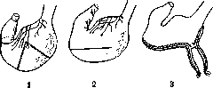
Figure 22. 1-incisions for the Heidenhain stomach pouch; 2-incision for the Pavlov stomach pouch with preservation of innervation; 3-diagram of the Pavlov stomach pouch.
and the division of the large and small Stomach occurred. If a metal cannula is placed on the large Stomach, then juice from the large Stomach can be collected through a fistula, and from the small stomach through the opening made, and thus the amounts of juice obtained under different conditions (irritation, experience of so-called 'imaginary feeding', introduction of alcohol into the rectum) can be compared. In the history of the question of the conditions of gastric juice secretion, a cardinal moment is the experiment of so-called 'imaginary feeding' (I. P. Pavlov and Shumova-Simanovskaya; 1889). The authors proceeded from observations by Beaumont on a Canadian hunter in the early 19th century. These observations led to the creation of a gastric fistula in a dog by Basov in 1842 and by Blondlot in 1843. With these fistulas, it was still impossible to obtain pure juice. In view of this, the authors added the severing of the esophagus and sewing its ends into a skin wound on the neck. Thus, during eating, food fell out of the esophagus incision and did not enter the Stomach. In such dogs, an alkaline reaction of the fundic part could be established on an empty stomach. After 5-7 minutes from the start of eating, the reaction changed, and then abundant secretion began with an acidity of 0.5-0.55% HCl and with a high content of pepsin. On these dogs, it could be established that mechanical irritants cause secretion not of acid, but only of alkaline mucus. The question of the mechanism of secretion immediately arose. How are impulses transmitted from the oral cavity to the glands of the Stomach? Severing both vagus nerves immediately destroyed the effect of imaginary feeding. Thus, the nn. vagi are the secretory nerves of the Stomach. Later, Ushakov confirmed this in the conditions of acute experiments by direct irritation. The reflex from the oral cavity to the glands is an unconditioned reflex. Zeleny saw similar secretion in a dog without hemispheres. On the basis of this unconditioned reaction, conditioned reflexes to sight, smell, etc., can also arise. It is these reflexes that often confuse researchers. If a dog is constantly fed in a stall, then simply placing it in the stall becomes a signal for food and causes 'conditioned' secretion. All these factors can also cause constant secretion, which many authors consider even normal. After severing the vagus nerve, Stomach secretion continues, although the act of imaginary feeding no longer yields a drop of juice. It is true that recently Volbort and Kudryavtsev have shown the secretory role of the splanchnic nerves by direct irritation after preliminary severing. But in any case, even complete denervation does not destroy secretion. Edkins in 1906 indicated the humoral nature of secretion under the influence of gastrin (a hormone extracted from the pyloric mucosa). However, these works were criticized, since many different kinds of extracts caused secretion of acid juice. The works from Pavlov's laboratory by isolating different parts of the Stomach through complex operations were much more important for understanding. These experiments led to the conclusion that hormones are formed in the pylorus, which determine secretion, since even extensive nerve destruction has little effect on it. Schmidt's data agree with this, who observed the disappearance of the second phase of secretion after resection of the pyloric part. Ivy and Farrell gave new proof of humoral impulses by transplanting an isolated stomach under the skin with severing of all vessels and nerves. Such a stomach begins to secrete juice after eating. The data of Razenkov are in complete agreement with this, who observed juice secretion in a dog with a Heidenhain stomach when blood was injected from a dog in a state of digestion; however, when blood from a starving dog was transfused, there was no gastric juice secretion. Thus, in the secretion of gastric glands there is a phase of reflex juice secretion and a phase of chemical secretion, with the most probable secretion of hormones from the pylorus under the influence of various irritants, such as: water, extractive substances of meat, fish, vegetables, products of protein digestion (peptones), soaps, salts, etc. The total amount of juice secreted for equivalent portions of food by nitrogen content varies by type; thus, for 250 g of bread, 42 cm³ was secreted, for 100 g of meat 27 cm³, for 600 g of milk 34 cm³. Each type of food has its own secretion curve (Fig. 23) and its own enzyme content. When the amount of food is doubled, the amount of juice also doubles, and the times of digestion are approximately as the square roots of the masses (weights) of food. The role of fat in the secretory work of the fundic glands should be noted. Fat, entering the duodenum, inhibits the secretion of acid and enzymes and delays peristalsis. If a Stomach is completely separated from the duodenum, then fat has no inhibitory effect. On the other hand, breakdown products of protein-peptones and fat-soaps, acting on the pylorus, cause secretion of acid juice. Mechanical irritation of the pylorus causes increased secretion of pepsin, so the consistency of food also affects the quantity and quality of the secreted juice. - The pyloric juice is a transparent, thick liquid of alkaline reaction, containing pepsin secreted in a zymogenic form. Juice secretion is caused by local irritation by acid, and along with an increase in the amount of acid, there is an increase in the amount of enzymes. If a drain is introduced into an isolated pylorus, juice is secreted more or less constantly; eating significantly reduces this constant secretion; after severing the splanchnic nerves, this suppression after eating disappears. The role of the vagus nerve cannot be considered fully clarified. Small doses of pilocarpine, arecoline cause an increase in voluntary secretion, while large doses, on the contrary, cause suppression. Atropine sharply reduces secretion, while at the same time decreasing the pepsin content. It is curious that after atropine, acid no longer causes increased secretion and an increase in the amount of enzymes.
V. Savich. Motor function. The stomach itself has the ability to perform peristaltic movements. Peristaltic waves, which occur in the pyloric part of the Stomach, are usually not observed in the cardiac part; the latter is a more or less passive reservoir of food, from which it is gradually directed into the active antrum pyloricum. During the process of digestion, the content of the antrum pyloricum remains almost constant in volume, while the content of the cardiac part continuously decreases after the end of a meal. Mixing of the contents of both parts of the Stomach does not occur. In its mechanical action, the Stomach to a large extent resembles the heart. Just as the cardiac part of the stomach represents an expanding reservoir collecting food material for the rhythmically contracting strong pyloric press, so the atria of the heart represent a stretchable reservoir collecting blood, which then fills the strong, rhythmically contracting pump-ventricle. Positional function predominates in the atria and the cardiac part of the Stomach, while the motor function predominates in the ventricles of the heart and in the pyloric part of the Stomach. Numerous experiments and observations on the stomach of the frog, cat, dog, and human have shown that in these animals the pyloric part, with its well-developed muscular wall, performs strong, more or less rhythmic movements, while the cardiac part, with its relatively weakly developed musculature, represents a reservoir from which (in the expression of Sherrington) the pyloric mill receives its product for grinding. The maintenance of low pressure in the Stomach when it is filled is achieved not only by the positional activity of its walls, i.e., the ability to respond to an increase in its content with a kind of active diastole proportional to the volume increase, but also by the positional activity of the muscles of the abdominal wall. With its positional activity, the diaphragm also participates in the regulation of the capacity of the abdominal cavity. As the Stomach fills to varying degrees, it adapts by changing the tone of its musculature to the amount of its content, so that the intragastric pressure remains constant, and the level of the upper boundary of the gastric contents in the vertical position does not change with the change in the amount of mass contained in the Stomach. The main functional task of the cardiac reservoir of the Stomach is that it must easily adapt to rapid and abundant food intake without a noticeable increase in pressure within it. High pressure in the cardiac part would present an obstacle to the further entry of food from the esophagus. Every passage of swallowed food through the cardia is accompanied by rapid, slight expansion of the fundi. The intragastric pressure at this time slightly decreases, but then returns to what it was before. This expansion occurs reflexively through the vagus nerve. In the human Stomach, not only does mixing of the contents of the cardiac and pyloric parts not occur, but also in the cardiac part itself, sequentially ingested portions of food do not mix with each other. When a new portion of food enters a partially filled Stomach, it penetrates into the central part of the food mass located in the cardiac region, and thus moves the central portions of this mass to the periphery. Grützner, testing the reaction of different layers of food in the Stomach, found that while the content of the pyloric part was acidic everywhere, in the cardiac part only the outer layer had an acidic reaction, in which presumably digestion with the help of gastric juice occurred. The central layers, however, had an alkaline reaction—there presumably digestion with the help of saliva continued. It is very probable that in humans, as in other animals, the cardiac part of the stomach in the early stages of gastric digestion serves as the site of action of ptyalin on the food mass. In the upper part of the cardiac part (fundus), swallowed air and gases formed during gastric digestion accumulate. The size of the gas bubble on average: diameter of the base 8-10 cm, height 3-5 cm. Peristaltic contractions of the pyloric part, forming a reverse flow when the pyloric sphincter is closed, contribute to the best grinding of food masses and their proper mixing with gastric juice. When the pyloric sphincter is open, peristaltic contractions gradually empty the pyloric part, moving its contents into the duodenum. Insufficient chewing of food enhances the peristalsis of the pyloric part and thus forces this part to perform the work that the jaws should have done. The pyloric opening of the Stomach is surrounded by a very strong ring of circular muscle fibers (sphincter pylori). In life, this opening, located at the end of the pyloric canal, is always tightly closed, except at individual moments of the digestive act, when it opens intermittently with irregular intervals to allow the appropriately processed food material to pass from the stomach into the duodenum. Cannon mixed flour food with bismuth nitrate and observed the movements of the Stomach with the help of X-rays on cats. 5 minutes after food intake, near the duodenal end of the antrum pyloricum, a slight circular contraction is noted, which moves peristaltically toward the pylorus; it is followed by several repeated waves with irregular intervals. 2 or 3 minutes after the first movement is noted, a very slight contraction appears near the middle of the Stomach and, sinking deeper into the greater curvature, slowly moves toward the pyloric end. Not every wave of contraction pushes food through the sphincter pylori. The first area to noticeably decrease in volume is the pre-antral part of the pyloric part. As the process continues, the middle part of the stomach takes the form of a tube with a rounded cardiac sac on one side and an active antrum on the other. Along the tube, very shallow contractions can be noted going one after another toward the pylorus. By the contraction of the longitudinal, circular, and oblique muscles of the spherical cardiac sac, its contents are squeezed into the tube-shaped part. The contractions of these muscles apparently occur in such a way that they as it were wash off the superficial food layer, altered by the gastric juice secreted by the walls of this part, and squeeze this layer into the tubular part, placing a new food layer in the cardiac part under the action of gastric juice. When the cardiac sac is emptied, the waves of contraction carry the contents of the tubular part into the antrum. Cannon summarizes the results of his research as follows: 'The Stomach consists of two physiologically different parts: 1) the active pyloric part, along which during digestion waves of contraction pass with a continuous rhythm, and 2) the cardiac part, which is an active reservoir that expels its contents in small portions from time to time, as the mechanism of the pyloric part is prepared to receive them.' In the cat, the food in the cardiac part, when examined 1/2 hour after the appearance of peristaltic waves, had the same mushy appearance as it had when it entered the Stomach. The content of the pyloric part had a completely different appearance: it resembled thick soup. The contractions observed in the pyloric part perform three functions: 1) mixing, 2) grinding, and 3) pushing food into the duodenum. The regulation of the entry of food in small portions from the cardiac part into the pyloric part, according to authors who have studied this question (Shemyakin), depends on the sphincter located at the boundary between the cardiac and pyloric parts, which, at its maximum contraction, completely separates the pyloric part, so that not a drop of liquid passes from the fundal part. Kathcart considers that the normal activity of the prepyloric sphincter has a twofold purpose: 1) to prevent the mechanical pressure of food from the cardiac part into the pyloric part, which could hinder digestion, and 2) to prevent the backflow of digested material, i.e., to form a chamber or cavity in which digestion or grinding could be carried out most economically. During the 4th month of embryonic life, the mucous membrane of the pyloric part of the Stomach, in its villous appearance, in the form of its glands, and in the character of its epithelium, much more resembles the mucous membrane of the duodenum than the mucous membrane of the cardiac part of the stomach. Deshin, on the basis of literary data (Cunningham, Paterson, Batuev) and his own observations, believes that the key to resolving the question of the diversity of stomach forms in humans must first be sought in the rhythmic movements of the Stomach, noted in the process of gastric digestion. Connecting the forms of the human Stomach observed on the corpse with the data of physiology and radiology, one must conclude that the normal human Stomach, due to its motor activity, cannot have a constant shape during life. The largest group of stomach forms observed on the corpse undoubtedly represents only individual phases of gastric digestion, fixed at the moment of death.
If cardiac activity had ceased in the given subject several minutes earlier, then the shape of the Stomach on the corpse would also have been different. Since the part of the Stomach adjacent to the pylorus plays a particularly important role in the motor activity of the Stomach, it becomes obvious that on corpses antrum pyloricum (canalis pyloricus) should be found particularly often in a contracted state. In an expanded state, antrum pyloricum is not clearly demarcated from vestibulum and under certain circumstances can completely disappear as an anatomically separate part from vestibulum. If in the last hours of life the Stomach becomes atonic and expanded, the corresponding picture will also be observed after death. Since the introduction of formalin into anatomical practice, the motor phases of gastric digestion have more often been encountered on the dissecting table. This was due, firstly, to the fact that formalin, being an excellent fixative, directly fixed individual motor phases, and secondly, to the fact that, by preventing putrefaction processes, it hindered the development of gases in the Stomach that stretched its walls and thus changed the shape of the Stomach postmortem. With maximum uniform relaxation of the walls, for the same Stomach the shape should be constant and have the appearance which is inherent to the Stomach due to its own anatomical structure and which is the starting point for its transitional contractions. Wernstedt calls this form, from which all others can be produced, the basic form of the Stomach, while he calls all transitional forms functional. A removed Stomach, placed in appropriate conditions, performs movements (Hofmeister, Schütz). Magnus believed that the rhythmic movements of the Stomach and intestines are caused by the presence of Auerbach's plexus, however Alvarez, based on his experiments as well as observations by other authors, decisively speaks in favor of the myogenic nature of these movements, since strips devoid of nerve plexuses could produce rhythmic movements. In the Stomach, peristaltic contractions are observed, beginning in the esophageal region and extending to the pylorus, with some waves disappearing while others cause corresponding contractions of the pylorus. The peristaltic wave thus spreads in such a way that the irritated area above contracts, while relaxation occurs below; in the next moment, the contraction wave passes to the muscles that were in a state of relaxation, while the areas lying even further relax. This creates conditions for the movement of semi-liquid food masses. Both peristaltic and antiperistaltic waves, approaching the corresponding sphincters, cause their relaxation. If the animal is fasting, periods of strong motor activity of the gastrointestinal tract can sometimes be observed. From time to time, after 1 hour-1 hour 20 minutes, periods of strong movements of the Stomach and intestines begin, accompanied by the expulsion of bile from the bile ducts—the so-called 'periodic activity' of the digestive tract, to which Boldyrev (1904) paid special attention, but which was also noted much earlier (Morat, 1882; Rossbach, 1890 and others). Rossbach already associates the sensation of hunger with movements of this type. Irritation of the oral cavity with solid food (for example, eating bones) causes a reflex contraction of the prepyloric sphincter, so that the fundic part is separated from the pylorus. In addition to the oral reflex, the prepyloric sphincter can also close due to irritation from the duodenum, for example by fat (Orbeli and Khozroev). In addition to the independent motor functions of the Stomach and intestines, their work is regulated on the one hand by the vagus nerve (motor nerve), and on the other by the splanchnic nerve (inhibitory nerve). The study of the motor function of the Stomach can be conveniently carried out using X-rays after administering porridge with bismuth or barium salts. Finally, the motor function is studied by means of balloons connected to a recording device. In dogs, fistulas are applied in various places for this purpose, through which these balloons can be conveniently introduced; in humans, thin probes with a bulb at the end are inserted into the Stomach and duodenum, which records the pressure changes. Some questions can be conveniently studied after applying fistula tubes to the Stomach and duodenum. Infusion into the Stomach may be accompanied by infusion into the intestines, and then there is a significant slowing of the emptying of the Stomach. By this method, the delaying effect of fat (Lintvarev) and acid (Serdjukov) on the transition of Stomach contents to the intestines was demonstrated. High acidity in the duodenum similarly not only delays the transition from the Stomach to the intestines, but usually causes the contents of the duodenum to be thrown back into the Stomach. Generally, with fatty food, throwing back is a constant phenomenon. When 0.5% HCl is poured into the Stomach, it is partially neutralized by the throwing back of alkaline intestinal juices, and only then does it begin to quickly leave the Stomach; when the pylorus is ligated, the change in reaction occurs very slowly and incompletely, since here alkaline juices cannot be thrown back into the Stomach. It is curious that an isolated stomach according to Ivy and Farrell, devoid of nerve connections, nevertheless has independent movements that disappear after fat is introduced into the large Stomach. Here there seems to be some kind of humoral mechanism of delay. It should be noted the symptom of a coated tongue in disorders of the Stomach. Kost gave such patients licopodium in wafers and on the next day could find its spores on the tongue, so that the coating is a consequence of reverse movements of the Stomach. The mixing of food from different meals in the Stomach occurs far from perfectly. If a rat is given food colored in different colors three times, and then killed, frozen, and a saw cut made through the Stomach, it will turn out that the first portion surrounds the periphery of the Stomach, the third near the esophagus, and between them the second (Grütner).
A. Deshin, V. Savich. III. Methods of stomach research. Methods of research of the St. can be subdivided into the following sections: 1) logical conclusions from the patient's complaints and history, 2) physical methods of examination - inspection, palpation, percussion, auscultation, 3) functional research using thick and thin tubes, 4) chemical, physico-chemical and microscopic examinations of stomach contents, 5) gastroscopy (see), 6) X-ray diagnosis. A thorough analysis of the patient's complaints and history often makes it possible, if not to establish a diagnosis, then to approach it very closely. Always keeping in mind the physiological scheme of the St.'s work, an experienced physician will correctly evaluate vomiting on an empty stomach of food taken the day before, bloody vomiting, sour belching, the nature of pain and other symptoms and on the basis of these data can often make a presumptive diagnosis. Inspection of the epigastric region in the lying and standing positions often yields valuable results: with thin abdominal coverings, mainly in asthenics, as well as in early childhood and in persons who have become very emaciated, with a prolapsed St. one can see the contours of the distended St. as a whole or the contour of the greater curvature or peristaltic waves. With an empty St. during intractable vomiting or esophageal obstruction, a characteristic diagnostic depression is observed in the epigastric region. In the standing position of the patient with gastroptosis and enteroptosis, a depression of the abdominal area above the navel and a protrusion and sagging of the area below the navel are observed. Pathological movements in the area of the St. are usually directed from the bottom of the St. to the pylorus and more rarely - reverse antiperistaltic movements (e.g. with high degrees of pyloric narrowing). Pathological movements of the St. are especially clearly visible in neuropathic children and most often in pyloro-spasms, stenoses in early childhood. This also includes the peristaltic restlessness described by Kussmaul and a special type of spastic contraction of the St. according to Boas (see below). Palpation. Superficial (orienting) palpation of symmetrical areas can reveal painful points, tension of a certain area of muscles or a tumor of the St. (with a thin, flabby abdominal wall). Deep palpation (with bent legs or with the pelvis elevated and the head and upper part of the body in deep position) has been especially developed by Russian clinicians (Obraztsov, Gausman, Strazhesko). The method of sliding palpation with four large or medium bent fingers of one hand, with a double hand or, in bilateral palpation, with two hands makes it possible to feel the greater curvature in the form of a 'step'. With a prolapsed St. on the spine itself in the form of two steps, both curvatures can be felt, and 10-12 cm to the left and right of it during deep breathing of the patient in the form of an 'arch-shaped roller' the greater curvature can be traced (Gausman in 45%, Strazhesko in 50-60% of all examined patients in general). The normal pylorus is often accessible to palpation (Obraztsov, Gausman, Strazhesko) immediately behind the right rectus muscle at various heights from the level of the navel; sometimes the antrum pyloricum is also palpable. Especially with a prolapsed St., the pylorus is often palpable in the form of an induration, sometimes round in size like a forest nut, sometimes elongated like a bullet. When the contracted pylorus relaxes during palpation, a fine rumbling is felt under the fingers, resembling the squeaking of a mouse (Obraztsov). The pylorus periodically relaxes and then becomes indurated again. The induration lasts 40-50 sec., relaxation 15-30 sec. (Strazhesko). In spasms (ulcer, erosions, increased tone of the vagus), the periods of induration are prolonged. In atonies, contractions of the pylorus occur less frequently. Percussion. Despite the great importance of X-ray examination, percussion has not lost its significance in determining the position, size and shape of the stomach. The lower, upper, right and left boundaries are determined with the patient lying on their back. The lower boundary, where the low, loudly resonant sound of the St. changes to a higher and less resonant sound of the colon, is determined by quiet percussion. Obraztsov recommends performing it with one finger (Fingerperkussion nach Boas). If the percussion boundary is unclear, 1 liter of water is introduced into the stomach on an empty stomach (Piorry, Penzoldt) or sequentially 1/4 liter and then twice more with small intervals of 1/2 liter (Dehio). In the latter case, the dull strip formed at the boundary of the St. and colon will expand upward if the tone of the St. musculature is in good condition; in atonia the dull strip will shift downward. In some cases, the St. should be inflated through a tube introduced into it with the help of a compression bulb or by making the patient successively drink a solution of Ac. tartarici (2.0 in half a glass of water) and immediately after that Natr. bicarb. (2.0 in half a glass of water). Often in this case, with thin abdominal walls, the contours of the St., its shape can be seen and the boundaries checked by percussion. In normal conditions, according to Obraztsov, the lower boundary of the St. in the lying position is above the navel, at the boundary of the lower and middle thirds of the distance between the navel and the xiphoid process. Auscultation plays no essential role in stomach examination. Still, in some cases it can be applied with some benefit. If one listens during swallowing under the xiphoid process or behind at the level of the X thoracic vertebra, 2 sounds are heard: the 1st, immediately after swallowing, has the character of a sound formed by forcefully pushing liquid through a space containing air [Durchspritzgerausch according to Melzer, primares Geräusch according to Ewald]; after 12 sec. the 2nd sound is heard, less clear and resonant. The second sound is more constant than the first. In some cases, both may be absent. In strictures of the esophagus, the 2nd sound may be delayed by 50-60 sec. and has the character of a thumping sound observed when liquid passes with rumbling through a narrowed opening. Auscultatory percussion. Already Laennec in the study of demarcation of cavities in pneumothorax recognized the value of combining auscultation and simultaneous percussion. At a later time, auscultatory percussion again attracted attention (Henschen, Runeberg) and was applied for demarcating cavities containing air or liquid and parenchymatous organs, in particular for demarcating the stomach from the large intestine, demarcating the part of the stomach covered by the liver from the free part, etc. The technique is as follows: to demarcate the lower boundary of the St. from the colon, the stethoscope is placed below the left costal arch, at the anatomical projection of the part of the St. not covered by the liver, then one finger is quietly percussed (according to Obraztsov) or the tip of the index finger is rubbed (Friktionsauskultation) downward to a line across the navel and to the right of the midline. The sound heard when passing from the area of the St. to the colon instead of a clear tympanic sound becomes duller. The method is applicable in persons with thin abdominal walls. Large fat deposits, markedly tense musculature often exclude the possibility of its application. Functional examination of the stomach with thick and thin tubes. Examination with a thick tube aims to determine the state of the secretory and motor functions of the stomach. For this, the stomach contents are first examined on an empty stomach, and then after a test breakfast of Boas-Ewald, consisting of 35 g of bread and 2 glasses of tea or 400 g of water. Thus, in each examination, the tube is introduced into the patient twice (see below - stomach pumping). This method does not guarantee that the contents of the St. have been completely extracted. And yet the amount of contents can be judged both about the secretory function of the St. and about the motor, more correctly - evacuatory function. One hour after the Boas-Ewald breakfast (and according to other indications after 45 min.), the stomach contents are pumped out, obtaining 120-150 cm³. Figures above and below this indicate pathological deviation. Among the indirect methods for determining the amount of stomach contents is a whole series of methods, of which it is necessary to dwell first on the method of Mathieu-Remond. One hour after the test breakfast, the stomach contents are pumped out (portion 6), and immediately through a funnel connected to the tube, 200 cm³ of distilled water is introduced; the funnel is repeatedly lowered and raised in order to thoroughly mix the water with the remaining contents. The resulting mixture constitutes the second portion of stomach contents. The amount of HCl (a) in the first portion (b) and in the second (a') is determined with Tepfer's reagent. The second portion consists of x [remaining stomach contents after pumping out the first portion (b)] plus q - the amount of water introduced. The acidity of the 2nd portion x+q will be inversely proportional to the amount of water added, hence from the formula: a : a' = (q+x):x one can determine x, which equals a'q/a; to obtain the entire stomach contents, one must add b, i.e. the amount of juice extracted the first time, and the formula will take the form a'q/a + b. Example: one hour after the test breakfast, 50 cm³ with an acidity of 40 was extracted. After introducing 200 cm³ of distilled water and rinsing the St., contents with an acidity of 20 were obtained.
If the figures are substituted into the above formula, the total content of the Stomach will be expressed by the figures: The Mathieu-Raymond method has received fairly wide distribution and especially in combination with the Eisner method (see below) and taking into account the acidity of the gastric contents makes it possible to evaluate the motor function of the Stomach, although of course quite approximately. A whole series of other methods (Sahli, Boas, Fuld, Strauss, Martinet, etc.) for a whole series of reasons has not come into general use. The Eisner method consists of the following: after determining the contents of the Stomach by the Mathieu-Raymond method, the Stomach is washed until a transparent liquid is obtained. All the wash waters are collected in several graduated cylinders and the liquid is allowed to stand for 24 hours. Normally the total amount of residue after one hour after the Ewald test breakfast is subject to significant fluctuations (30-100 cm3). Residue exceeding 100 cm3 undoubtedly indicates motor insufficiency. If the increase in food residue does not go parallel with the increase in total residue, then motor disorders are not accompanied by hypersecretion. On the other hand, an increase in gastric contents without an increase in food residue indicates hypersecretion. The most acceptable from a physiological point of view and practically-clinical is the examination of the motor function of the stomach (resp. pylori) and even the degree of its impairment by the Riegel method (Riegel) or by the method of Bourget and Kemp (Bourget, Kemp). Riegel gives 400 cm3 of soup, 150-200 g of beefsteak, 50 g of potato puree and 35 g of bread. Normally after pumping out after 7 hours the Stomach should be empty. Kemp gives the patient on an empty stomach the following lunch: 250 cm3 of oat soup, 50 g of chopped boiled veal, 4 pieces of bread with butter, 8 boiled plums and 1 tablespoon of cranberry compote. If after 6 hours food residues are found in the stomach, there is a decrease in motor function. Examination with a thin tube. In the last 16 years some clinics have begun to widely use a thin tube (of the Bondi-Einhorn duodenal tube type) for moment-to-moment examination of the work of the gastric glands, since in a number of works it has been proven that the maximum acidity may not occur after 1/2-1 hour after the test breakfast, but later or (more rarely) earlier. The thin tube gives a moment-to-moment "cinematographic picture" of the work of the glands of the Stomach (fig. 24), while the thick one gives a "photograph of one moment's work" (Ehrenreich). The presence of the tube itself in the Stomach is not a reason for the manifestation of secretory and change in motor function of the stomach. Zimnitsky, based on experiments in I. P. Pavlov's laboratory on dogs, tries to establish types of secretion in the clinic: 1) normal, 2) asthenic, or labile, 3) inert, or sluggish. For this purpose Zimnitsky on an empty stomach pumps out with a syringe (in 10-20 cm3) as much as possible of the contents of the Stomach through a thin tube. Through the same tube 200 cm3 of warm broth is introduced. Every 15 minutes a sample of 5-10 cm3 of gastric contents is taken, in which the total acidity is determined by titration with a p/10 solution of NaOH (indicator - 1% alcoholic solution of phenolphthalein) and free HCl by the same method (only using the indicator - 0.5% alcoholic solution of dimethyl-amidoazobenzene). After an hour all the contents of the Stomach are pumped out and 200 cm3 of broth is introduced again. Then every quarter hour the same is done as in the 1st hour. The figures expressing the total acidity and separately free HCl for the 1st and 2nd hour are added together. 1st type (normal) - the total acidity for the 2nd hour is greater than for the 1st, but not more than by 20-30 units. 2nd type (asthenic, or labile) - the sum of acidity for the 2nd hour is less than for the 1st hour. 3rd type (inert, or sluggish) - the figures for the 2nd hour are much higher than those for the 1st hour (e.g. 60 and 165, 100 and 214). Some clinical forms of stomach diseases have a certain pathological type of secretion, which returns to normal upon recovery. Age deviations should also be taken into account (see Infant). The study of "subsequent secretion", when it is assumed that the stimulus that caused the maximum work of the glands has been removed and the glands by inertia continue to secrete pure gastric juice, has been the subject of many works. In 1903 Gurvich proposed using the method of determining "subsequent secretion" in the clinic, establishing clinically that the intensity of "subsequent secretion" depends on the degree of previous secretory stimulation of the glands and on the quality of the stimulus (broth, 2% solution of Liebich's extract, Boas-Ewald breakfast, water). Leporsky first (1918) introduced "cabbage juice" into clinical practice as a test breakfast and with its help began to study the chemical phase of juice secretion. After introducing 200 cm3 of cabbage juice into the Stomach and a preliminary sample after 10 min., complete pumping out of the Stomach with a syringe through a thin tube is performed after 25 min., then pumping out is repeated every 15 minutes until juice secretion ceases. Each portion is measured, mucus is measured, and chemical examination of total acidity and free HCl is performed. In normal conditions the first is considered to be 60-40, the second 40-20 cm3 of p/10 solution of NaOH. Since the introduction of the thin tube for moment-to-moment examination of the work of the Stomach, the problem of different types of secretory curve in different individuals and in various diseases depending on a certain stimulus has arisen (Ehrenreich; 1912). As test breakfasts were used: the Boas-Ewald breakfast or oatmeal (Rehfuzz), the alcoholic breakfast according to Ehrmann (Ehrmann) (300 cm3 of 5% ethyl alcohol is introduced and pumped out after 30-45 minutes) or the caffeine breakfast (Katsch and Kalk) and others. Despite many objections, besides the Boas-Ewald breakfast, some others deserve attention, such as the alcoholic and caffeine breakfasts (Coffeini puri 0.2, Aq. destill. 300.0 and 4 drops of 0.5% solution of Methylenblau). Extraction of contents in 10 cm3 portions after the test breakfast is performed every 10 minutes (2-21/2 hours), as long as secret is obtained. Rehfuzz gives different types of secretory curves: 1) normal curve, if maximum secretion is observed after 60 min., and in the following 60 minutes secretion ends, 2) hyperacidic form, if after 60 min. secretion reaches a higher acidity than in normal conditions (above 60 p/10 solution of NaOH), 3) persistent hyperacidic form, if secretion reaches maximum after 60 min. and remains at high figures for a long time, 4) masked hyperacidic form ("getarnte Hyperaciditat"), when the curve gives a maximum rise after 30 minutes and quickly falls, 5) creeping up with remissions hyperacidic form (Klettertyp), characteristic of duodenal ulcer (figure 25). Sometimes gastric juice secretion is observed in cases of anaciditas only after the introduction of 0.5 mg of histamine (Katsch and Kalk) intramuscularly. The study of types of secretion was greatly helped by the method of chromoscopy, proposed in the clinic by Glassner and Wittgenstein. Intragluteally on an empty stomach 4 cm3 of 1% aqueous solution of neutral red is introduced. A normal Stomach excretes the dye through the mucous membrane after 12-15 minutes with gastric juice. With increased HCl secretion, neutral red is excreted after 8 minutes, with decreased secretion after 25-45 minutes, with achylia neutral red is not excreted at all. Luria and Mirkin, using a combined method of studying curves after a caffeine breakfast and chromoscopy, deepened the differential diagnosis of various forms of secretory disorders of the Stomach. Examples of typical curves of gastric secretion with a caffeine breakfast and with a caffeine breakfast followed by the introduction of histamine can serve as the sample curves presented (figure 25). Pumping out after the breakfast was performed every 10 minutes. Levin proposed a method that makes it possible to take into account the amount of secreted juice and the amount of gastric contents evacuated into the intestine. Phenolrot is introduced into the test breakfast.
Every 15-20 minutes, the amount of gastric contents is measured by this method of its complete extraction and reinfusion through a tube. By colorimetric examination of all the portions left, the residue of the test meal and the secretion in the stomach at different moments is determined quantitatively. Formulas are used to calculate secretion, evacuation, and absolute acidity. - Despite the extensive development of methods for moment-by-moment examination with a thin tube, the thick tube and the Boas-Ewald test meal have not lost their significance. Macroscopic, chemical, and microscopic examination of gastric contents. In addition to accounting for gastric contents on an empty stomach and after a test meal, in certain cases macroscopically one can see a number of properties (layering of gastric contents) and pathological impurities: mucus, blood, bile, pus, pieces of tissue, food residues. In some cases, an unpleasant odor of decomposing contents (H2S) is mixed in. All these signs are characteristic of one or another clinical form of stomach disease. Macroscopically visible bread residues, poorly crushed, with a small amount of liquid secretion, indicate a violation of the secretory function and poor chemicalization of the test meal. Normally, on an empty stomach, the stomach is empty or contains a small amount of juice and mucus, amphoteric or weakly acidic to litmus reaction. A large amount of mucus (more than 40 cm3 on an empty stomach according to Kuttner) indicates catarrhal changes in the gastric mucosa. Mucus is a means of protection. The inability to form it gives a picture of disease of the gastric mucosa - "amyxorrhoea gastrica" (Kaufmann). Mucus is characterized by certain external signs and its viscous quality when pouring gastric contents from one vessel to another. In "myxorrhoea gastrica", or "gastromyxorrhoea", an especially large amount of mucus comes to the fore. Vomiting of bile and large amounts of bile in the gastric juice can constantly occur with narrowing of the descending part of the duodenum. Bile, often thrown into the stomach along with duodenal contents during a test meal, distorts the picture of secretion, sharply reducing the acidity of the gastric secretion. Chemical examination of gastric juice obtained with a thick tube is performed on an empty stomach after a test meal and in some cases 3 hours after a test dinner (at the height of digestion). When examining with both thick and thin tubes, the main importance is the chemical determination of total acidity and the amount of free hydrochloric acid. HCl is first determined qualitatively: the blueing of Congo paper moistened with gastric juice, cherry-red coloring when heated on a porcelain lid where drops of juice and reagent come into contact, the reddening of gastric juice when 1-2 drops of a 0.5% alcoholic solution of dimethyl-amido-azobenzene are added, Töpfer's reaction, and finally the intense reddening, ruby-red coloring of the yellowish-brown solution of Tropeolin 00 when gastric juice containing free HCl is added to it - all these signs characterize a positive reaction for HCl. From the intensity of the Congo paper coloring, one can approximately judge the amount of HCl: a small amount of HCl gives a gray-blue color, a normal amount gives a violet-blue color, and an amount exceeding the norm gives an intense blue color. All the above reagents, which characteristically react to the presence of free HCl, do not react indifferently to other components of gastric juice, such as acid phosphates in normal conditions, lactic and acetic acids in pathological conditions. According to Boas, the reagents that take on a characteristic color only from the presence of free HCl include the Gunzburg reagent and the Boas reagent (Resorcini resublimati 5.0, Sacchari albi 3.0, Spirit, diluti ad 100.0). When 5-6 drops of gastric juice containing free HCl are dried by heating on a small flame in a porcelain dish with 3-5 drops of Boas reagent, a pink and even more intense - up to the color of cinnabar - coloring is obtained after complete drying, which gradually disappears upon cooling. Before determining the amount of free and bound HCl, the presence of organic acids (lactic, butyric, and acetic) formed in the stomach during fermentation, most often with motor insufficiency or with stagnation due to impaired pyloric passage (new growths, catarrhs), should be excluded. To exclude lactic acid entering the stomach with meat food, milk, sour milk, sauerkraut, etc., Boas recommends (especially if food from the previous day is found in the portion of gastric contents on an empty stomach) to wash out the stomach in the evening and give a soup from Knorr's oatmeal with salt (up to 1 liter), in the morning pump out the contents and examine it for lactic acid. For practical purposes, it is sufficient to examine after a usual test meal. The simplest way to determine lactic acid is to use the Boas or Uffelmann test. Other organic acids, such as butyric, acetic, and valeric, formed in sharply expressed decomposition processes in the stomach, most often with prolonged food stagnation, are easily driven off by heating, and in the vapors of the test tube, litmus paper is colored red. The isolated qualitative determination of these acids has no clinical significance. The next stage is the quantitative analysis of gastric juice. In the absence of organic acids, as indicated by qualitative analysis, the total acidity of gastric juice depends on free and bound HCl and to a small extent on acid phosphates. The introduction of physicochemical principles into the concept of acidity showed that true acidity, i.e., the amount of free hydrogen ions (pH), can be determined only with the help of the physicochemical method of concentration chains or the simpler and more applicable in clinical work indicator method proposed by Michaelis and Davidson (Michaelis, Davidsohn). The method developed by L. Michaelis specifically for gastric juice is as follows: as an indicator, crystal violet is used, from which a standard solution is prepared in a dilution of 0.03 in 150 cm3 of water. In 7 test tubes, labeled with pH values, n/10 HCl is poured in the following amounts:
2 3 4 5 6 7 p/NS1 (SM?) . . 6.35 4.0 2.5 1.6 1.0 0.63 0.4 pH (labels) . . 1.1 1.3 1.5 1.7 1.9 2.1 2.3 Then in each tube the contents are topped up with distilled water to 10 cm3. In the 8th tube, and if 2-3 gastric juices are being examined, then in the 9th and 10th, 10 cm3 of gastric juice giving a positive congo reaction are poured in. Then 5 cm3 of a standard solution of crystal violet are added to all tubes; in the Walpole comparator, the tube with gastric juice is compared with each of the 7 tubes; observation in the comparator is made only with the frosted glass. With a small amount of gastric juice, it can be taken in half the amount, but should not be diluted. Identity in color of the tube containing gastric juice with one or another of the 7 tubes indicates the pH of the gastric juice (marked on the label). This method determines the active H-ions, "true acidity". Titration with indicators congo, phenolphthalein, dimethyl-amidoazobenzene and others determines the so-called "potential H-ions". In the gastric juice obtained after a test breakfast, part of the HCl is in a bound state; it is neutralized by salts, proteins, peptones, and alkaline content thrown in from the duodenum. During titration with n/10 NaOH, the incoming OH-groups cause the release of H-ions and reveal the bound HCl. Thus, titration gives an idea of the sum of "active" and "potential" ions, i.e., what was formerly called total acidity. The amount of so-called bound HCl equals total acidity minus "true acidity". In the chemical analysis of gastric juice, one should first perform qualitative reactions for HCl and lactic acid and determine total acidity by titration with n/10 NaOH using phenolphthalein as an indicator. If in the absence of lactic acid, 5 cm3 of n/10 NaOH were used for 10 cm3 of gastric juice, then for 100 cm3 of it 50 will be used, i.e., its total acidity is 50. And since 1 cm3 of n/10 NaOH = 3.65 mg HCl, then 5 cm3 = 3.65×5 = 18.25 mg, i.e., 10 cm3 of gastric juice contains 18.25 mg, or 1.825°/00 HCl. In the absence of free HCl in the stomach, the "deficit" of HCl is determined: 10 cm3 of gastric juice are titrated with n/10 HCl using dimethyl-amidoazobenzene as an indicator until a red color appears, i.e., until traces of free HCl are present. The amount of n/10 HCl used in percent indicates the HCl deficit in this juice. Next, the so-called free HCl can be determined by the indicator method of Michaelis as described. The use of other indicators during titration, for example dimethyl-amidoazobenzene, will give a slightly lower figure for total acidity, since dimethyl-amidoazobenzene changes its color at pH < 7, i.e., in an acidic reaction, phenolphthalein - at pH > 7, i.e., in an alkaline reaction. Of the three stomach enzymes - pepsin, rennet, and lipase - the methods for determining the first two are of clinical importance. - Method of Mett: into glass tubes with a diameter of 1-2 mm, raw chicken egg white is carefully sucked in without air bubbles, the tubes are immersed in a beaker of boiling water, the egg white coagulates, and the tubes can be used for the experiment. They are kept in a cylinder with glycerin. In experiments, pieces 2 cm long are immersed in a small beaker with 3-5 cm3 of gastric juice (acidified with HCl if the congo reaction is negative) and after adding 3-5 drops of toluol, are placed in an incubator for 24 hours. After 24 hours, it is measured how many millimeters of the egg white have been digested. In healthy individuals, 6 to 12 mm are digested. There are also other methods (Gross with casein, Fuld-Levison with edestin, Jacobi-Solms with ricin, etc.). Labferment can be determined qualitatively by Leo's method: 5-10 cm3 of milk (raw or boiled) are mixed with 3-5 cm3 of gastric juice and heated in a water bath at 37° for 10 minutes; if the enzyme is present, coagulation occurs after 10 minutes. A quantitative method was also proposed by Fuld and Blum. - Blood in the gastric juice is often already visible macroscopically. A bright red color or one resembling coffee grounds is characteristic of stomach ulcers on the one hand, and of an ulcerating carcinomatous tumor on the other. Microscopy allows one to judge small, invisible to the naked eye hemorrhages by the presence of erythrocytes. The determination of hidden stomach hemorrhages by chemical reactions to blood first requires a milk or vegetable diet. The examination of gastric contents after a test breakfast and vomiting should be supported by repeated, careful examinations for blood in the feces. The main chemical methods for determining hidden hemorrhages in gastric contents are the same as in the examination of feces: guaiac test (see) Weber, its modification according to Schumm, the same according to Boas, benzidine test (see) and others. Microscopic examination of gastric contents on an empty stomach and after a test breakfast. In this and other cases, one may encounter varying amounts of mucus, epithelial cells, erythrocytes, leukocytes (or their nuclei), food residues, tissue fragments, bacteria, yeast fungi. The data obtained by this method and summarized with the overall picture of the stomach's condition can be of very great importance. For example, the finding of Boas-Opler rods in hypo- or achlorhydrias, the finding of specific cells in neoplasms, the detection of sarcina are very characteristic of certain clinical forms. The so-called long Boas-Opler rods in cases of stagnation in the stomach (pyloric obstruction, atony in carcinoma) produce lactic acid, causing lactic acid fermentation of sugar. They are characterized by their shape and intense light refraction. Sarcina in the stomach are either in the form of regular bales or in the form of amorphous clumps of individual balls. They belong to plant organisms (reaction to cellulose). They are found in the stomach in cases of stagnation, but in the presence of HCl, both in hypo- and hyperacidoses. They are absent in carcinomatous achilia (Boas). Along with sarcina, yeast fungi can be found in every gastric juice: shiny, oval, double-contoured, either as individual specimens or in chains connected to each other. There are particularly many of them in cases of delay in gastric contents. - Urine and stomach functions. Indirect methods for assessing the motor and secretory functions of the stomach include accounting for the amount of urine and determining pepsin in the urine. In case of water retention in the stomach (dilation, pyloric obstruction, atony), the amount of urine may be below 500 cm3 per day (severe impairment of the stomach's evacuative capacity), with an amount of urine from 500 to 1,000 cm3 per day - moderate impairment (Boas). The absence of pepsin in the urine is characteristic of achilia and often of stomach cancer. The examination for pepsin is performed by the method of Fuld and Giraym with edestin.
A. Korkhov. Evacuation of the stomach, removal of gastric contents by means of a tube. The method of evacuation with a soft tube was introduced into medicine more than 50 years ago by Oser and Ewald, while the hard tube is considerably older, but is not used at present. Technique: a thick rubber tube with a diameter of 12-14 mm (see above about the thin tube) with a moistened end is inserted into the mouth to the root of the tongue; the patient is asked to hold the head slightly bent down and make several swallowing movements, during which the tube is passed past the epiglottis, which at this time closes the entrance to the larynx; further the tube is pushed forward without the patient's help to a depth of 50-60 cm,
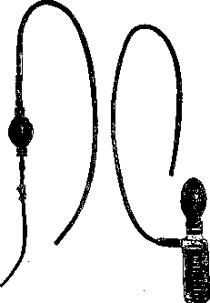
Figure 26.
and with the help of slight movements of the tube, the siphon is established. Before inserting the tube, the patient's dental prostheses should be removed (if they are present). With sufficient skill, the entire procedure takes only a few minutes and is easily tolerated. At

it is necessary to resort to cocaineization of the pharyngeal mucosa only in exceptional cases - with sharp irritation of the nasopharynx or phenomena of tetany. Sometimes when withdrawing the probe, a strong spasm of the esophageal or pharyngeal musculature is observed - in this case, a little warm water should be introduced through the funnel to relax the spasm. Usually, after introducing the probe, it is sufficient to have the patient tense the abdominal press so that the contents of the Stomach begin to exit through the probe ('expression'). To completely empty all contents, a glass jar with an aspiration balloon can be attached to the probe (fig. 26). Too vigorous aspiration can lead to suction of the gastric wall and even slight injury to the mucosa. The rubber balloon proposed by Kuttner, attachable to the probe after introduction into the stomach (fig. 27), is very appropriate. Application: 1) for diagnostic purposes to examine the stomach contents on an empty stomach or after a test meal, 2) therapeutically: a) in cases of stagnation in the Stomach, completely replacing the more tedious for the patient lavage or preceding it, b) in acute poisonings to remove the poison from the stomach followed by lavage. - Contraindications: gastric bleeding, severe heart diseases, aortic aneurysm, severe hypertension, angina pectoris, severe arteriosclerosis and severe weakness of the patient. However, in acute poisonings, the serious condition of the heart cannot be an obstacle to removing the poison by probe.
X-ray examination.-The stomach does not differ from surrounding parts in its ability to absorb X-rays. When examining the abdominal cavity without preparation, only the air bubble in the upper part of the Stomach and random gas accumulations in the intestine are visible. When the Stomach is inflated with air, its image can be obtained as a light area. This method, however, is rarely used, as it gives relatively little information. The systematic development of stomach examination with X-rays began with the introduction by Rieder of filling the Stomach with porridge mixed with bismuth (Bismuti subnitrici 30.0 per dose). Cases of poisoning forced a switch to harmless bismuth carbonate, and at present, porridge mixed with barium sulfate (Barium sulfuricum) is mainly used - an insoluble, extremely stable compound that passes unchanged through the entire gastrointestinal tract without being absorbed. Due to the high toxicity of soluble barium salts, absolutely pure preparations are necessary; the usual dose is 50.0-100.0 or more per dose. When using these heavy metal salts, a dark image of the organ filled with them is obtained on examination. The advantage of Rieder's method is its approximation to physiological conditions. With this technique, the image obtained is not of the organ itself, but of the contrast mixture filling its cavity, and is therefore bounded not by the serous, but by the mucous membrane. When the subject takes the contrast mixture, one can gradually observe how it first accumulates in the upper part of the Stomach in the form of a triangle with its base upward and located directly under the gastric bubble (fig. 28-1). This triangle gradually elongates downward and to the right, and later the rounded contour of the lower part of the stomach begins to gradually appear. According to the works of Forsell, the shape of the stomach is determined by the architecture of its musculature, forming above the vault, below the body of the Stomach, the pouch and canal: fornix, corpus, sinus and canal. Corresponding to the muscle fibers of the pylorus, a light strip is visible on examination between the accumulations of contrast medium in the Stomach and in the duodenum. Since the shape of the Stomach is determined by smooth musculature with variable tone, it cannot be constant. Even in the same subject, the silhouette of the Stomach in the shape of a bull's horn (normal type of Stomach according to Holzknecht; fig. 28-2) can change to the shape of a hook (normal type of Stomach according to Rieder). Between the latter and the elongated Stomach in ptosis (fig. 28-3), there is a whole series of transitions. The position and shape of the Stomach are influenced by its filling, the tone of the musculature, the organs surrounding the Stomach, the position of the diaphragm, intra-abdominal pressure, and peritoneal ligaments. According to roentgenological data, the Stomach of chest infants is located horizontally due to the wider than in adults, lower transverse diameter of the chest cavity and the abundance of gas in the intestine under the Stomach. Since the Stomach itself contains a lot of air, it is well visible even without contrast medium. In children over one year, the vertical position already predominates (see separate table, fig. 1). Functional moments are no less important than purely anatomical ones. For this reason, constitutional types of Stomach - a high transversely located Stomach in the shape of a horn in a picnic, an elongated, vertically located Stomach in the shape of a hook in an asthenic - are not constant. The influence of the autonomic nervous system on the shape of the Stomach has been proven experimentally both by direct irritation of nerve trunks and pharmacologically. Irritation of the vagus nerve increases the tone of the Stomach, irritation of the sympathetic nerve decreases it. A hypertonic Stomach appears as a narrow tube, with the cardiac part somewhat dilated and the pylorus gaping (fig. 28-4). This condition is observed in neuroses, in lead colic, sometimes in tabetic crises. Conversely, with reduced tone of the Stomach, it appears as a flaccid sac (see separate table, fig. 5). Food taken in such a Stomach immediately sinks to the bottom, arranging itself in a semicircle with the upper horizontal level below the navel (fig. 28-5). This condition may be a sign of impaired compensation of the musculature in pyloric stenosis, but can also be caused by a number of functional factors; for example, it is observed after taking large doses of morphine. A valuable sign of Stomach dilation is the displacement of the pyloric part to the right, with the described semicircular shadow located in the middle, and not on the left, as in pure ptosis. With simple prolapse of the Stomach, there are no disturbances of motor functions. However, intra-abdominal pressure also affects the tone of the Stomach, so with relaxation of the abdominal press, both prolapse of the Stomach and decrease in its tone are observed. With both hyper- and hypotonia of the Stomach, there are gradual transitions from barely noticeable disturbances of tone to a pronounced, bright picture (fig. 28-4 and 5).
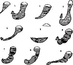
Fig. 28. Diagram of stomach shapes: 1-gradual filling of the stomach of the Rieder type; 2-stomach of the Holzknecht type; 3-prolapse of the stomach; 4-increased tone of the stomach; 5-atony of the stomach; 6-segmental peristalsis; 7-ulcer of the lesser curvature; 8-cancer of the lesser curvature; 9-cancer of the stomach.
Peristalsis is expressed in the appearance of wave-like constrictions along the greater and lesser curvatures, which slide toward the pylorus (fig. 28-6). One wave follows another; with the pylorus open, they lead to the passage of food into the duodenum, with the closed pylorus they are important for mixing food. When peristalsis is enhanced, these constrictions are particularly deep. They begin near the cardia and follow one another quickly, resulting in true segmentation of the Stomach. With sharply increased tone, peristalsis is weakly expressed, with hypo- and atony it is often increased. This combination of enhanced peristalsis with reduced tone is often observed in pyloric stenosis and should be considered as a compensatory phenomenon (see separate table, fig. 6). After 3-4 hours, when a normal Stomach would have already emptied, in such cases a significant residue with the described signs of atony is still determined in it. Another valuable sign of pyloric stenosis is
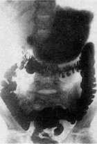
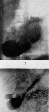
Ml «i
Figure 1. Normal stomach of a child. Figure 2. Folds of the mucous membrane of a normal stomach.
Figure 3. Mucous membrane of the stomach (folds deformed). Figure 4. Gastrospasmus. Figure 5. Atony of the stomach (delay). Figure 6. Stenosis of the pylorus (segmented peristalsis). K" st. Shemdok. Peristaltic waves going in the reverse direction occur, the so-called antiperistalsis. Both the tone and the peristalsis and the associated emptying of the St. are influenced by a number of purely functional factors. Peristalsis can be strengthened by massaging the area of the St., by repeated tension and relaxation of the abdominal press. Morphine sharply delays the emptying of the St. With increased acidity, evacuation of the stomach is slowed down, with decreased acidity it is accelerated. A direct sign of hypersecretion is the so-called intermediate zone between the upper level of the contrast medium and the air bubble in the cardiac part. It must be borne in mind that a similar zone is also obtained in the absence of secretion if a rapidly settling contrast powder with liquid is used. In the diagnosis of organic diseases of the stomach, it is necessary to distinguish direct signs caused by anatomical changes and indirect signs associated with functional impairment (fig. 28-#). The latter includes the limited persistent spastic retraction of the greater curvature at the level of the location of the ulcer on the lesser curvature. A direct sign of ulc-er is the protrusion of the wall, the so-called niche symptom. A sign of cancer is a filling defect (fig. 28-8). A new growth protruding into the cavity of the St. partially fills it, so that the contrast medium cannot be located in this place. A tumor infiltrating the wall leads to the cessation of peristalsis in a limited area or, with extensive spread, transforms the shadow of the stomach into a narrow, irregularly bounded strip (fig. 28-9). After taking a small amount of contrast mixture, if it gradually flows down along the folds of the mucous membrane or is distributed in a thin layer over the surface due to kneading of the stomach area, an image of the relief of the mucous membrane can be obtained. The most distinct pictures are obtained after taking a small amount of barium sulfate in a thick solution of Gummi arabici. Here, too, rather inconsistent pictures are obtained in connection with the motor function of the muscularis mucosae (see separate table, fig. 2 and 3). Under normal conditions, regular longitudinal folds are visible; in some cases of catarrh of the St., a special spotted pattern is obtained (etat mamelonne); with atrophy of the mucous membrane, uneven distribution of barium over the surface without clearly expressed folds. This new method will apparently play a major role in the early diagnosis of ulcers and cancer of the St.-For a complete radiological examination of the St., both fluoroscopy and X-ray pictures are needed; the first is mainly for studying the motor function (for which roentgenokymography was also used with complete success), the latter for a more detailed image of details. In the clinic of stomach diseases, the radiological method at present is indispensable; the signs of organic diseases obtained by it have great diagnostic significance, although radiological data do not always coincide with patho-anatomical ones. Data on the motor function have opened up new broad horizons. Thus, they pointed to the importance of local spasms (see separate table, fig. 4) in organic diseases of the organ itself (cancer, ulcer-the spasmogenic theory of ulcer of the St., according to Bergmann) or neighboring organs (spasm of the St. in biliary colic, enhanced peristalsis of the St. in ulcer of the duodenum, etc.). Radiological observations are of great importance for the doctrine of vegetative neuroses (sympathico- and vagotonia or disturbed equilibrium between both systems according to the latest views), for their connection with organic diseases of the central nervous system (tabes), with intoxications (opium, lead), with psychogenic factors, with constitutional features (hypotonia with ptosis in asthenics), o. Den. IV. Motor and secretory disorders of the stomach. Pathology of the motor function of the stomach. Corresponding to the two main types of motor function of the stomach-the so-called peristaltic function and the emptying function (motor function in the narrow sense), the pathological disturbance of them can also be twofold. The first group of disorders includes: atony, resp. hypotonia, hypertension of the stomach and its spasm. The second group includes those disorders in which the movement of the stomach suffers due to damage to the motor mechanism of the entire stomach or its parts-cardiae, body and pylorus.-Under the name of atony, resp. hypotonia of the stomach is meant the state of absence or weakening of the peristaltic function of the St., resp. the tonic function of its musculature (Stiller), i.e. the state of inability of the gastric wall to tightly grasp the contents present in the St., while the emptying and motor ability of the St. is reduced (more often), but may remain normal, and in a number of cases even increased. Atony of the St. can be caused by, resp. accompanied by, weakening of the supporting apparatus of the St., as observed in gastoptosis and splanchnoptosis, as well as by decreased elasticity of the gastric wall; atony of the St. can also develop due to disturbance of the innervation of the stomach in general nervous weakness, in states of general hypotonia, in the presence of general constitutional weakness or without it, in general nutritional decline; it can also be observed in infectious diseases (as for example in typhoid, dysentery) and in other exhausting diseases accompanied by adynamia, as well as in a number of organic diseases of the stomach. Since atony of the St. is more often observed in gastoptoses, the clinical picture is similar to that in gastoptosis. Atony of the St. is established exactly only by X-ray examination, in which in case of atony of the St. it is determined that the contrast food collects in the lower parts of the stomach; the St. is not only elongated in length, but its sinus part is wide and low-lying; the walls of the St. in the middle part approach each other, forming a "waist", and the gas bubble has the shape of an elongated pear. The filling of the St. occurs in such a way that the first portions of food quickly descend to the lower pole of the St. and it fills "from bottom to top". The determination of the splashing sound is a rather unreliable method for determining atony of the St. The treatment of atony is similar to that in gastoptosis and in asthenic states. The main thing is: general strengthening treatment, elimination of dyspeptic phenomena and constipation, prescription of enhanced nutrition, treatment by lying down, general strengthening of the nervous system. The state of increased tone of the gastric wall is called hypertension of the St. Hypertension of the St. can be observed with increased excitability of the nervous system, both general and in particular of the nervous system of the St. or other organs of the abdominal cavity. It occurs in biliary and renal colics, in ulcers of the St., in general nervous increased excitability, in lead and nicotine poisoning, etc. In the presence of hypertension of the St., during X-ray examination the St. takes a more transverse position, is more displaced to the right, its greater curvature approaches the anterior abdominal wall and has a sharply expressed horn shape, the pyloric part appears narrower than the cardiac part, and when filled with contrast food, greater resistance is observed than is usually the case with normal tone of the St. Often in the presence of hypertension, a picture of increased motor excitability of the St. is observed in the form of enhanced peristalsis, enhanced emptying and especially spastic phenomena. To these can be added phenomena of altered secretion (hypersecretio, hyperchlorhydria, etc.). Some forms of organic lesions of the St. (tumor, linitis plastica, shrunken St., for example after poisonings with acids, in syphilis) during radioscopy can give a picture in many respects similar to simple hypertension of the St. In such cases, prolonged observation, repeated X-ray examination and the use of atropine make it possible to differentiate organic reduction of the organ from the functional state of increased tone of the stomach. The highest degree of enhancement of the tone of the St. is the state which is called spasm of the St. Spasm of the St. is observed in biliary colic, in the incarceration of a stone in the ureter, in tabes, in poisoning with morphine, both acute and chronic, in intestinal colic, in pancreatitis; spasm of the St. can be observed especially often in ulcers of the St. and duodenum, as well as in inflammatory processes in their vicinity, in perigastritis; thus, spasm of the St. can arise both as a result of local irritation of the St., and as a result of the so-called "viscero-visceral reflex" (Bergmann). Spasm of the St. can be 1) partial, 2) regional, 3) complete. In partial spasm, only a small limited part of the musculature of the St. is involved, as observed for example in ulcer of the St., in perigastritis. An example of regional spasm can be the complete contraction of the entire area of the pylorus, which is observed with superficial ulcers either in the pylorus itself or near it. Regional spasm can also involve the entire body of the St., which is observed when the ulcer is located in corpus ventriculi. Complete spasm, involving the entire St. as a whole, has been observed in poisonings, in tetany (Walvogel, Schwarz).
In terms of duration, stomach spasm can be either prolonged or intermittent. It often occurs without any subjective sensations, but more frequently it is accompanied by attacks of pain of varying intensity. The diagnosis of spasm of the St. is established by X-ray examination. Treatment of spasm of the St. consists of treating the underlying disease that caused the spasm, for example, ulcer of the St., gallstone disease; symptomatically, atropine (0.0005-0.001) or Papaverine (0.04) is administered subcutaneously or intravenously. Motor disorders in the strict, narrow sense can vary depending on the nature of the affected function and the location of the lesion in the St. In terms of localization, the disorder can affect the entire St. or only one of its parts, for example, the cardia or the pylorus. The main types of motor disorders of the St. are as follows. 1. Disorders related to impaired activity of the cardia. The most important type of such disorder is cardiospasm. 2. Impairments of the motor capacity of the stomach can manifest as changes in the nature of gastric peristalsis. Such disorders include: decreased or increased peristalsis, general contraction of a hypertrophied and dilated stomach (Magensteifung), and its antiperistalsis. - Weakening of peristalsis can occur in infectious diseases and states of cachexia, in states of paresis or complete paralysis of the St. (see below), and often in its atony. In a number of diseases of the St., such as cancer of one or another part of the St., callosal ulcer, inflammatory disease around the St. (perigastritis), various conditions leading to fixation of the St., only local, focal weakening and decrease in peristalsis are observed. - With increased gastric peristalsis, increased frequency of peristaltic waves, increased size of the waves up to the formation of functional constrictions ('segmenting peristalsis'), and acceleration of wave movement are observed on X-ray examination. Increased peristalsis of the entire St. can be observed in healthy individuals during strong hunger, in neuropathic states, but mainly in the presence of an obstacle to emptying the St., for example, in pyloric spasm, ulcers of the St. or duodenum, cancer of these organs, in pericholecystitis, etc. - When the obstacle to emptying reaches a high degree, disruption of coordination and a sharp increase in peristalsis occur with increased tonic contraction of the musculature of the entire St., accompanied by an attack of pain - the so-called Magensteifung appears. In this condition, which is most often associated with dilation and significant hypertrophy of the gastric musculature, significant distension of the area of the St. of firm consistency is observed, with slowly passing peristaltic waves over it. - In severe stenoses of the pylorus, antiperistalsis can be observed, i.e., the peristaltic wave begins at the pylorus and passes along the greater curvature toward the cardial region. 3. The most important from a practical standpoint are the motor disorders related to impaired activity of the pylorus. Such disorders include: a) spasm of the pylorus, b) its insufficiency, c) its stenosis. - Under the name pyloric spasm (pylorospasm) is meant a condition of more or less prolonged closure of the pylorus with simultaneous absence of emptying of the St. The duration of this spasm can vary - from several minutes to several hours; in the latter case, delayed emptying of the St. up to 12 hours or more is observed. A pronounced spasm of the pylorus of the St. is accompanied by attacks of pain, which sometimes take on a colicky character; these attacks disappear after vomiting or after removal of the stomach contents. Pyloric spasm is mainly observed in ulcers located in the pylorus itself or near it, as well as in ulcers of the duodenum and inflammatory processes of it. In exceptional cases, pyloric spasm can be caused by foreign bodies, as well as under the influence of morphine. Treatment of pyloric spasm consists of treating the underlying disease, and symptomatically - in the administration of atropine and Papaverine. - The opposite condition to spasm of the pylorus, when its ability to hold back food is weakened and it cannot close, is called its insufficiency. Insufficiency of the pylorus can be functional and organic. The former occurs in achylia gastrica, in stenoses of the duodenum with simultaneous dilation of its bulb, in paralysis and paresis of the St., and in the so-called 'spastic insufficiency of the pylorus' (Schlesinger), caused by tonic contraction of the longitudinal musculature of the pylorus. Organic insufficiency of the pylorus is observed in its infiltration: inflammatory, cancerous, ulcerative; in its tuberculosis and syphilitic lesions. Narrowing of the pylorus [previous designations: 'dilation of the St.' (Riegel), 'motor insufficiency' (Rosenbach), 'mechanical insufficiency' (Boas), 'ischochymia' (Einhorn)] develops as a result of inflammatory conditions of the pylorus with the formation of erosions and cracks of its mucous membrane, ulcer of the pylorus; it is also observed as a result of development of a cancerous tumor, tuberculosis of the pylorus, sclerosing pyloritis, in some poisonings (acids, alkalis). Inflammatory processes around the pylorus and duodenum leading to adhesions in their vicinity, pericholecystitis and other local inflammations of the peritoneum also often lead to the development of pyloric stenosis. From a functional point of view, the following forms of pyloric stenosis are distinguished: a) Compensated stenosis, when in the presence of stenosis the time of emptying remains either normal or only slightly delayed. In compensated stenosis, gastric peristalsis is increased due to the resistance to the passage of food through the pylorus, and this increase is strong enough to achieve emptying of the stomach in a normal or slightly increased time; the musculature of the stomach is usually hypertrophied in this case. b) Decompensated stenosis, when the increase in peristalsis is insufficient to empty the stomach in time, or when weakening of peristalsis occurs due to overexertion of the hypertrophied musculature of the St. - in other words, when the ratio between the strength of the musculature of the St. (the amount of its emptying), on the one hand, and the degree of its stenosis, on the other, is disrupted. Transitional states exist between these forms. - In the first degree of stenosis, subjective phenomena may not be observed at all; in a number of cases, slight restlessness (the so-called 'peristaltic') in the upper part of the abdomen after eating is noted; in a number of cases, belching, heartburn, increasing after eating, especially of acidic food, are observed; in the presence of ulcer, cancer, symptoms characteristic of these diseases are present. Objectively: in examination with a test breakfast - often normal secretory condition, more frequently hypersecretion, superacidity, prolongation of the secretion period. Examination with a test meal gives prolongation of the period of food remaining in the St. (more than 5 hours) with an abundant amount of gastric juice. On X-ray examination, a hypertonic form of the St. is noted, more often in the form of a horn, increased resistance with the first portions of food, lively, deep, high-starting in the cardial part peristalsis, jaggedness of the contours of the greater curvature, unevenness of emptying - first rapid emptying, then significantly slowed; complete emptying of the St. after taking a usual contrast meal is delayed up to 6-8 hours. - For differentiation of the diagnosis of pyloric stenosis from diseases that can give a similar picture and in which only functional spasm can be observed (for example, in nervous conditions, in hypersecretion), attention should be paid to phenomena that can indicate the cause of the development of stenosis (ulcer, cancer, etc.); the use of a test meal, as well as atropine, is also recommended. In decompensated stenosis of the pylorus, subjectively observed are: a feeling of fullness in the area of the St., attacks of spasmodic pain; appetite, although preserved and even often increased, but a feeling of rapid satiety is observed; 'peristaltic restlessness' is observed to a great extent, often in attacks; sensation of movements of the St., rumbling, belching of sour or disgusting taste, vomiting of masses of various kinds and properties. Often the vomited masses contain a significant amount of remnants of food taken many hours and days before the vomiting occurs; often a large quantity of light gastric juice (hypersecretion) is vomited, which resembles attacks of Reichmann's disease. In severe stenoses, thirst develops, the amount of urine decreases, stool is delayed despite the use of laxatives, but often persistent diarrhea, decline in nutrition, loss of strength, tendency to fainting, sharp emaciation, and sometimes tetanic phenomena develop. On objective examination, the following are observed: emaciation, decrease in skin turgor, often complete disappearance of subcutaneous fat, thinning of the skin; on inspection - strikingly clear outlines of the stomach, visible peristaltic waves on it, contraction of the entire St., accompanied by attacks of pain, appearance of belching and vomiting.
Upon palpation: splash fasting sound, slight palpability of the greater curvature. Upon examination with a tube in the morning on an empty stomach - significant amount of contents and food residues, especially after a丰盛 dinner the previous day, formation of three layers in the contents: upper - mucous, middle - secretory, and lower - food; upon chemical examination of the contents in some cases - increased total and free acidity, in others - decreased acidity and even achylia; with decreased secretion of gastric juice - often large amounts of organic acids, with cancerous stenosis of the pylorus - lactic acid. With increased acidity in the stagnant contents upon microscopic examination - abundant yeast cells, sarcinae; with decreased secretion - undigested muscle fibers, Boas-Oppler rods. X-ray examination reveals irregular filling, irregularities in the silhouette (shape of an ellipse, egg, cup), the lower pole is often descended to the pubis, significant secretory layer giving a weak shadow, wide shadow of the body of the Stomach without formation of a "waist", significant displacement of the body to the right, enhanced peristalsis, deep frequent waves, alternation of phases of enhanced muscular work and rest due to complete fatigue of the Stomach musculature. Depending on the degree of stenosis and the size of the obstruction, X-ray examination determines delayed emptying of the Stomach: after 8-12-24 hours or more, residues of contrast food can still be seen in the Stomach. - Diagnosis of pyloric stenosis is established if a splash sound is determined in the morning on an empty stomach, if there are signs of Magensteifung and etc.; upon probing - signs of stagnation are determined, vomiting of food taken long before the onset of vomiting, belching of gases, slowing of emptying of the second degree (see below), distinct peristalsis. Complications of pyloric stenosis are tetany and Reichmann's syndrome (see below). Treatment of stenosis can be a) surgical, b) dietary and c) medicinal. Surgical treatment is indicated in cases where the stenosis is not compensated, when improvement of the process is impossible, when there is indication of progressive scarring increasing the stenosis, and if there is suspicion of development of a malignant tumor of the pylorus. - Dietary treatment is conducted in two directions: 1) selection of food in terms of nature and consistency is made according to the size of the stenosis and 2) selection of food is made according to the state of secretion in this or that case of pyloric stenosis. In the first case, one can use Boas's scheme, which consists in establishing the degree of passage of various food substances through the pylorus by prescribing in the evening various food substances with subsequent morning retrieval of the Stomach contents. Boas recommends the following tests. 1. Passability for rice. In the evening before, 200 g of milk rice porridge is given, bread with butter, one egg and a little soup. The next morning the Stomach is washed to determine the amount of food remaining in the stomach. 2. Passability for vegetable purees, potato puree, porridges. 3. Passability for white bread, biscuit, crackers. 4. Passability only for liquid food. With the help of this kind of diet, the degree of tolerance, resp. the degree of stenosis in relation to the consistency of food is established; at the same time, the tolerance of the stomach in relation to the amount of food taken is also established, since the degree of food retention in the stomach depends on the absolute amount of food taken. When with the help of these trial diets the degree of difficulty of emptying is established, then a diet is prescribed in accordance with the degree of stenosis. When prescribing such a diet, it is necessary to care 1) that the food in terms of calories is quite sufficient and can ensure the increase in weight of the patient and 2) that the food can pass through the pylorus without being retained in the stomach. Finally, the food must be selected taking into account the nature of secretion in this or that case of stenosis (super-aciditas, subaciditas, hypersecretio - see below). In addition to these basic rules, the following must be taken into account: quantitatively - in terms of volume - the food must be limited, food intakes should not be frequent; and the intervals between them should be quite long, - i.e. small and infrequent food intakes are necessary. - Boas recommends such approximate diets for pyloric stenoses. For mild cases of pyloric stenosis: in the morning - 200 g of milk soup, seasoned with wheat or oat flour, to this a lot of butter or cream, one egg, soaked crackers, cookies; at 12 o'clock in the day - 200 g of thick milk-flour soup, pureed vegetable soups with butter, 100 g of potato puree or vegetable puree with a large amount of butter or cream, 100 g of finely chopped meat, 50 g of pureed compote from apples, plums and 50 g of pudding; in the evening at 7 o'clock - scrambled eggs, potato (50 g) or vegetable puree, white bread (50 g), butter, tea with milk or cream. - With moderate degree of pyloric stenosis the following diet is recommended: in the morning - the same as in the above diet; at 1-2 o'clock in the day - 100 g of thick milk-flour or vegetable soup, 50 g of potato or vegetable puree with a large amount of butter and 50 g of pureed meat; in the evening - one soft-boiled egg with butter, 50 g of potato puree, 50 g of white bread, butter, pureed cheese, tea with cream or milk. - In severe cases of stenosis is prescribed: in the morning - 200 g of milk-flour soup; at 12 o'clock in the day - 200 g of the same or vegetable soup with butter and egg, 50 g of jelly from calf's feet or meat aspic; in the evening - 200 g of milk-flour soup with butter and egg, 100 g of tea with cream. The last diet due to its low calorie content and insufficiency in vitamins cannot be prescribed for a long time. - Diet taking into account the amount of secretion is prescribed according to the degree of these changes (superaciditas, subaciditas, hypersecretio). - Medicinal therapy in pyloric stenoses has only symptomatic significance: in the presence of spasm, atropine and papaverine preparations are recommended; to improve and increase tone in an atonic stomach - strychnine; if pyloric stenosis occurs with signs of decreased acidity, prescription of HCl is recommended; in the presence of increased acidity or acidism phenomena, therapy is directed against these latter conditions (see below). Systematic washing of the stomach is of essential importance both in the morning and in the evening hours. 4. Motor disorders of the Stomach can also manifest in violations of the time of its emptying. Emptying can be accelerated or slowed down or finally may not occur at all. To determine the speed and character of emptying, various methods are used. Of such methods, the most generally accepted are: the method with Boas-Ewald breakfast with determination of the solid residue according to Eisner and of the total amount of gastric contents according to Mathieu-Remond, the method of fractional evacuation of gastric contents after a colored test meal, the method with a test meal according to Riegel and the method of X-ray examination. All these methods to a significant degree have conditional value according to the irritant applied, and therefore cannot be considered precise methods. When determining motor ability with Boas-Ewald breakfast, Riegel's meal, in X-ray examination, they are guided either by establishing the time when complete emptying of the Stomach occurs after taking a certain food, or by the amount of gastric contents obtained over a certain period of time, more precisely - of the residue of the test food taken. For example, if when giving Boas-Ewald breakfast upon evacuation of contents after 60 min. nothing is found in the Stomach, this may indicate accelerated emptying; if the amount of evacuated gastric contents reaches 100-300 or more cm3, this may indicate both slowed emptying and disorder of motor ability (insufficiency of the second degree in pyloric stenosis, paralysis of the stomach, with nervous disorders of the motor function of the stomach and etc.). The amount of contents within the limits of 20 to 100 cm3 is considered a value indicating normal emptying. If after giving Riegel's meal food residues are found after 7 hours, they speak of insufficiency of motor ability of the first degree; if this time is extended to 12 hours - then of the second degree. Since in its properties the test meal presents a significant demand on the emptying mechanism, in a number of cases its use makes it possible to discover less pronounced motor disorders than when using Boas-Ewald breakfast. The most valuable method for determining motor disorders is X-ray examination, which makes it possible to determine many details of the violation of movement at individual moments and stages, as well as acceleration or slowing down in various degrees, or such a phenomenon as the Kreuzfuchs phenomenon, consisting in that at first with enhanced peristalsis rapid emptying of the stomach occurs (in the first hour up to 2/3 of the barium mixture leaves the Stomach, while the other part leaves slowly and in small portions).
The last symptom is characteristic of pyloric spasm. On X-ray examination, rapid and emptying is observed in achylia, sometimes in cancerous infiltration of the pylorus (incontinentia pylori). If the duration of emptying is 2h/2-3 hours, the motor function is considered normal; such duration in the presence of enhanced and expressed peristalsis can also be observed in some cases of compensated stenosis. If the duration of emptying reaches 6-8 hours, this indicates a decrease in the motor function of the St. When the time of emptying increases to 12 hours, they speak of insufficiency of emptying of the 2nd degree. The 3rd degree of this disorder is determined when emptying lasts 24 or more hours. Various liquid trial irritants (broth, vegetable juices, solutions of alcohol, caffeine, etc.), with or without the addition of coloring substances, with the use of moment-by-moment research with a thin probe, are also widely used in determining the nature of motor disorders. A valuable property of this method is that with it the motor properties, resp. disorders of the St. are determined with simultaneous consideration of the regulating role of acidity in the process of emptying. The degree of motor disorders of the stomach is determined by the course of the acidity curve or by the time of disappearance of the coloration of the contents (when a coloring substance is added to the irritant). According to Katsch, there are several types of secretion curves, by which it is possible to judge the nature of motor disorders. These types are as follows. 1) Type of early appearance of significant acidity: rapid and steep rise of its curve, rapid emptying, as can be observed in ulcers of the St., duodeni with signs of compensated stenosis or without them; in a number of cases, hypersecretion, hyperchlorhydria are observed. 2) The acidity curve of normal height with a normal rate of rise, as observed in cases of normal motor function of the stomach, or slightly weakened. 3) The type of curve when acidity appears with delay: the curve is stretched, which indicates a slowing down of emptying caused by organic damage to the pylorus or by fnkts. its state («pyloric spasm»). 4) Low type of curve with rapid disappearance of coloration: indicates acceleration of emptying with insufficient acidity.-Each of the above-mentioned methods for determining the motor function of the St., having its positive and negative sides, can give valuable practical results for judging the nature of motor disorders of the stomach, but, taken individually, none of them can be considered a method that fully exhausts the subject, and each of them serves as a supplement to the other in understanding the phenomena of these disorders. 5. In the clinic of motor disorders of the St., there are two forms of the disease in which there is a sharp weakening of the musculature of the St., as a result of which, even in the absence of a mechanical obstacle, there is a significant violation of emptying of the St. and stagnation of food in it. These forms are named: 1) gastroparesis and 2) paralysis of the St. In gastroparesis, chronic muscular weakness of the St. is observed. Unlike atony of the St., in which there is mainly a violation of peristalsis, and emptying can be normal, in gastroparesis emptying suffers mainly. According to Schlesinger, chronic «gastroparesis is the cause of severe motor insufficiency with a free pylorus». Gastroparesis, being a manifestation of fatigue of the musculature of the St., can occur in a state of malnutrition, during fasting, due to reflex influences, in fright, fainting, in lesions of the bile ducts, the vermiform appendix, in cancerous, tuberculous cachexia, in atony of the St. after infectious diseases. It can be observed in organic stenosis of the pylorus and, which is important diagnostically, in ulcers of the stomach in the region of the lesser curvature. The cause of gastroparesis is assumed in this last case to be a lesion of the conducting system of the St. (Keith) or Auerbach's plexus. Clinically, gastroparesis manifests itself with symptoms similar to those in decompensated stenosis of the pylorus; importance is attached to the sign consisting in that the vomit contains an extremely large amount of bile; essential data is given by X-ray examination: increase in the size of the St., sharp slowing down of emptying in the absence of | narrowing or obstacle in the region of the pylorus. It should be borne in mind that gastroparesis also occurs in organic stenosis of the pylorus in its decompensated form.-Treatment consists in the selection of food, nutritious in composition and not burdensome in volume, restriction of the introduction of liquids per os and replacement of this with enemas, in evening washing of the stomach, administration of strychnine; in the presence of organic stenosis of the pylorus, surgical treatment is indicated. The condition of sharp weakening of the musculature and complete cessation of movement represents acute paralysis of the stomach. In this disease, the tone of the St. disappears, its movements cease, the St. stretches and fills with an abundant amount of secretion («acute dilatation of the stomach»). It is observed more often after operations, especially in the abdominal cavity, under the influence of anesthesia, in severe infectious diseases (typhus), in damage to the central nervous system, in excessive intake of food, in particular liquids, in contusion of the abdominal region, etc. Clinically, the disease usually begins suddenly and develops quickly and violently; facial features change sharply, the pulse becomes small, sharp pains appear in the upper part of the abdomen, severe thirst, abundant vomiting of liquid (in a larger quantity than the patient takes in); there is a sharp swelling of the region of the St. with simultaneous retraction of the lower part of the abdomen; at the same time, unlike peritonitis, there is no reflex muscular tension. Very abundant vomiting is observed, often reaching a volume of 30 liters. The result of this is severe thirst, decrease in skin tur-gor, sharp loss of weight, decrease in diuresis to complete anuria. The disease can last from several hours to several days and in 2/3 of cases ends in death. The cause of this lesion is considered to be severe atony of the stomach and stenosis of the duodeni due to its bending over the vascular-mesenterial bundle or due to irritation and damage of extragastric nerves, as well as the intramuscular nerve apparatus of the St. The diagnosis of the disease is established on the basis of the clinical picture described above. In differential-diagnostic respect, one must keep in mind ileus (fecal vomiting), acute stenosis of the pylorus or duodeni (preserved sharp peristalsis), acute pancreatic necrosis (attacks of most severe pains). - Treatment: washing of the St., complete abstinence from food, position on the right side (Kussmaul) or knee-elbow («a la vache»), drop enemas, administration of strychnine and physostig-mine, as well as choline. The use of morphine, adrenaline and atropine is contraindicated. Pathology of the secretory function of the St. Pathological disorders of the secretory activity of the St. can be expressed in changes in gastric secretion both in quantitative and in qualitative relations. To changes in gastric secretion of a quantitative nature belong: hyperchylia (hypersecretio) and hypochylia (subsecretio), achylia; to qualitative changes in secretion belong superaciditas, resp. subaciditas, and hyperchlorhydria, resp. hypochlorhydria. I, Gastric hyperchylia (supersecretio, hypersecretio)-such a state of gastric secretion when, in the absence of a food irritant, a significant amount of gastric juice is separated and accumulates, or when to a certain irritant the secretion continues abnormally for a long time and in large quantities. Clinically, several forms are distinguished: 1) Reichmann's syndrome, or gastrosuccorea, syn. chronic constant secretion of gastric juice (Riegel), parasecretion (Ewald); 2) periodic intermittent hyperchylia, observed in migraine, in Quincke's edematous disease and mainly in the so-called gastric crises in tabes dorsalis, and 3) hyperchylia as a symptom of a number of organic lesions of the St., mainly in ulcers of the St. and 12-perstnoy intestine.-1. In the R e i h m a n o v s k o y f o r-m there is excessive secretion of gastric juice, accompanied by the onset of sharp pains in the stomach in the evening; the pains continue all night, reach their maximum by morning and spread throughout the abdomen; during the day they subside, they also soften after taking food, the next night the attack is repeated. There is severe heartburn, increased appetite, stool is delayed, eructation is never observed. On objective examination, up to 1 liter of gastric juice with a strongly acid smell is pumped out of the stomach. Reichmann's syndrome is considered either as a secretory neurosis of the St., and then they speak of «genuine» outflow of gastric juice, or as a concomitant symptom in ulcer of the pyloric region and benign stenosis of the pylorus and ulcer of the 12-perstnoy intestine. The latter form of Reichmann's syndrome is observed more often.
Regarding the mechanism of Reichman's syndrome, it is assumed that either under the influence of an ulcer, a special state of excitation of the stomach's nervous system develops, especially in the presence of vegetative disharmony, or else that this syndrome represents a symptom of a stagnant stomach, in which the retained food residues, even microscopic ones, are capable of maintaining prolonged and strong secretion of gastric juice. 2. In periodic gastric hyperchylia, there occurs periodically excessive secretion of gastric juice, with the periods occurring irregularly, usually lasting no more than 24 hours, rarely (in hysteria) a week. In the intervals between attacks, patients feel healthy. In the group of periodic gastric hyperchylia, several separate subtypes are clinically distinguished. The first subtype can include cases that represent variants of chronic constant secretion of gastric juice, as observed in stomach and duodenal ulcers. The periodicity of hyperchylia in this case is an expression of an exacerbation of the ulcerative process. Periodic hyperchylia is most pronounced in the second subtype, in tabes dorsalis, and is called tabetic crises. The attack of pain begins suddenly, quickly reaches the degree of convulsive pains in the stomach area and is accompanied by severe, constantly recurring vomiting, almost intractable—at first with food masses, and then with gastric juice together with bile and mucus. The general condition sharply deteriorates. The attack ends as suddenly as it began, and the next day the patient feels completely healthy. It is characteristic of these conditions that in the intervals between attacks, no deviations from the norm are found in the stomach, and that ordinary means for reducing the symptoms (stomach lavage, alkalis) do not have a mitigating effect here.—The third subtype of periodic gastric hyperchylia is called gastroxynsis (see) and is described by Rossbach.—As a special subtype of periodic hyperchylia, periodic gastric hypersecretion occurring in Quincke's angioneurotic edema is also considered. In this condition, attacks are observed, accompanied by vomiting, attacks of pain in the stomach area, in the left hypochondrium, nausea; these phenomena are intensified with food intake. The attack usually ends after 10-12 hours. The diagnosis of this form is based on the alternation of these periods of attacks and periods of edema. 3. Hyperchylia is most often a symptom of a number of organic diseases of the stomach. In these lesions of the stomach, a form of the constant type (hyperchylia continua) is distinguished, when gastric juice is secreted for a long time, quickly, and, most importantly, independently of a physiological irritant, and in significant quantities; in another form of hyperchylia, so-called food, or alimentary, in response to one or another food irritant, gastric juice is secreted in a much larger quantity than is observed in normal cases, and for a long time. In constant hyperchylia, gastric juice is usually obtained on an empty stomach with a significant degree of acidity in quantities from 100 to 500 cm3. In food hyperchylia, the stomach on an empty stomach is either empty or contains a small amount of gastric juice; when an irritant is introduced, a significant amount of gastric juice is obtained, more than 60-100 cm3 per hour. Both of these forms are clinically close to each other, and transitions from one to the other are observed: food hyperchylia during exacerbations passes into constant and vice versa—constant hyperchylia during periods of improvement passes into food. In a number of cases, food and constant hyperchylia are expressed only in an increase in the duration of gastric juice secretion, with the amount of juice not being sharply increased. Food and constant hyperchylia represent a functional disorder, which is caused by greater irritability of the secretory apparatus and the presence of significant irritation. They are observed in stomach ulcers, duodeni, in stenosis of the pylorus and in stagnation of contents in the stomach, in some forms of gastritis and finally in various food and toxic strong irritations of the gastric mucosa. Gastric hyperchylia is often observed in lesions of other internal organs besides the stomach: in cholecystitis, cholelithiasis, appendicitis, as well as in some forms of metabolic diseases. Clinically, hyperchylia is often subjectively expressed by pains in the stomach area, but it often occurs without any symptoms. Objectively, the diagnosis is made mainly on the basis of the results of the study of the magnitude of secretion on an empty stomach and after the application of a food irritant, using a probe, especially by the fractional method. In X-ray examination, a high secretory, so-called intermediate layer is found in the stomach. The treatment of hyperchylia, resp. hypersecretion is identical with the treatment of stomach and duodenal ulcers and amounts to: 1) treatment of the underlying disease that caused the large secretion of gastric juice; 2) treatment with diet—prescribing food capable of reducing secretion; fatty diets (Yarotsky and others) and dry eating with a restriction of NaCl content in food are used; 3) treatment with systematic stomach lavage, especially in the presence of stagnation; 4) treatment with mineral waters (Slavyanovsky spring, hot narzan in Pyatigorsk and others); 5) prescription of atropine preparations, Magnesium-perhydrol and others. II. Gastric hypochilia—reduced secretion of gastric juice both in the sense of decreasing its quantity (subsecretio) and in the sense of lowering its acidity (subaciditas). In the latter case, the total acidity is reduced, and often free HCl is completely absent; when examined with a liquid irritant (vegetable juice, alcohol, Liebig's extract or broth), low values of free HCl are usually found. When examining simultaneously for chloride content in gastric juice in the state of hypochilia, in some cases a curve of secretion is found either normal (normochlorhydria) or slightly reduced; in other cases, the chloride curve is sharply reduced—these are cases of hypochilia with hypochlorhydria (hypochlorhydria). Hypochilia occurs as a functional disorder of the stomach in anemia, cholelithiasis, as well as a symptom of organic lesions of the stomach—in chronic catarrhs, mild dyspeptic conditions, cancer and sometimes even stomach ulcers. In the diagnosis of hypochilia, it must be borne in mind that hypochilia can be caused by delayed emptying of the stomach, and the acidity curve can reach its maximum very late, only after 60 and even 120 minutes; this course of the curve is the more pronounced the more sharply the secretory ability of the stomach is reduced. In most cases with hypochilia, the pepsin content in the gastric juice is reduced. To accurately identify various degrees and types of hypochilia, the fractional method is used when applying breakfast: vegetable, fish or meat broth, coffee, alcoholic, or else a test with histamine (0.0005 subcutaneously) is made. Clinically, in its significance and picture, hypochilia stands very close to achylia, which is the highest degree of hypochilia. In relation to the pathology of such qualitative changes in gastric secretion as hyperchlorhydria, super-aciditas, it must be borne in mind that the very question of the possibility of these changes in secretion cannot be considered finally resolved. Until recently, there has been a discrepancy between the physiological understanding and the clinical view on this question, consisting in the following: according to the view of physiologists (Heidenhain, Pavlov, Bickel and others), the concentration of HCl in gastric juice is a constant quantity—there is isochlorhydria. The variation in the degree of acidity and the magnitude of HCl concentration depends only on the speed of secretion, resp. on its magnitude, as well as on the state of the leveling apparatus (amount of mucus, etc.) and the motor mechanism of the stomach. Accordingly, this view, the concepts supersecretio and hyperehlorhydria, resp. superaciditas, are synonymous concepts. This view is also shared by many clinicians (Einhorn, Modrakowski and others).—According to another view, the concentration of HCl in gastric juice can change both physiologically and under pathological conditions; the magnitude of the concentration depends both on the degree of irritation and on the individual excitability of the stomach's secretory apparatus. Changes in irritability in the direction of its increase, the resulting increased tendency of the stomach to give the maximum concentration of HCl, necessitate the term 'hyperchlorhydria' as a concept of a state not equivalent to the concept of hypersecretion (Katsch, Gregersen and others).—Finally, according to a third view (Roseman), both physiologically and pathologically, there is poikilochlorhydria, i.e., fluctuation in the concentration of gastric juice depending on the strength of the irritant.—In the current state of this question in the clinic of gastric diseases, two forms of violation of HCl secretion are distinguished: 1) an increase in the concentration of HCl in gastric juice (hyperehlorhydria) and 2) an increase in HCl secretion (superaciditas).—Under the name hyperehlorhydria (according to Katsch) is meant increased irritability in relation to the concentration of HCl; accordingly, hyperehlorhydria represents a symptom indicating the degree of reaction, the magnitude of irritability, analogous to the concept, for example, 'increased dermographism.'
Accordingly, hyperchlorhydria can be both a gastric symptom and a symptom of general excitability of the autonomic nervous system. In the first case, it is a symptom of increased irritability of the stomach's secretory apparatus. Practically, hyperchlorhydria includes those cases when the acidity of gastric contents is determined to be above 100 (0.365% HCl). The causes of increased irritability of the stomach's secretory apparatus, resp. hyperchlorhydria, are considered to be: habitual use of sharp food irritants (spices, vodka, abundant food in general and meat in particular), further the presence of stomach ulcers, acute and subacute gastritis, a state of increased excitability of the nervous system in general or its individual parts, changes in the general hormonal setting, in the state of acid-base balance, general metabolism, etc. Under the name superaciditas is meant such a pathological condition of gastric secretion when a larger than normal amount of HCl is secreted and when the total acidity exceeds 70. The increase in HCl secretion can occur due to an increase in the acidity of gastric juice, i.e., due to hyperchlorhydria, or due to an increase in the amount of gastric juice secreted, i.e., due to hyperchylia. Thus, the concept of superaciditas is broader and includes both of the just-mentioned forms. In the origin of superaciditas, the following are significant: 1) individual peculiarities (constitutional in the broad sense of the word) of the stomach's secretory apparatus, expressed in the presence of a certain proportionality between the strength of irritation and the degree of response; 2) variability in the concentration of HCl in the secreted gastric juice (see above); 3) in the presence of the latter, the state of the stomach's neutralizing apparatus (amount of mucus secretion, prepyloric juice, and reflux of duodenal juice); 4) the amount of secretion; 5) the nature of emptying, depending on the state of the stomach's tone, the activity of the pylorus, and other factors affecting the motor and emptying capacity of the stomach; 6) the chemical composition of the blood (hyperacidemia, Foldes), disturbance of acid-base balance (Balint, Arnoldi) may also have an influence. -This multiplicity of conditions affecting the origin of superaciditas indicates that we are still far from understanding the causes of its occurrence. Superaciditas is often encountered (from 30 to 51% of all cases of stomach diseases) in all ages, more frequently after 40 years. It is especially common in stomach and duodenal ulcers, in acid gastritis, in the initial stages of pyloric cancer, in chronic appendicitis, cholecystopathies, in vagoneuroses, in general and particularly gastric neuroses; finally, superaciditas can also occur in healthy individuals (Boas, Kuttner, etc.).-Clinically, superaciditas varies in its course: 1) without the presence of any painful symptoms ('constitutional, or latent hyperchlorhydria' of Riegel; 'non-pathological hyperaciditas' of Einhorn); 2) superaciditas exists for a long time by itself, and sometimes together with hypersecretion, but manifests only periodically, in attacks; 3) superaciditas can manifest in a special symptom complex, which is called acidism (Bergman, Kach, and others). In the case when a state of 'acidism' is observed, the latter is expressed by special attacks, which consist of the following. Usually 2-3 hours after eating, a feeling of pressure and discomfort in the epigastric region appears, a feeling of heat in the stomach, heartburn, subjectively very sharply felt by the patient; a need for belching develops, and when it occurs, a feeling of sharp acidity in the mouth and an unpleasant sensation in the teeth (roughness), often with profuse salivation. The attack sometimes disappears after belching, sometimes after defecation, often after taking soda. There are cases when the attack intensifies despite the occurrence of belching or taking soda and lasts until the next day. The degree of expression of the attack can vary greatly in different patients-from barely noticeable in strength and duration to the degree of attacks similar to those in gallstone colic. 'Accompaniments' of acidism are constipation, increased thirst, most often increased appetite. It should be borne in mind that 'acidism' can be observed in the presence of only superaciditas, but often also in superaciditas+hypersecretio, very frequently in stomach ulcers, in its neuroses; in normal individuals-in violation of diet or consumption of food in large quantities or containing a number of substances belonging to the group of strong irritants of gastric secretion. Objectively, superaciditas is expressed by the finely granular appearance of the gastric contents obtained with the Boas-Ewald breakfast, a large Strauss coefficient; tests for free HCl are sharply positive (Kongo, Gunzburg); upon titration, total acidity is above 60, free HCl above 50, pH equals 1.5-1.7. The maximum acidity usually occurs after 60 minutes. Research by the moment-to-moment method with liquid irritants (alcohol, caffeine, vegetable juices) gives secretion curves, which essentially clarify the details of the character of secretion and acidity in terms of their magnitude, duration, course, etc.-Treatment of superaciditas coincides with that for hypersecretion; strengthening of the nervous system, regular food intake is recommended; the latter should be predominantly fatty-carbohydrate and achloric; among medicinal substances-atropin, hydrogen peroxide (1/4%), alkalis. Hypochlorhydria and subaciditas do not exist from the point of view of physiological concepts of isochlorhydria and depend only on the decreased rate of outflow of gastric juice, resp. decreased secretion (Leo). Clinically, however, cases are observed where gastric juice with a decreased concentration of HCl is secreted-these cases can be classified as hypochlorhydria. Under the name subaciditas is meant a condition in which a decreased content of HCl is found in the gastric contents, resp. juice. In the origin of subaciditas, a number of factors play a role that reduce the acidity of gastric juice. Such factors are decreased secretion, decreased concentration of HCl in the juice, the state of the motor, resp. emptying mechanism of the stomach. Subaciditas is encountered in various general conditions (anemia, avitaminoses, infectious diseases, lesions of the nervous system) as well as in various diseases of the stomach (gastritis, cancer, in some cases stomach ulcers, etc.). Objectively, upon examination, a decrease is found in both total acidity (below 30) and free HCl (below 0.08%), pH-2.0-2.9. When studying states of decreased acidity, it is necessary to use the moment-to-moment method with strong secretory irritants; the use of histamine (0.0005 subcutaneously) is of great service here. The emptying capacity in subaciditas is often increased, but not infrequently normal or even decreased. Subaciditas very often combines with hypochylia. Clinically, in its signs and course, subaciditas is similar to achylia gastrica.-Einhorn described a disturbance of gastric secretion which consisted in the abundant entry into the stomach of a fluid containing neither HCl nor enzymes and representing a transudate. This disturbance was named by the author 'gastrorrhea.' It was observed in cases where there was simultaneously narrowing of the pylorus and atrophic cirrhosis of the liver. As a rare disease, it has no practical significance.
n. Leporsvia. V. Prolapse of the stomach (gastroptosis). To give a precise definition of the concept 'gastroptosis' at the present time does not seem possible. To what extent in pronounced cases the picture of a 'prolapsed St.' causes no doubts, to that extent in a number of other cases the boundaries between a moderately prolapsed and elongated St. or a St. in the shape of a hook in women appear unclear. Neither the navel nor the linea biiliaca can orient in this regard, since their height, as well as the shape of the St. itself, depend on the structure of the entire organism and the ratio of its parts. Gastroptosis often represents a partial manifestation of splanchnoptosis, but it is also observed in isolation, and in such cases it is necessarily combined with coloptosis, while the liver and other organs may remain non-prolapsed. Radiologically, three degrees of St. prolapse are distinguished: 1) the lesser curvature lies above the linea biiliaca, 2) at its level, 3) below it. The St. in this case is often not only prolapsed but also atonic and dilated [see separate table (art. 75-76), fig. 5]. Two main forms of organ prolapse and in particular of the St. are distinguished: 1) acquired and 2) constitutional. Acquired prolapse is the result of prolonged mechanical stretching of the abdominal walls, causing a decrease in the tone of the abdominal muscles as a consequence of pregnancies, tumors, ascites, significant fat deposition in the abdominal cavity, followed by sharp weight loss (Landau type). The constitutional form of gastroptosis is observed mainly in persons of the so-called asthenic type (Stiller's type), but in relatively rare cases also in persons with normal build. Since the abdominal coverings and the ligamentous apparatus of the abdominal viscera in asthenics are much more susceptible to stretching than in picnics and normosthenics, under conditions that cause acquired forms of ptosis, the latter are more likely to develop on the favorable asthenic soil. Thus, a third, so-called 'mixed form' of stomach prolapse is obtained. The pathogenesis of acquired forms of gastroptosis, which are based purely on mechanical causes, has been outlined above and appears sufficiently clear. The pathogenesis of constitutional forms of prolapse is much more complex, for the explanation of which numerous theories exist. The oldest of them is the so-called 'corset theory', according to which the cause of prolapse was considered to be the compressive effect of a corset or tightly lacing (Hertz, Meinert and others). That prolonged wearing of a corset or tight lacing, especially in young age, can alter the shape of the skeleton and disrupt the function of the abdominal muscles and diaphragm, is beyond any doubt, and this mechanical factor must be recognized as having certain pathogenetic significance, but the fact that gastroptosis also occurs in men and among women of such tribes who never knew either corsets or lacing (Becher, Lennhoff) deprives this theory of its generalizing value. The second theory in time sees the cause of prolapse in the weakness of the ligamentous apparatus (Glenard and others). But since the ligamentous apparatus (in the fair opinion of Quincke) only regulates the position of organs and is not their carrier, the relaxation of ligaments must be considered as a secondary phenomenon or, rather, as a partial manifestation of asthenic constitution, in which weakness of the entire connective tissue is noted as a rule (Tuffier, Vir, Strauss and others). With such an understanding of the question, Gredel's doctrine of primary pyloroptosis as a mandatory prerequisite for gastroptosis falls away by itself. Braytsev draws attention to the relationship between the stomach and the transverse colon. In gastroptosis, the transverse colon is always fixed on an excessively long (high) mesentery (mesocolon). In accordance with the height of its mesentery, the intestine appears elongated and descends deeply into the pelvis, describing an upward-opening arc and forming acute angles in the region of the hepatic and splenic flexures, of which the right one is lower than the left. In contrast to this, the gastrocolic ligament is usually short, sometimes so much so that the transverse colon almost comes into contact with the greater curvature of the St. With such a relationship, any movement of the transverse colon, especially when filled with fecal masses, must be transmitted to the stomach and stretch its musculature, mainly in the cardiac part, since due to the nature of the anatomical relations, the traction from the intestine must be transmitted precisely to this part of the St. Consequently, the St. primarily prolapses due to its stretching in length, while its dilation occurs later, when its musculature can no longer cope with the obstacle that has arisen in the region of the bend of the duodenum and the ascending part of the St., when its atony sets in and its motor function falls. The described relationships are congenital, but the clinical manifestations of gastroptosis develop only with age due to the progressive decrease in the elasticity of tissues and the effect of unfavorable factors created by life, among which the peculiarities of labor and nutrition are of great importance. At present, the theory that considers prolapse of organs as an act of their adaptation to primary changes in the shape and capacity of the abdominal cavity, which are the result of asthenic constitution, enjoys the greatest recognition. The latter is characterized by a number of signs that are not always all fully present in the same person, but which even in their partial manifestation make it easy to grasp the peculiarities of this constitution. The reduced tone of the abdominal coverings accompanying asthenic constitution, due to the congenitally weak development of the abdominal musculature or its reduced tone, is one of the most essential moments in the development of organ prolapse in asthenics. Such coverings easily yield and stretch under the influence of the gravity acting from within (pregnancy, tumor, ascites, etc.). The narrowed and elongated abdominal cavity in asthenics forces their St. to assume a vertical position. Not meeting sufficient resistance in the weakened abdominal coverings, it descends downward, following in this respect the descending intestine, on which it lies, as on a bedding. A contributing factor is also the hypotonic state of the musculature of the stomach itself and the entire ligamentous apparatus. The elongated shape of the prolapsed St., resp. its vertical position, depends mainly on the fact that its upper pole is fixed at the cardia and mainly at the diaphragm, so that under these conditions only the caudal part of the St. descends. Taking into account that asthenic constitution imprints its mark not only on the muscular and ligamentous apparatus but also on the nervous system, it is easy to understand the resulting reduced functional ability on the one hand and increased excitability on the other. No wonder among this category of persons there are so many with disorders of the nervous system in general and the vegetative in particular, and in the description of the clinical picture (see below) of gastroptosis it will be seen that the nervous element sometimes predominates in the picture of the disease over the stomach. The described pathogenesis of gastroptosis also explains most of the encountered clinical symptoms: antropiloroptosis leads to a bend between the upper horizontal part of the duodenum and its vertical part, hence the delayed emptying of the stomach contents and the frequent colicky pains; the transverse colon descends downward for the reasons mentioned above (Braytsev) or under the weight of the prolapsed St. and forms acute angles in the region of both flexures, hence often accompanying constipation and abdominal pains; the stretching ligaments cause irritation of the nerves embedded in them (Rovsing) and thereby cause unpleasant sensations and pains in different places of the abdomen. Kach believes that all the static deviations described here in the abdomen depend on the altered nervous setting in asthenics, due to which the synergy between the tone of the abdominal musculature and the organs located inside the abdomen is disrupted. According to the fortunate proposal of Volkov and Delitsin, the entire abdominal musculature together with the pelvic floor is called the circumstrictor abdominis. Passing to the symptomatology of gastroptosis, it should first be pointed out that both the pronounced constitutional Stillerian type of gastroptosis and the acquired Landau type can proceed without any painful sensations and functional disorders. Hence follows the basic position that organ prolapse is only a predisposing factor to the disease. The disease itself arises only when life imposes on such an organism requirements that are beyond its strength. The complaints of the sick relate mainly to the digestive tract and the nervous system. Heaviness, pressure and distension in the epigastric region even after a light meal, rapid satiety, belching - such are the most frequent complaints of gastroptotics. All these unpleasant sensations last for 2-3-4 hours, until the St. is freed from most of its contents. Patients try to alleviate their condition by stretching and loosening their constrictive clothing, lying on their back or on their stomach, etc. Quite often patients complain of a feeling of pressure in the region of the St. on an empty stomach. Appetite in them is very variable, most often reduced, but can also be normal. Some patients experience a certain fear of food intake (sitophobia).
Frequent companions of gastroptosis must include chronic constipation, which often significantly contributes to the deterioration of the general condition of patients. Pain in the area of the Stomach and in different places in the abdomen, in the lower back and sacrum, especially after intensified walking or physical work, pass quite quickly or at least ease after the patient assumes a horizontal position. Characteristic should also be considered those sudden, sharp, quickly passing pains that appear in a limited place in the abdomen, e.g., when jumping, when lifting heavy objects, when suddenly raising the hands, etc. In addition, patients most often complain of pulling pains in different parts of the body, headaches and dizziness, mild excitability, rapid fatigue, reduced work capacity, variability of mood, etc. The painful signs listed here equally apply to both Still's type of gastroptotics and Landau's type, but in the first case, deviations from the nervous system play a particularly prominent role. Here, deep neurasthenic and psychasthenic phenomena often join, due to which all complaints of patients acquire special brightness and variety. According to the apt expression of Strauss, the asthenic constitution transforms the central nervous system into a resonator of bodily sensations. - Upon objective examination of persons with the acquired form of gastroptosis, the first thing that strikes the eye is the pendulous abdomen (venter propendens), depending on the strong relaxation of the abdominal muscles and often the accompanying divergence of the rectus abdominis muscles. In the constitutional form, however, there are present one or another signs of asthenic habitus; the abdomen appears flat, but in the standing position of patients, its lower part is somewhat protruded - especially in comparison with the sunken area of the Stomach and the flattened chest. The beating of the abdominal aorta is quite prominent; the abdominal walls are very yielding, which makes it possible to easily feel the large intestine throughout its entire length (boudin coecal, corde colique, cordon sigmoi'dal Glenard's), while the low position of the transverse colon is determined. It is also possible to feel other prolapsed organs or parts of organs: kidneys, liver, as well as one or two curvatures of the Stomach according to Gausman. The splashing sound determined by palpation in the area of the Stomach has no pathognomonic value; of course, in gastroptosis it is easier to evoke due to the relaxed abdominal covers and the reduced tone of the walls of the Stomach itself. The position of the Stomach before the X-ray era was determined by inflating the Stomach, palpating a probe introduced into the Stomach, and gastrodiaphany. All these methods have now been replaced by X-ray examination of the Stomach. When examining a prolapsed Stomach, the first thing that strikes the eye is the elongated Stomach, whose lower pole is far below the navel, often at the level of symphysis ossium pubis, and sometimes even in the pelvis. In this case, the antropyloric part does not shift to the right (difference from an enlarged Stomach); the pylorus is more or less prolapsed, often mobile, which can be confirmed when the patient is on the right side; the sinus is somewhat widened, while in the area of the body of the Stomach the greater and lesser curvatures are brought closer to each other; the upper pole remains in the same position as in normal, due to its attachment to the diaphragm; only the antropyloric part is prolapsed and often the pars horizontalis duodeni, which appears oblique, going from left-bottom to right-top. In very many, but far from all cases, a decrease in the tone of the Stomach (hypotonia, atonia) is found; in this case, the contrast mass accumulates mostly in the sac, and the left knee of the Stomach is only partially filled. Disorders of the function of the Stomach may be completely absent. The chemistry may be normal. Superaciditas is often noted, but subacid conditions also occur. The motor function is either normal or slightly slowed down. Significant slowing of the movement of stomach contents (eight-hour residue) according to K. Faber is noted only in 10% of cases. Due to the bending of the duodenum, the outflow of bile may be difficult - and jaundice may occur (Pertes); stagnant gastric hemorrhages (Pertes) are observed, which may give rise to thoughts about a stomach ulcer. Differential diagnosis. Recognition of gastroptosis does not present particular difficulties if X-ray examination of the stomach is used, but it should be remembered that gastroptosis not only does not exclude other very serious organic diseases - ulcers, cancer, pylorostenosis - but also often has many common manifestations with them. Pain associated with food intake and sometimes reaching considerable degree, sitophobia, at the same time the presence of increased acidity - may lead one to think of an ulcer, all the more so that, according to the teaching of Still, supported by Biro and others, gastroptosis is a moment predisposing to the development of an ulcer. In these cases, the question is decided by repeated examination of feces for hidden blood and repeated X-ray examination of the Stomach. Sometimes the onset of bloody vomiting immediately dispels all doubts. In older persons with unclear gastric complaints and progressive emaciation, one should not forget about the possibility of a malignant tumor, and in order not to miss the moment for surgical intervention, it is important to keep this possibility in mind and not try to attribute all unclear complaints to the existing ptosis. It is also clear that the narrowing of the pylorus, associated with the expansion of the Stomach and its low position, can give rise to confusion with gastroptosis. Here the question is resolved on the basis of signs of motor insufficiency with a sharp slowing down of the evacuation of food masses. The differential diagnosis between the constitutional and acquired forms of gastroptosis is decided on the basis of the anamnesis and the general appearance of the patient. Treatment. Only those cases of gastroptosis that cause any suffering require treatment. But preventively, it should be kept in mind that the asthenic constitution predisposes to the so-called mixed forms of ptosis with all its sufferings and that therefore, on the one hand, it is necessary to spare the neuromuscular apparatus in such people, above all avoiding everything that can weaken it, and on the other hand - it is desirable to systematically strengthen it with reasonable physical culture. It is also necessary to monitor the state of general nutrition, not allowing any significant loss of body weight. One should by all means fight against the fashionable craze for weight loss in women with asthenic constitution. Children with asthenic constitution should be accustomed to reasonable, accessible to them sports (swimming, rowing, games in the open air) and to hygienic gymnastic exercises. Attention should be paid to non-restrictive clothing, especially in girls, etc. Preventive measures also include hygiene of pregnancy (wearing a bandage) and the postpartum period (bandaging the abdomen and strengthening the abdominal muscles). - In the therapy of acquired forms of gastroptosis, a properly fitted bandage plays a dominant role. The latter should not be higher than the most protruding point of the abdomen (up to the navel or slightly higher) and should not slip, for which it is provided with strengthening straps of rubber tubing or ties attached to stockings. Modern women's rubber corsets, tightly gripping the lower abdomen and reaching only to the navel, are quite suitable for this purpose. Such a bandage should be put on in a lying position, in which the entire intestine is drawn to the posterior wall of the abdomen and with the help of the bandage is lifted upward. To check whether the bandage is correctly applied, one can do so in front of an X-ray screen. In purely constitutional forms of gastroptosis, a bandage does not bring significant benefit, and its application is considerably hampered by the fact that it is almost impossible to adapt it to the flat abdomen of asthenics. In these cases, one can use a kidney-shaped pelt, which exerts uniform pressure on the lower part of the abdomen. - To combat the asthenic habitus, general strengthening of the body is necessary. For this purpose, first of all, enhanced nutrition is appropriate, the brilliant results of which, especially during periods of exacerbation of suffering, have been proven in a huge number of cases. It should be carried out according to general principles, with frequent and small food intakes, calculated for high caloricity. Here plays a role not only the accumulation of intra-abdominal fat, but also the improvement of general nutrition of the body, which has a favorable effect on the nervous system and on the internal secretion organs, functioning insufficiently in asthenics. Therefore, the auxiliary action of insulin as a means of increasing appetite and improving the assimilation of carbohydrates is sometimes especially appropriate here (5-10 units daily 1/2 hour before meals). All other methods of therapeutic action should be directed at toning the body and strengthening muscles, which is achieved by a number of physical methods of treatment: massage, gymnastics (special gymnastics of the abdominal muscles), hydrotherapy, electrization, reasonable sports, staying in a mountainous area or on the seashore. In cases of very pronounced pendulous abdomen with divergence of the rectus muscles, gymnastic exercises are inappropriate for obvious reasons. In these cases, attempts have been made to reduce the abdominal covers surgically and often with success.
Chronic constipation is treated according to general principles. All the physical methods of treatment mentioned above, in connection with the proper choice of food, undoubtedly have a favorable effect on constipation. Arsenic, iron, and especially strychnine are widely used in the form of subcutaneous injections (solutio "Triplex") or in pills. As for the surgical methods of treating gastoptosis, even surgeons do not have a unified view on this matter. In any case, surgical intervention is permissible only in cases where therapy proves helpless, but even here one has to think about the methods proposed so far (see below), since most of them are based on incorrect pathogenetic concepts. Liek completely denies the usefulness of surgery for gastoptosis, finding that in many cases it gives no more than a simple bandage, and in mentally unbalanced individuals it even worsens the condition. In general splanchnoptosis, surgery should not be performed, as it offers little chance of cure. The following surgical methods are used. 1. Gastroenterostomy with a 15-16 cm loop. It is indicated for atonic expansion of the Stomach with persistent stagnation of gastric contents, and for mechanical obstacles to emptying (flexure of the duodenum). Its use is limited because it leads to complications (circulus vitiosus). 2. Gastroplasty (see above); it cannot be considered a physiological method (see figures 1, 2 and 3, volume VI, article 321). 3. Gastropexy (see above); the question of its perfection is subject to doubt (Rovsing's method) or remains not fully resolved (Pertes and Vogel's method, Hammesfahr's method, etc.; see figures 1-4, volume VI, articles 319-320). 4. Beya-Bira's (Beya, Bier) method - suturing of the lesser omentum; the purpose of the operation is to raise the prolapsed Stomach to its normal place and straighten the flexure of the pylorus and duodenum. However, the lesser omentum is often very thin, difficult to suture, and the stability of the Stomach's new position is problematic. There are modifications of this method proposed by Rote and Malushev (Rote, Maluschew). According to Rote, onto the omentum sutured by Beya, three 5x1.5 cm wide fascial strips are sutured along the body's axis; Malushev passes the sutures through the omentum not directly under the liver, but two fingers lower, so that its upper part remains free; after this, another row of sutures is applied, which on one side captures the omentum near the liver, and on the other side the Stomach below and parallel to the lesser curvature, with the surface of the Stomach being cross-hatched for strength of adhesions with the omentum. Douglas, after suturing the lesser omentum, sutures the gastrocolic ligament to the anterior abdominal wall above the navel for several centimeters. 5. The methods of Coffy and Braytsev stand somewhat apart. Coffy fixes the greater omentum in the upper third of the incision above the navel near the transverse colon to the anterior abdominal wall in a transverse direction, thereby creating support for the Stomach from the transverse colon and mesocolon. Braytsev, after opening the abdominal cavity with a median incision between the xiphoid process and navel, performs extra-mucous pyloroplasty; he incises the gastrocolic ligament transversely; sutures the mesocolon to the anterior abdominal wall in a transverse direction at the level of the 10th rib, with special attention to be given to suturing the left half of the mesocolon. Only 6-7-8 sutures are required. The incised gastrocolic ligament is captured by 2-3 sutures above and below to the point of attachment of the mesocolon. As a result of this method, the Stomach occupies its normal place, retains freedom of all its movements, empties quickly, decreases in volume, and after some time returns to normal; the transverse colon occupies a high position; its traction on the Stomach is eliminated. 6. Deryuzhinsky, Shlezinger, and Martin are advocates of Stomach resection, with Deryuzhinsky performing pylorus resection, and Shlezinger and Martin performing transverse resection. We can limit ourselves to these methods. Future EXPERIENCE will decide the question of the advantages of one or the other.
I. Bichunekiy, V. Braytsev. VI. New formations of the Stomach. Tumors of the Stomach are divided into benign and malignant. Among benign tumors, myomas, fibromas, angiomas, and lipomas are described; they are located mainly in the submucosal, muscular, or subserosal layers of the organ; in general, they are rare and may not give any clinical manifestations, being most often discovered accidentally at autopsy. In individual cases, when tumors are located on a stalk in the pyloric region, symptoms of intussusception or incarceration in the pyloric ring are noted. Among benign epithelial tumors of the Stomach, submucous adenomas can be mentioned, which apparently arise on the basis of developmental abnormalities or on the basis of regenerative processes in previous ulcerative changes. Such adenomas may ulcerate, cause stenotic phenomena, or serve as the starting material for new vegetations described by authors as displaced pancreatic buds, displaced Brunner's glands, adenomyomas of the Stomach, etc. Conditionally, polyps of the Stomach, both single and multiple (polyposis ventriculi), can be classified as benign tumors. Polyps are located mainly in the pyloric part, vary in size and shape, for example, as slightly raised round warts, as papillae, sometimes as mushrooms on a thin stalk (figure 29), or resemble cauliflower; the surface of polyps is sometimes ulcerated, bleeding - Figure 29. Multiple polyps of the Stomach (a); b-pylorus. In structure, they sometimes resemble the normal structure of the Stomach, being as it were an eversion of the submucosal layer with its covering mucosa; sometimes, however, they show considerable atypicality in structure. Depending on the content of blood vessels, glands, and granulation tissue in the polyp mass, they are referred to as angiomatous, glandular, and granulation polyps. Since a significant part of polyps has an inflammatory origin, being a product of excessive regeneration (polypous gastritis), and another part is based on abnormalities in the formation of the Stomach's mucosa itself, the classification of Stomach polyps as tumors is conditional; sometimes malignant degeneration of polyps occurs (for example, transition to cancer).
/ Cancer occupies first place among malignant tumors of the Stomach; cancer of the Stomach is also the most common cancer among others and the most common malignant tumor in general. According to the statistics of Anshütz and Konjetzny (Anschutz, Konjetzny), almost half of fatal cases
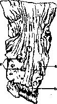
are multiple polyps of the Stomach (a); b-pylorus.
The percentage of deaths from stomach cancer falls on stomach cancers. According to data from Moscow morgues for 1923-27, stomach cancer accounts for 37.86% of all cancers. The same data show that when calculated for adults (over 16 years), every 19th-20th person dies from stomach cancer. The disease is observed significantly more often in men; thus, according to Kalb, mortality from stomach cancer in men in relation to the number of all deaths from cancer is: in France 60%, in Bavaria 85.9%, in Hungary 84.3%, in Italy 84.5%, in Sweden 84.8%, in Switzerland 90%, in England 73%; in tropical countries, stomach cancer occurs less frequently. The favorite age is between 40 and 60 years, but even before 40 years, diseases of stomach cancer are not a great rarity; cases are observed in youth and even in childhood. There are indications of different attitudes of races to stomach cancer; thus, among North American Indians it supposedly almost never occurs (Levin). There are no definite data indicating an increase in stomach cancer in recent years. Stomach cancers in the vast majority of cases are located in the area of the lesser curvature, more often closer to the pylorus than to the cardia. However, in contrast to stomach ulcers, the cardial location of cancer by no means represents a rare phenomenon. The size, shape, and character of growth of cancer vary greatly. Thus, mushroom-shaped (fungoid) cancers [see separate table (art. 115-116), fig. 3] or flat, barely rising above the level of the mucosa are observed; sometimes stomach cancer has no clear boundaries, manifesting as a diffuse thickening and hardening of the entire wall of the stomach from the cardia to the pyloric valve (see separate table, fig. 1); sometimes a strong decay of the tumor is observed, covering its main mass, which creates some difficulties in differentiation with stomach ulcers. On the other hand, some stomach cancers undoubtedly arise at the edges of chronic ulcers (in 2-10% of the latter); scar changes at the bottom, on the serous cover, and on the edges of ulcers, and on the other hand, the development of cancer somewhere on one side of the ulcer speak of the malignant transformation of the latter. The boundaries of stomach cancer are not always determined by its external appearance and contours: there is a tendency for many cancers to grow over a considerable extent under the mucous membrane, for a long time not giving particular changes from the latter; this should be remembered in the surgical excision of stomach cancers. Stomach cancers are distinguished: glandular (adenocarcinoma, or cylindrical cell cancer), colloid (mucous), medullary, fibrous (scirrhous), squamous cell (rare). Such a subdivision is not entirely correct in the sense that almost every stomach cancer, if not in form, then in origin, is a glandular cancer, and depending on its tendency to mucous transformation (colloid cancer), on the quantity and degree of predominance of the cancerous parenchyma (medullary cancer) or connective tissue stroma (scirrhous), the corresponding nomenclature changes. Soft white mushroom-shaped ulcerating, more or less clearly defined cancers mostly have the type of adenocarcinoma, diffuse tumors without clear boundaries more often have the character of colloid cancer and scirrhous. Regarding the latter, it should be borne in mind that sometimes there is a sharp wrinkling of more or less significant parts of the organ and even the entire stomach, and there may be cases where even with the help of microscopic research only with difficulty are individual groups of cancer cells found among the proliferated stroma and sharply hypertrophied bundles of the stomach musculature (linitis plastica maligna of French authors); some (organic) forms of pyloric stenosis apparently belong here, representing the theoretical interest that they demonstrate as it were the process of spontaneous healing of cancer, its outcome in gastrostasis.
Stomach cancer metastasizes through the lymphatic, circulatory system, as well as by implantation along the omentum, peritoneum, especially mucous cancers; in the first case, the glands of the lesser omentum, periportal, often upper retroperitoneal are affected, from where the spread of metastases can go downward along the aorta and upward into the mediastinal glands, the lower glands of the neck, in particular the so-called Virchow's gland behind the left clavicle (in connection with the lesion of the thoracic duct). Through the circulatory system, metastases first affect the liver (through the v. portae system); the quantity and size of nodes in it can be very diverse, up to simulating a huge primary liver cancer. Metastases to distant organs are also noted, such as: lungs, pleura (with hemorrhagic pleurisy), kidneys, more rarely - in bones, ovaries; the latter (especially with colloid stomach cancer) are often affected in pairs (retrogradely along lymphatic paths), while the contours of the organ can be preserved for a long time (Krukenberg's tumor of the ovaries, see). Growing through the entire wall of the stomach, cancer cells can implant along the peritoneum, omentum; often a continuous seeding of the abdominal organs with cancer nodes is observed with a simultaneous fibrinous-hemorrhagic exudate in the abdominal cavity; the omentum becomes greatly thickened, shortened; subsequent stenoses of parts of the intestine are also possible. Perforation of stomach cancers is not a frequent phenomenon due to the early development of adhesions. Occasionally, complications with phlegmonous processes in the stomach itself are observed. The remaining, not affected by cancer part of the stomach mucosa is usually in a state of more or less pronounced atrophy.
Secondary stomach cancers are observed relatively rarely; thus, cancer of the pancreas, large intestine, liver can grow into the stomach and subsequently, ulcerating from within, simulate primary stomach cancer; metastases in the stomach are observed in individual cases of cancer of the bronchi.
-I. Davydovsky. The etiology and pathogenesis of cancer of the Stomach remain unclear. Hereditary predisposition undoubtedly plays a certain role. In 10-15% of patients with stomach cancer, hereditary predisposition to neoplastic diseases can be found. Families have been described where most members died from stomach cancer. The clinical view has been established that the development of cancer is promoted by prolonged irritation of the stomach mucosa (the irritation theory). Konetsny's observations (in 90% of the operatively removed stomach cancers he studied, he found changes characteristic of stomach catarrh and atypical proliferation of glandular epithelium) created the theory according to which chronic gastritis is a prerequisite for stomach cancer. The symptomatology and clinical manifestations of stomach cancer are quite diverse, depending on the nature of the tumor and mainly on its location. Soft forms of stomach cancer give rise to metastases more readily and pronounced symptoms of cancer poisoning; localized at the exit and entrance, causing narrowing of the entrance or exit orifice of the stomach, they promote rapid development of difficulty in food intake and, together with intoxication—cancerous and putrefactive-fermentative—rapidly undermine nutrition and cause the development of secondary anemia and cachexia. The most favorable form in terms of symptoms and course is the scirrhus form of cancer of the stomach body. Developing slowly, without causing bleeding and intoxication, and without hindering the passage of food into the intestine, the scirrhus of the stomach body remains hidden for a long time without symptoms or is characterized only by the usual clinical signs of ordinary chronic gastritis. But even the fungoid-polypoid cancer of the stomach body, prone to disintegration and bleeding, still does not give such severe phenomena as cancer of the entrance and exit of the stomach. Cancer of the stomach body creeps up slowly and imperceptibly. At first, patients complain of loss of appetite, mild dyspeptic phenomena expressed in the form of an indefinite feeling of pressure under the xiphoid process after meals, at times belching and nausea. They readily eat spicy, piquant food, but simple healthy food is not to their taste; For many, meat and fatty food become repulsive. In other cases, until the tumor disintegrates and secondary acute stomach catarrh and putrefactive and fermentative processes develop, a satisfactory appetite is maintained for a long time; dyspeptic phenomena do not attract attention due to the preceding chronic gastritis with achylia or hypochylia for a number of years. After a considerable period (4-8 months), pain often joins these phenomena, however, it does not have the same character as in ulcer. The pain is dull, in the form of pressure or heaviness, increasing after meals, but not reaching the intensity of pain in stomach ulcer. Only in the case of the tumor spreading to the pancreas or its penetration into the solar plexus do the pains become very intense, sharp, radiating to the back, spine and sides, preventing patients from lying and sleeping. Vomiting may be absent for a long time. But when putrefactive and fermentative processes develop in the stomach, foul-smelling, disgusting belching and vomiting with a foul odor (smell of carrion), coffee-colored masses due to the admixture of blood, appear. Vomiting in most cases is not frequent (not every day) and depends mainly on the quantity and quality of food, appearing several hours after dinner, and in other cases immediately after eating. Women suffer from vomiting more often than men. Bowel movements in the first period are normal, but then constipation may appear, alternating with diarrhea. Diarrhea usually occurs at the same time as vomiting and is caused by the accompanying catarrh of the intestines, caused by irritation by food entering the intestine, which has rotted and not been processed in the stomach. The patient, already sufficiently emaciated and pale, at this time begins to lose weight rapidly, acquires an earthy facial color, loses strength; finally edemas appear, which predict imminent death. Examination of the patient in the first period of the disease for a long time gives indefinite results. Only progressive pallor and emaciation are noted. But there is a certain group of patients with cancer of the stomach body who, belonging to well-nourished people with good facial color and an athletic build, for a long time, even with an already developed and palpable tumor, maintain a flourishing appearance. Then these patients also do not escape the common fate of cancer patients—they begin to lose weight, become pale, and often develop general edemas, depending, on the one hand, on the hydremic state, and on the other—on the accompanying weakness of the heart [watery type of stomach cancer (Ivanov)]. Most patients with stomach cancer, losing their subcutaneous fat, become mummified. The body temperature in most patients is below normal, but cases of stomach cancer are observed in which it is elevated and has a septic character, accompanied by chills and sweats. The tongue, until secondary stomach catarrh and vomiting develop, is often clean, smooth, and shiny, resembling Günther's tongue in pernicious anemia. At the end of life it becomes coated, dirty. On examination of the abdomen and its palpation, no deviations are found for a long time. Methodical deep palpation, even in experienced hands, with still good nutrition in this first period, rarely gives positive results. But as the patient emaciates and the tumor grows, the conditions for palpation improve; first something indefinite in the form of 'resistance' begins to be palpated, and then in the standing or lying position, better on an empty stomach and after evacuation of the intestines, a node or ridge can be palpated in the stomach area. At the moment of palpation, the patient feels pain. 'The diagnosis is ready, and the verdict is almost ready' (Obraztsov). With further growth of the tumor and progressive emaciation, the tumor sometimes becomes visible, and if there are no adhesions around it, its respiratory excursions are noticeable. Examination of the stomach contents often from the very beginning already gives characteristic changes. Free HCl disappears; with reduced pepsin content, a significant amount of lactic acid appears (in 80-93% of cases), then—blood, Boas-Oppler rods, pus, particles of the tumor. The absence of free HCl, however, is by no means necessary—in 24.5% according to Boas's statistics, HCl is preserved. The presence of gastric bleeding, determined by 'examination of fecal masses for the so-called hidden blood,' is an early and constant sign of stomach cancer (94.5%, Boas). On X-ray examination of the stomach, a filling defect is found at the site of the tumor [see separate table (pp. 75-76), Fig. 12], distortion of the stomach silhouette, which is especially clear in the shrunken, as if crumpled stomach in scirrhus [see separate table (pp. 75-76), Fig. 11]; peristalsis at the site of wall infiltration is absent (Groedel), evacuation of food occurs at an abnormal rate. The filling defect is better detected on the X-ray than on fluoroscopy, even in cases where the tumor involves the lesser or greater curvature. Small tumors of the posterior or anterior wall of the stomach are not revealed by X-ray. In this case, gastroscopy more likely gives a positive result. Blood examination reveals the presence of hypochromic secondary anemia, occasionally taking on the features of malignant or severe aplastic anemia. Sometimes anemia comes to the forefront among other symptoms. The percentage of Hb falls to 20 and below, poikilocytosis, polychromatophilia, normoblasts, myelocytes and myeloblasts appear; a hemorrhagic diathesis can also develop. Cancer of the pyloric part of the stomach, characterized by the same symptoms, differs, however, in that dyspeptic phenomena, due to the rapid onset of narrowing of the exit and stasis of stomach contents, occur more quickly. Signs of stomach expansion appear quite rapidly: a feeling of heaviness and fullness under the xiphoid process, belching, nausea and periodic vomiting, increase in stomach volume, visible peristalsis. On examination of the stomach contents, in addition to the absence of HCl, presence of lactic acid, blood, Boas-Oppler rods, etc., stasis and presence of fermentation acids, which increase the total acidity of the stomach contents (30-40-50 by Ewald) in achlorhydria, are soon found. Due to the rapidly developing emaciation and better conditions for palpation of the tumor, the pyloric tumor is diagnosed at a smaller volume than the tumor of the stomach body. Simple cancerous infiltration of the wall, due to the development of narrowing and hypertrophy of the musculature, already allows the painful, uneven pylorus to be palpated and the development of the tumor to be diagnosed at the very beginning of the disease. X-ray examination gives a filling defect in the pyloric part [see separate table (pp. 75-76), Figs. 10 and 12], enhanced peristalsis, later secondary atony and delay in evacuation. Cancer of the cardiac part of the esophagus, if it does not affect the cardia itself, proceeds for a long time with the indefinite symptoms of hidden cancer of the stomach body and is diagnosed by X-ray [see separate table (pp. 75-76), Fig. 11]; the latter gives a clear picture in high localization of the tumor, often only when taking the X-ray with the patient lying down. When the cardia itself is involved, the symptom of cancerous, low-lying stricture of the esophagus develops (see Esophagus).
Course. Non-operated patients with stomach cancer in the very early period die within 1-2-3 years. Cancer of the body proceeds more slowly; cancer of the pyloric and cardiac parts proceeds more quickly. The duration of life for a patient with scirrhous stomach cancer is from 2 to 5 years. Diagnosis of stomach cancer is difficult at first, when there are only indefinite dyspeptic phenomena and increasing anemia and cachexia, but no palpable tumor and no blood in the stomach contents and in the feces. Diagnosis is based on three cardinal signs: 1) repeated detection of hidden blood in the excretions and stomach contents with impeccable technique for its determination (after appropriate diet), 2) a palpable tumor in the area of the stomach, and 3) a filling defect on the X-ray. Although these signs may also be observed with other stomach tumors (sarcoma, polyps, syphilis, etc.), this has almost no practical significance due to the rarity of these tumors (1-1.5%). The most difficult is to differentiate stomach cancer from callous peptic bleeding ulcer, since all the indicated cardinal symptoms may be observed with it. The proposed biological reactions for cancer: Abderhalden, Maragliano, Bothelo, etc., and reactions of Zalkovsky (in urine), Davys, etc., are not specific for cancer. One can use the Bothelo and Davys reactions due to the simplicity and convenience of their performance, but conclusions must be made with caution. In case of doubt, it is better to resort to exploratory laparotomy, and microscopic examination of the resected portion establishes the diagnosis. Therapy. A thorough suspicion of stomach cancer already requires surgical intervention - exploratory laparotomy, since internal treatment does not cure cancer, and early operation gives a prolonged effect in 33% of cases (Schmieden-Kallmann). But cancer of the dome and cardia of the stomach is inoperable, and just like large tumors of the body and pylorus with obvious metastases or far-advanced cachexia, it is not radically operated. With cancer of the entrance part, only gastrostomy is performed; with cancer of the pylorus, gastroenterostomy is performed. With internal treatment, one must first care to maintain the patient's hope for recovery and try to ease him through the inevitable death. It is necessary to prescribe a digestible, light, nutritious diet with high caloric value and large amounts of protein, as for example with a less strict diet for stomach ulcer, with the addition of light salted snacks, cream ice cream, a little cognac, good wine, fruit juices. From medications, bitters, hydrochloric acid, gastric juice, anti-fermentation substances and analgesics are prescribed. It is recommended not to spare the patient morphine, pantopon, narcophine, etc. for suffering, and subcutaneous cardiac drugs in the development of dropsy and asystole. With narrowing of the pylorus by the tumor and in the absence of bleeding, systematic washing of the stomach with lukewarm water with the addition of sodium salicylate (1:1,000) is beneficial. With vomiting and cachexia - nutrient enemas of glucose. X-ray and radiotherapy do not help. Sarcoma of the stomach is a rather rare disease (1% of all stomach tumors); it is more often observed in the pyloric region and on the greater curvature; microscopically sarcomas have different characters: round-celled, spindle-shaped, polymorphic, angioplastic, lymphosarcomas, myosarcomas. A slight tendency of stomach sarcomas to ulceration is noted; they grow slowly but can reach giant sizes, which usually does not happen with cancer; stenoses are rare; metastases are not observed in most cases. Sarcoma more often than cancer develops in the stomach secondarily. This is usually a lymphosarcoma infiltrating the stomach wall. Metastases soon appear in the glands, and the spleen enlarges. Stomach sarcoma is observed at a young age (between 15 and 30 years) and differs in rapid course (6-7 months), ending in death. The symptomatology of stomach sarcoma differs in no way from that of stomach cancer and is determined mainly by the localization of the tumor. The difference is the fairly frequent enlargement of the spleen and lymph glands and leukocytosis with lymphocytosis. To recognize stomach sarcoma and distinguish it from cancer is possible only with microscopic examination of pieces obtained by probing or excised metastases from the skin, from the glands. Therapy in the early period is surgical, in the late period - dietetic-symptomatic; X-ray and radiotherapy. The technique of surgical intervention comes down to partial and circular resection, as with stomach cancer.
N. Strazhesko. Operative treatment of stomach tumors. Benign tumors of the stomach (adenomas, polyps, fibromas, fibromyomas, adenomyomas, myomas, lipomas, angiomas and lymphangiomas) sometimes, by their location, disrupt the activity of the Stomach to such an extent that operative intervention is necessary. Depending on the case, the operation usually consists of partial or circular resection of the Stomach. In cancer of the Stomach, the main task is to remove the neoplasm at the earliest possible stage of its development. The operative technique of today makes it possible to resect cancer of the Stomach with any of its localization and with any extent of the Stomach's own lesion. Nevertheless, in reality, the number of cases subjected to radical operation, in relation to the total mass of diseases from cancer of the Stomach, constitutes only a small percentage. There are several reasons for such discrepancies between reality and theoretical possibility. First of all, neither the size of the tumor nor the duration of the disease are directly related to the dissemination of the neoplasm; the involvement of the glands begins in most cases early, and in some cases, dissemination by other routes (peritoneum, liver) proceeds even faster than through the lymphatic pathways. The tumor itself remains free from adhesions in only a minority of cases, but mostly fuses with the enlarged glands into one mass, and then grows into neighboring organs. The possibility of radical operation depends mainly on the stage of the disease. Meanwhile, many forms of cancer, especially of the body of the Stomach, for a long time do not manifest themselves with any symptoms, and the first thing that attracts the attention of the patient, those around him, and even the physician is the developing causeless cachexia; in most cases, the early stages of cancer are overlooked either due to the absence of any clinical manifestations or because the symptoms are attributed to other forms of diseases of the Stomach. On the other hand, patients themselves seek medical help too late or do not dare to undergo the operation offered to them in time. But even in cases that seem early, with a small tumor, dissemination to the glands sometimes manages to go beyond the limits accessible for removal. Resection of the Stomach in cancer can be considered more or less radical only when the neoplasm has been widely excised within completely healthy walls. From the diagram of the location and course of the lymphatic pathways of the Stomach itself, it is seen that from the pyloric part, lymph and therefore possible spread of the tumor, goes mainly along the lesser curvature toward the cardia; therefore, excision along the lesser curvature must be made 5-6 cm away from the visible borders of the neoplasm; practically this leads to the fact that resection almost always must extend almost to the cardia itself. On the contrary, toward the duodenum, the neoplasm is not prone to spread and rarely crosses the boundaries of the pylorus; this makes it possible here to resect 2 cm away. However, the main difficulty of radical operation lies in the removal of the affected lymphatic glands. The first to be affected are the glands lying along the lesser curvature; their chain extends to the point where the esophagus empties. From there, lymph flows to the glands surrounding the a. coeliaca, and further to the glands lying near the aorta. The glands of the lesser curvature often fuse with the tumor mass into one whole, and through them the tumor adheres to the liver and diaphragm. From the greater curvature, the flow of lymph goes to the glands located between the layers of the omentum gastro-colici, from there to the glands at the head of the pancreas. The efferent ducts of these latter glands go to the glands lying along the upper edge of the pancreas and along the a. hepatica. Removal of these glands sometimes presents an insoluble task, as well as that of the aortic glands. Moreover, in the presence of involvement of these glands, one can be sure of the involvement of even more distant glands, and resection becomes pointless. Thus, the contraindication to excision is not so much the size of the tumor itself as the degree of involvement of the lymphatic glands, dissemination, and finally, adhesions and immobility of the tumor. Absolute contraindications are ascites and dissemination to the peritoneum, metastases in other organs, infiltration of the tumor into the liver (unless the underlying part of the liver can be resected together with the tumor). Infiltration of the tumor into the mesentery of the transverse colon and its spread to the colon itself allow resection on condition of simultaneous excision of the colon. Similarly, partial excision of the head of the pancreas is also possible. Adhesions of the tumor to the abdominal walls and other organs make the tumor immobile and usually exclude resection; however, here much depends on the peculiarities of the case and the individual assessment of the possibility and difficulty of the operation. The details that decide the question of the removability of the tumor, in most cases, even seemingly very favorable, can be established only when the abdominal cavity is opened, and therefore the question of exploratory laparotomy should be widely raised. Of the patients admitted to surgical departments, radical operation is accessible, as large statistics show, to barely a quarter (24.6% according to Weil, 30% according to Spasokukotsky). However, these figures have relative significance; some surgeons go further in indications for resection, operating even under conditions that obviously do not allow hope for radical removal of the tumor, and, giving preference to resection over gastroenterostomy in all conditions, they raise the number of resections to 62% of patients admitted to surgical departments (Finsterer). As for the outcomes of resections of the Stomach for cancer, the immediate postoperative mortality, if compared with data from previous years and recent times, has significantly decreased, however, it still remains high, as can be seen from the following data: in Pauchet, out of 260 resections for cancer, fatal outcomes constituted 27%, in Persson, out of 361 cases-28%, in Petrov, out of 48 cases-35%, in Velikoratsky, out of 110 cases-42%. Any complications, expanded indications for the use of resections, significantly worsen the prognosis. If all cases are divided into groups depending on the stage of the disease and the complexity of local relations, the figures of immediate mortality fluctuate within wide limits-from 7% to 37%. In total and subtotal resections, mortality is not less than 50%. In the future, the vast majority of those who have undergone the operation die from recurrence, originating mainly from the glands; the average duration of life after resection, if large statistics are taken, equals 1½ years. Only about 1/6, in the best case 1/3 (Finsterer, Weil, Hartmann, Mayo) of those who have undergone the operation remain without recurrence for more than 3 years. 16% live more than 5 years, 14% more than 10 years, but in individual cases, recurrences occurred even after 13 years. After total extirpations in the cases of Wrede and Moyninger, patients remained alive after 3½-4 years. For most cases, resection is a palliative operation, but even with obviously non-radical removal of the tumor, patients after resection are in a more favorable position than after gastroenterostomy, precisely because they are freed from the disintegrating or bleeding tumor. Thus, the results of radical operation at present still remain far from satisfactory, and further successes should be expected not so much from the development of surgical technique as from the earliest possible diagnosis and the earliest possible operation. With the current state of the question of diagnosing cancer of the Stomach in its early stages, often very uncertain, patients should be inclined toward operation at the first suspicion of cancer, without hesitating to perform exploratory laparotomies. In these stages, exploratory laparotomy does not present great danger and cannot be equated with trial operations relating to the late period, when even such an innocent intervention as exploratory laparotomy presents great danger in view of subsequent complications from the heart, lungs, etc. The operative methods used for excision of cancer of the Stomach always consist of circular resection. In cancers occupying the pyloric area and not spreading further from there to the body of the Stomach, resection begins from the duodenum and includes the body of the Stomach, depending on the spread of the neoplasm, over a greater or lesser extent, and as indicated above, from the side of the lesser curvature, the boundaries of excision must extend far toward the cardia, often to the point where the esophagus empties. With extensive involvement of the Stomach, subtotal or total resection is performed. In exceptional cases, when the tumor occupies the middle of the greater or lesser curvature and moreover has limited size, circular resection of the body of the Stomach is applied, and both segments of the Stomach are sutured end-to-end. The usual methods of resection are the first and second methods of Billroth (see Billroth's operations on the Stomach). (For other modifications of resections see below - operations on the Stomach). In cancer occupying the cardial part of the Stomach, resection of the cardia is possible, but the technique of the method is still so undeveloped that to date, case histories count only several dozen cases with mortality of about 70%).
(see cardia resection). - Palliative operations for stomach cancer. In cases of inoperable stomach carcinomas, the most important palliative operations on the stomach are: gastrostomy and gastroenterostomy. Both are used in cases accompanied by narrowing of the cardia, resp. the pylorus. Gastroenterostomy in cases of pyloric closure by a tumor can give a brilliant, sometimes rather prolonged effect, but is completely useless for those cases where one deals with carcinoma of the stomach body without manifestations of motor insufficiency; therefore, if during laparotomy in such cases it is found that the tumor is inoperable, it is preferable to refrain from creating an anastomosis and close the wound. (For outcomes of gastroenterostomy in cancer - see Gastroenterostomy.) In very extensive involvement of the stomach walls, sometimes there is no place to create a gastroenterostomy; in such cases one has to resort to jejunostomy. In cardia cancers, palliative intervention in the form of gastrostomy is indicated only in the stage when the patency of the cardia is impaired to such an extent that a sufficient amount of semi-liquid food can hardly be introduced and patients begin to experience hunger and lose weight. Only in those cases where the patient has already undergone a trial laparotomy and the tumor has been deemed inoperable, should the operation still be completed by creating a gastric fistula in case of the very probable future obstruction; the creation of the fistula itself will only slightly complicate the trial laparotomy and will not worsen the patient's condition.
V. Dobrotvorskiy. VII. Stomach ulcer. Stomach ulcer was first identified as a separate clinical unit by the Frenchman Cruveilhier between 1829 and 1838. Stomach ulcer is one of the most common diseases of the stomach. It is characterized by a tendency to recur. The etiology and pathogenesis of stomach ulcer have not been finally clarified. Stomach ulcer is not equally prevalent in all countries. Statistical data. There are indications that stomach ulcer rarely occurs in Negroes and southern Chinese, as well as in some parts of Brazil. According to combined data from Moscow morgues for 1923-27, chronic stomach ulcer as the primary disease appears in 1.17% of all adult deaths from 16 years of age (251 cases out of 21,388 autopsies), with the percentage being lowest in recent years (1926-1927) (0.9 compared to 1.3 for 1923-25). The question of the frequency of stomach ulcer in its entirety cannot be considered clarified, in particular the significance of nutritional conditions, starvation, and various general diseases is not fully clear. The significance and participation of vegetative neuroses are more or less considered clarified; apparently the mass development of such neuroses influences the increased incidence of stomach ulcer; on the other hand, one cannot forget about the better diagnosis of ulcers in recent years. In women, the disease occurs approximately twice as often as in men; the age is usually from 20 to 40 years, but children also get sick (both newborns and older ones). Pathological anatomy and pathogenesis. A distinction is made between acute and chronic stomach ulcers. These in turn can be single and multiple. Acute ulcers [see separate table (p. 103-104), fig. 3] are a very common phenomenon; their size varies from a barely noticeable point to several cm in diameter; the shape is most often round or oval. Usually these ulcers are of b. or m. superficial nature, for which reason they are also called erosions; in rarer cases the defect affects the entire thickness of the mucosa or extends beyond it, and finally in individual cases acute ulcers can be perforative in nature. The bottom of the ulcer is either clean or (more often) brown-black in color; the latter is associated with hemorrhage and the formation of hydrochloric acid hematin under the influence of gastric juice (hence the term "hemorrhagic erosions of the stomach"). Hemorrhage should probably be considered both as a factor directly traumatizing the mucosa and as a factor producing the ulcer; the subsequent digestive action of gastric juice at the site of such tissue destruction creates a defect; in other cases, the possibility of primary necrosis of the mucosa followed by hemorrhage cannot be excluded. Acute erosions are observed in various areas of the stomach, although they predominate in the body and the prepyloric region; point erosions are more often located closer to the greater curvature, while larger ones are closer to the lesser curvature. The stomach contents in the presence of erosions are colored b. or m. distinct brown, especially the mucus covering the defects. By the end of the first day, reactive (inflammatory and regenerative) processes already appear at the bottom and edges of the ulcer, and in most cases after 3-6 days, depending on the size of the ulcer and other circumstances (see below), the ulcer heals without a trace or (in larger ulcers) leaves a small star-shaped scar, sometimes covered by a not entirely typically constructed mucosa. Experiments on animals have shown that even very large defects of the mucosa heal in this way. Chronic stomach ulcers have the greatest practical interest. Usually they are single, more rarely 2-3 or more ulcers are observed; the shape is usually round (ulcus rotundum), sometimes oval-elongated, irregular; often along the line of the lesser curvature, ulcers are observed in the form of a butterfly with wings located on the anterior and posterior walls. As a rule, chronic ulcers lie close to the lesser curvature or directly on the latter along the "stomach road" (fig. 30) or closer to the anterior wall; the pyloric region, as well as the vestibule, isthmus of the stomach ("narrow places" of the stomach) give the main number of chronic ulcers. These ulcers are distinguished above all by the significant density of their bottom and edges, as if calloused (ulcus callosum); scleroses can involve the entire thickness of the stomach and extend beyond it (perigastritis), which often causes fibrous adhesions of the stomach with the liver, spleen, pancreas, transverse colon. The bottom of the ulcer is smooth or rough, sometimes distinctly granular, e.g. when parts of the pancreas are underlying; sometimes a bleeding or thrombosed vessel is visible on the bottom (see separate table, fig. 1). The coronary arteries of the stomach (more often the superior), as well as the splenic and gastroduodenal, are particularly often subjected to erosion; the significant caliber of these arteries and the well-developed anastomoses determine the special danger of these hemorrhages and their frequent fatal outcome. The depth of the ulcer is not the same in different plac

Figure 30. Magenstrasse. (After Aschoff.)
edge; it is considered characteristic when the edge of the ulcer directed toward the cardia is deeper, steep, somewhat undermined; the opposite edge, on the contrary, is low, gradually descending in terraces. Such relationships give the ulcer profile an irregular funnel shape with an acute angle directed toward the cardia. This phenomenon is explained by the fact that the food taken, on the one hand, and strong peristaltic movements, on the other, produce a purely mechanical shift of the mucous membrane in the pyloric direction, in which the cardial side forms as it were an overhang, while the pyloric side is flattened, smoothed, and the muscle beneath it is partially exposed (see below). The steep and deep cardial edge of the stomach ulcer is well detected by X-ray examination in the form of the so-called niche formation [see separate plates (pp. 75-76), figs. 7, 8 and 9]. In an already formed ulcer, and precisely under the steeper cardial edge of it, there occurs some stagnation of the acidic stomach contents, which also contributes to the corrosion and deepening of precisely this part of the ulcer base, enhancing its funnel shape. The former opinion, explaining the funnel shape and undermining of the cardial edge by the peculiarities of the vascular supply of the wall (according to the infarction type), is now abandoned. The terraced (stepped) nature of the pyloric side of the ulcer is determined by the layer-by-layer corrosion of the muscle bundles of the stomach wall and precisely by the fact that this corrosion, as it deepens, covers an increasingly limited area of muscle. Strong peristaltic movements here also produce some displacement of the muscle layers as their integrity is disrupted. Microscopically, the base of the ulcer is represented by granulation tissue of varying density and maturity; the most superficial layer of tissue is often in a state of necrobiosis or covered by a scab, exudate, due to the action of stomach contents, as well as possible secondary infections, including fungal; thus, in the question of the progressive growth of ulcers, Askanazy attributes special importance to the thrush fungus. In long-existing stomach ulcers, their base is completely devoid of muscle, a more or less dense scar tissue with perivascular infiltrates is visible, which usually extends to the serous covering and adjacent parts of organs. When studying the edges of the ulcer, it turns out that muscle bundles exist; at the cardial (steep) edge they are located very high, as if directly merging with the mucous membrane; at the opposite (pyloric) end, the muscle layers lie deep under the mucosa, appearing on the surface of the ulcer at the steps of the "terrace". The vessels of the ulcer base are often sclerosed, which however is explained by the presence of a chronic process in the surrounding tissue and cannot be interpreted as a factor determining the development of the ulcer. It is not excluded, of course, that such a condition of the vessels reduces the regenerative capacity of tissues and contributes to the progressive development of the ulcer. In
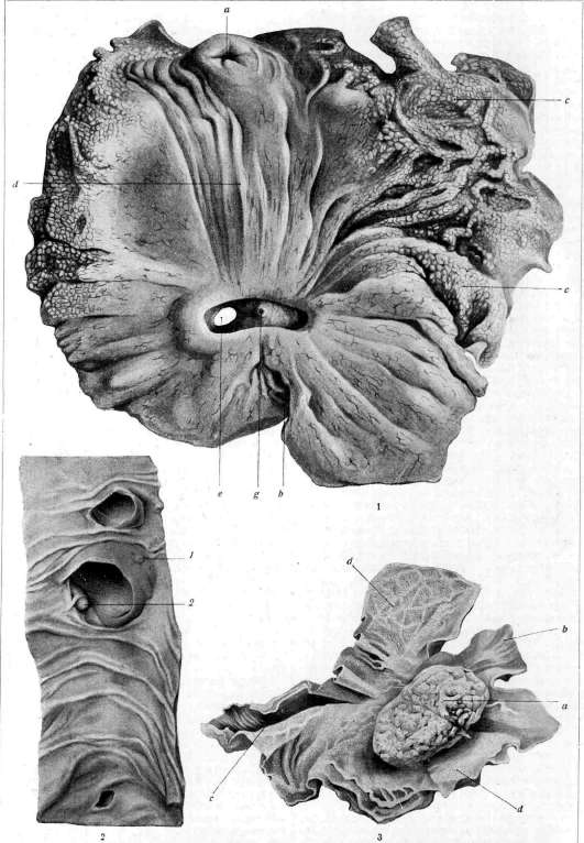
Figure 1. Round stomach ulcer with perforation (e), with corrosion and thrombosis of vessels (g); a - cardia; b - pylorus; c - greater curvature with phenomena of etat mamelonne; d - lesser curvature. (From specimen of the Pathological-Anatomical Institute of 1st Moscow State University). Figure 2. Three diverticula of the duodenum: 1 - duct, pancreat. accessor.; 2 - papilla Vateri. (From museum of 1st City Hospital named after N. I. Pirogov). Figure 3. Fungous (mushroom-shaped) stomach cancer (a) near cardia (b); c - pylorus; d - so-called etat mamelonne in the area of the greater curvature. The tumor has an uneven, in places ulcerated surface, of soft velvety appearance in other parts. (From specimen of the Pathological-Anatomical Institute of 1st Moscow State University).
To the article Diverticulum, Stomach. When in the depth of the ulcer base a strong proliferation of nerve fibers is found according to the type of amputation neuroma - a circumstance representing certain interest in explaining the particularly intense pains in this condition. It can be considered established that chronic stomach ulcer develops from an acute one, and the only question is under what conditions the latter heals and under what conditions it passes into a chronic one. A completely precise answer to this cannot be given; only the following is known. 1. The depth and width of the erosion have very relative significance; those erosions and ulcers caused experimentally as a rule heal well, and only very significant defects with simultaneous severe circulatory disorders (as for example when introducing adrenaline into the vessels) could sometimes be converted into a chronic ulcer. All other conditions being equal, the size and depth of the ulcer will have significance for example with increased acidity, with corresponding general predisposition (vegetative neuroses, etc.). 2. The most important factor influencing the transition of an acute ulcer into a chronic one is the fact of its localization: acute ulcers can occur everywhere, but transition into a chronic one is observed only along the lesser curvature, i.e. the "stomach track", and in the pyloric area. This depends primarily on functional-mechanical factors, and precisely the constant traumatization of the formed ulcers by the food passing here; such traumatization is especially facilitated by the presence of narrow places in the stomach (isthmus, vestibulum, pyloric area), which are actually the most frequent carriers of ulcers. In the same unfavorable direction in terms of healing of acute ulcers act: weak mobility, some rigidity of the folds of the mucous membrane of the lesser curvature, relatively weak anastomosis of the vascular network in the area of narrow places (not shared by all); the small thickness of the mucous membrane itself, the small production of mucus which would soften the traumatization, the significant duration of irrigation with gastric juice of this area (even with an empty stomach), and finally the structure of the glands of this area, their special sensitivity and weak regenerative ability. All this turns the entire question of the transition of an acute ulcer into a chronic one into the question of the localization of the ulcerative process itself. Nauwerk and Konetzny have put forward their theory of the development of chronic stomach ulcer, pointing to the significance of primary chronic ulcerative gastritis. One of the usual outcomes of chronic stomach ulcer is its healing with a scar; the latter always has a star-shaped character, resembling a scar from a purse-string suture; microscopic examination reveals in the scar atrophic and often atypically constructed mucosa lying directly on the scar tissue, very rich in elastic fibers. Scars of the pyloric area often give stenoses and symptoms of obstruction; the latter can however arise also with small stenoses and apparently even in their absence depending mainly on accompanying spastic moments caused by the ulcer itself. Deep disfiguring scars of the vestibulum area can give changes in the shape of the stomach in the form of the so-called hourglass. The transition of a stomach ulcer into cancer apparently does not represent great rarity (according to various authors, the percentage of such ulcers varies greatly, most accept figures from 2% to 10%). In each such case the transition must be verified by histological examination (see above - stomach tumors), but even then significant difficulties may arise. Perforation of the ulcer is one of the most frequent complications in its course; it can occur into the abdominal cavity, the greater omentum, more rarely - into the large intestine, with symptoms of the so-called enteritis, into the liver, into the spleen. Rupture into the abdominal cavity does not always end with acute general fatal peritonitis; the acidic, poor in bacterial flora stomach contents in stomach ulcer sometimes gives a picture of adhesive peritonitis with encapsulation of food particles, which during operation or autopsy can simulate tuberculosis of the peritoneum. From the above-mentioned classical ulcers of acute and chronic character, ulcerative processes in the stomach observed in acute and chronic gastritis, in tuberculosis, in poisoning with caustic substances, as well as ulcers arising during surgical interventions, for example when applying clamps to the stomach and etc., should be distinguished in principle.
I. Davydovsky. Etiology. In the USSR, stomach ulcers, as is evident from the data presented, were previously rare, especially among the rural population. In recent times, the number of patients with stomach ulcers has increased significantly; however, there are no precise statistical data on this issue. The increase in the number of patients with stomach ulcers in the USSR is explained by the changed nutritional conditions of the population, the massive development of vegetative neuroses, as well as improved diagnosis of ulcers St. St. ulcer is a disease predominantly of adults-between 20 and 40 years; it also occurs in children. Heredity and constitution play a significant role in the etiology of St. ulcer. Families are observed where most members suffer from St. ulcer. Predisposing factors-chronic catarrh, arteriosclerotic changes in the stomach vessels, chronic inflammatory processes in other organs of the abdominal cavity (such as: appendicitis, cholecystitis, adnexitis in women, etc.) and mainly changes in the tone of the autonomic nervous system, and possibly also the altered influence of trophic nerves on the tissues of the stomach wall. Many theories exist to explain the occurrence of St. ulcer. It must be thought that the occurrence of St. ulcer is conditioned by a whole series of complexes of conditions, which are effective in various combinations with each other and with a certain constitutional predisposition. The main moments contributing to the occurrence of St. ulcer and its transition to a chronic state, as well as recurrences, according to modern views are: a) disturbance of blood circulation, depending on functional anomalies in the vascular system (O. Miiller, Heimberger, Skulsky) or on arteriosclerosis and thrombosis of the stomach vessels and hemorrhage into the mucosa (Gruber, Virchow); b) decrease in the viability of the St. mucosa due to disturbed blood circulation in it and special environmental conditions in which its cells live (Vannotti, Zimnitsky), consisting in acidosis; c) increased secretion of gastric juice and d) mechanical damage to the mucous membrane by coarse and irritating food, especially in narrow parts of the St. (Aschoff). The manifestation of the influence of the above factors is facilitated by a disorder in the balance of the autonomic nervous system (Kraus, Bergmann), on the basis of which cellular and tissue exchange in the whole body, salt balance, acid-base equilibrium change, blood circulation in the stomach mucosa is easily disturbed under the influence of spasm of both the stomach musculature and the vessels themselves, hypersecretion occurs, especially in the presence of constant irritation in the ganglion coeliacum, which sends to the St. secretory, motor, vaso-motor and trophic nerves. Irritation in the ganglion coeliacum arises and is maintained due to the inflammatory process in it itself or reflexively-from the side of organs associated with it by nerve pathways (Rossle). Under these conditions, any bruise in the St. mucosa, resulting from trauma or mechanical irritation, ischemia from prolonged spasm or thrombosis of its vessels easily contribute to the formation of erosion on the mucosa, which due to hypersecretion and constitutionally reduced vital activity of tissues and special environmental conditions (acidosis) does not heal with a strong scar, but turns into an ulcer, especially if the ulcer surface becomes infected (for details on the pathogenesis of peptic ulcers-see Duodenum, ulcer). Clinical picture. The symptomatology of St. ulcer is extremely diverse. Sometimes a stomach ulcer proceeds without symptoms and heals with a scar or causes perforation of the stomach, without previously giving any sign of itself to either the patient or the attending physician. This is observed more often with localization of an uncomplicated ulcer on the lesser curvature or on the walls of the stomach body; however, with systematic examination of gastric contents or fecal masses, even in these cases, the presence of blood (hidden Boas' hemorrhages) can be periodically detected. The asymptomatic course of St. ulcer is nevertheless an exception. Most St. ulcers, in typical course, are accompanied by characteristic subjective and objective symptoms. Among them are typical: "ulcer pains", acidism, bloody vomiting, bloody stools (melaena), constipation, changes in sensitivity of the abdominal wall in the area of the St. and in the corresponding peripheral zones, changes in St. secretion (hypersecretion), presence of blood in gastric contents, and symptoms characteristic of stomach ulcer on X-ray. Only in rare cases are all the listed signs present-in most cases of St. ulcer, some or others are absent (atypical course of stomach ulcer). The most typical course is that of an ulcer in the pyloric region of the stomach and an ulcer of the lesser curvature. An ulcer of the pyloric part of the stomach is characterized by periodicity of pains, late onset of pains and often nocturnal hunger pains [syndrome pylorique Cyno (Soupault)], as well as, even in the absence of complications, a tormenting sensation of fullness and pressure in the area of the St. and a tendency to vomiting; these phenomena are explained by the longer duration of the period of gastric digestion, slow evacuation of food from the St. due to hypersecretion and spasm of the pylorus. With this localization of St. ulcer, periods of exacerbation in the form of paroxysms similar to tabetic crises alternate with periods of relative calm; this is explained rather by the restructuring of the whole organism and the autonomic nervous system, affecting the diseased organ, than by anatomical changes in the St. ulcer itself and around it (Bergmann). The restructuring occurs due to the law of cyclicality of the course of biological processes in the organism (Goldscheider, Pletnev). Pains in the form of either pressure or burning or purely cutting pain, radiating to the back and under the right scapula, do not occur immediately after eating, but after two, three, four hours, sometimes even much later. They often reach great intensity, at which the patient seeks to find a certain position in which the pain is less and remains motionless until they pass. These pains can be caused by various reasons-in most cases, especially with uncomplicated ulcers, they depend on spasmodic contractions of the St., in particular pylorospasm on the basis of an enhanced reflex from the duodenum when more acidic than normal gastric contents enters it. More rarely pains arise as a result of perigastritis complicating the ulcer and adhesions of the St. with other organs. Even normal movements of the St. in these cases can cause pain. The pains are accompanied by heartburn, belching, even vomiting of acidic masses, after which relief occurs. Late pains are explained by the highest rise of the secretion curve at this time and prolonged spasm of the pylorus. Then the pains pass, only to resume in five or six hours, usually at night. The patient, knowing about them, goes to sleep, placing a glass of milk beside himself or preparing to take soda. He knows that these pains will wake him up and that they can be relieved only by introducing food, soda, etc. These nocturnal pains are sometimes accompanied by abundant acidic vomiting without food, after which they pass. The pains are caused by accumulation of gastric juice in the stomach due to its constant secretion. On objective examination of the patient, emaciation and pallor are often noticed, depending on insufficient nutrition due to fear of causing pain by taking food, and sometimes from periodically recurring blood loss. Inspection of the St. area in most cases gives nothing. With significant emaciation, two to three hours after lunch, the tense St. is visible through the abdominal coverings. On palpation, especially in emaciated individuals, the pyloric part of the St. is easily palpable in the form of a tumor, constantly painful, sometimes nodular (in ulcus callosum). Palpatory mobility of the pylorus is limited in case of perigastritis. Percussion and palpation in the area of the entire stomach and pylorus in uncomplicated cases usually do not cause pain, although there are exceptions. With complication by perigastritis, hyperalgesia above the navel, in the area of the right rectus muscle of the abdomen, pain in this place on tapping (Mendel's symptom), tension in this area and limited tympanitis in the area of the right rib arch, between the mammary and parasternal lines (Strazhesko's symptom), and sensitive paravertebral points of Boas between the X and XII vertebrae on the right, sometimes painful IX-XII spinous processes of the vertebrae (Openchovsky's points) and more rarely horizontally located hyperesthetic zones of Head in the area of IX-XI intercostals on the right.-When examining gastric contents, hypersecretion is found in most cases, especially during periods of exacerbation, when abundant secretion of juice is observed with an empty St. If in St. ulcer hypersecretion is not more common than in half of all cases and ulcers are observed with subsecretion, then for pyloric ulcer hypersecretion is the rule, exceptions are rare. Hypersecretion is manifested during the Ewald-Boas breakfast, but even more sharply during fractionated long-term research after liquid breakfasts, as well as during repeated load according to Zimnitsky. The figures of total acidity reach 100-110, free HCl-80-90.
In this case, continuous secretion is often noted, and on an empty stomach, 80-100 cm3 of gastric contents with high acidity can be found in the stomach. At the same time, not only is the acidity increased, but also the digestive power of the gastric contents. In the contents, an increased amount of mucus is often found, which is an expression of the accompanying gastritis. The presence of blood cannot always be detected, but with repeated examinations it is usually possible. On X-ray examination, during fluoroscopy and simultaneous palpation, as well as on X-ray films (a series of films and in various positions is mandatory), not only completely clear symptoms of an ulcer are often found, but also its specific localization; the deeper the ulcer and the more complications it has given, the more definite signs it is characterized by. However, it must be remembered that not all ulcers of the Stomach, even with the best technique, are diagnosed by X-ray examination. On fluoroscopy, enhanced peristalsis is sometimes found, sometimes segmenting; with simultaneous palpation, tenderness in the pyloric region, limitation of its mobility, and its displacement to the right in the case of perigastritis, retraction on the opposite side in the contour, prolonged spasm of the pylorus; on X-ray films, a small niche with deformation of the contours of the Stomach is sometimes found. The intermediate layer is large due to hypersecretion; because of it, even before the introduction of contrast medium on an empty stomach, liquid contents can be detected in the Stomach by fluoroscopy. On repeated fluoroscopy, delayed evacuation is found, and after 5-6 hours, 2/3 of the contrast medium is often found in the Stomach, significantly diluted due to hypersecretion. An ulcer of the pyloric part is rarely accompanied by reflex spasm of the cardiac part of the Stomach; if this is observed, in most cases the spasm depends on the accompanying general vegetative neurosis or a second ulcer near the entrance to the Stomach. Also not characteristic of it is the radiation of pain to the left side. On the contrary, for an ulcer of the Stomach, localized on the lesser curvature near the entrance to the Stomach, pain during the very act of swallowing food, radiating to the left scapula and back, and symptoms of dysphagia, depending on spasm of the cardia musculature, are extremely characteristic. The closer the ulcer to the cardiac sphincter, the more intense the pain and dysphagia; an uncomplicated ulcer of the lesser curvature away from the sphincter may proceed without any symptoms. Otherwise, the case is with penetrating, so-called penetrating ulcer with perigastritis. In it, the pains are intense. Both painful points and zones are localized on the left, in the area of the X-XII vertebrae and corresponding segments. Hypersecretion is by no means mandatory, the presence of blood is detected on repeated examinations. For a cardiac ulcer, the easy vulnerability of the mucosa during probing, especially with a thick probe, is often characteristic. On X-ray examination, spastic stenosis of the cardia with subsequent dilation of the esophagus and retraction or niche on the lesser curvature may be found. With localization of the ulcer in the cardiac part of the Stomach, away from the cardia itself, a series of X-ray films should be persistently recommended: one fluoroscopy may not give indications. Of the ulcers of the Stomach, those of the body of the Stomach most often do not give symptoms, especially when localized on the anterior or posterior wall or greater curvature without complications. In this case, the characteristic pain syndrome may be absent, and the ulcer of the Stomach is accompanied only by the usual dyspeptic symptoms characteristic of catarrh of the stomach. Objective and X-ray examinations often give a negative result, and only the presence of blood in the gastric contents, resp. in the feces, gives reason to suspect an ulcer of the Stomach. In some cases, with a callous ulcer located on the posterior or anterior wall, it is possible to palpate an ulcer-tumor, and on fluoroscopy-to find at the site of the ulcer a persistent spasm of the musculature and a serrated contour of the Stomach, caused by spasm of the muscularis mucosae [see separate table (pp. 75-76), Fig. 7]. However, when a complication in the form of perigastritis or penetration occurs in the presence of previous adhesions, ulcer symptoms appear and gradually increase. Early pains appear (after 1/2-1-11/2-2 hours after eating) with radiation to the spine, to both sides of the chest, severe pains in the back if adhesions or penetration involve the pancreas or solar plexus; at the same time, hyperalgesia of the skin in the stomach area, Boas's and Opchensky's points in the IX-XII segments, and Head's zones on both sides are observed. Examination of the gastric contents does not always (in about half of the cases) reveal hyperacidity and hyperpepsia, as well as the presence of blood and mucus. X-ray examination often reveals a persistent spasm of the Stomach musculature in the form of retraction of the greater curvature [see separate table (pp. 75-76), Fig. 7] and conversely-on the lesser curvature-Gaudekowski's niche [see separate table (pp. 75-76), Figs. 7 and 8] or limitation of mobility in some part of the Stomach, sometimes also deformation of the Stomach in the form of an hourglass or cascade Stomach [see separate table (pp. 75-76), Fig. 9]. An ulcer located on the posterior wall, even complicated by perigastritis, is easily overlooked on X-ray-here sometimes a whole series of films in lateral positions is required. Complications in ulcer of the Stomach are observed quite often in the form of a) hemorrhages, b) perforation of the Stomach wall, c) perigastritis, d) various strictures developing as a result of ulcer scarring, and e) malignant degeneration of the ulcer. Various complications often combine with each other or are observed in the chronic course of ulcer of the Stomach in sequence. Many authors, for example Knud Faber, consider that hemorrhages in the form of profuse bleeding or a slight admixture of blood to the gastric contents, detectable only by chemically sensitive reactions to blood, are an obligatory symptom of ulcer of the Stomach. Not all clinicians agree with this. And if some, like Boas, attach great importance to hidden hemorrhages, considering them pathognomonic for ulcer and cancer, others, e.g. Bergman, think that 'hidden hemorrhages' can also occur with other diseases of the Stomach. Only profuse hemorrhages in the form of bloody vomiting (haematemesis) or bloody stool (melaena), which are observed in approximately 19% of all cases, have decisive diagnostic value for ulcer of the Stomach. Profuse bleeding may be the first and only sign of ulcer of the Stomach, sometimes threatening life (mortality from bleeding ranges from 1% to 3-5% with repeated bleedings). The first signs of bleeding* are not bloody vomiting or tarry stool, but indirect signs, such as pallor of the skin, dizziness, a feeling of weakness, sometimes loss of consciousness, rapid pulse, and signs of collapse; at the same time, the pains that tormented the patient daily often disappear. After some time (1-3 hours), vomiting appears. However, in other cases vomiting may not follow, and confirmation of gastric bleeding is then melaena and tarry stool several hours (5-12 hours) after the bleeding occurred. A more severe complication of ulcer of the Stomach is perforation of the Stomach wall, which can occur both with fresh ulcers several hours after their appearance, and with chronic ulcers as a result of gradual erosion of the Stomach wall. Perforation can occur with both a small and an enormous stomach ulcer. About 10% of all ulcers of the Stomach result in perforation. The clinical picture of acute ulcer perforation is characterized by: a) a feeling of severe pain in the upper abdomen, radiating to the back or shoulder; b) rapid decline in strength, appearance of deathly pallor (facies hippocratica) and cold sweat; c) increased respiration and pulse. The slightest touch to the upper abdomen causes severe pain. Vomiting often appears, temperature drops. The abdominal press is tense, especially in the area of the Stomach, and tympanitis appears over the liver. Peritonitis develops. 5% of patients with perforative peritonitis recover, half die on the second day (Schnitzler). However, if there were adhesions of the serous membrane of the Stomach with surrounding organs at the site of the progressing ulcer, then perforation may not lead to acute peritonitis, because the adjacent organs (liver, pancreas, omentum, intestines) close the defect. In this case, after perforation, only an exacerbation of the former inflammatory process occurs, accompanied by increased pain, elevated temperature, increased tension of the abdominal press, and other clinical phenomena of local peritonitis-perigastritis. The plastic fibrinous infiltrate that sometimes develops, accompanied by pain and elevated temperature, also involves neighboring organs in adhesions, e.g. the pancreas, and promotes the formation of an inflammatory tumor detectable by palpation. With the formation of such infiltrates and adhesions with neighboring organs (liver, pancreas, etc.), ulcerative destruction also spreads to these infiltrates and organs ('penetration' of ulcer of the Stomach).
In other cases, perigastritis takes on a purulent or putrefactive character in the form of a free abscess in the abdominal cavity or located under the diaphragm on the right or left, often containing not only pus but also food substances that have exited the Stomach during perforation, and foul-smelling gases that have developed due to putrefactive infection. In some cases, the abscess that has formed among adhesions carves a path into the Stomach, after which a separate cavity is formed next to the Stomach among the adhesions, communicating with the Stomach and giving a 'niche symptom' upon X-ray examination. The perigastritis that has formed, due to fibrous degeneration of the exudate, often contributes together with the scarring ulcer to narrowing of the Stomach at the site of the ulcer and formation of a Stomach in the shape of an hourglass or cascade Stomach [see separate table (pp. 75-76), Fig. 9], which, when sufficiently developed, impairs the function of the Stomach. In this respect, narrowing of the pylorus or cardia causes particularly serious consequences. Narrowing of the pyloric part is accompanied by clinical symptoms of Stomach expansion, such as: increase in its volume, stasis of contents, visible peristalsis after eating, and a characteristic X-ray symptom complex (see above - disorders of motor function of the stomach); narrowing of the esophagus is accompanied by dysphagic phenomena and symptoms of esophageal expansion (in detail - see Esophagus).- Complication of an ulcer with cancer, which develops at the site of a former scar or due to cancerous degeneration of the edges of the ulcer in ulcus callosum, is not encountered so often (see above - tumors of the stomach). With this complication of a stomach ulcer, free hydrochloric acid, despite the presence of stomach cancer, is often observed in a higher percentage and often remains until the very end. The course of a simple ulcer is extremely varied. In general, each ulcer that forms has a tendency to chronic course and recurrences. Some acute ulcers heal quickly. One cannot deny the possibility of complete healing of a chronic ulcer. In both cases, all the symptoms that have appeared with the ulcer can completely disappear not only with a simple ulcer but also with a chronic one complicated by a niche or callosally degenerated (Bergman).- In regard to prognosis, stomach ulcers can be divided into two groups: simple, uncomplicated ulcers, which heal well with appropriate regimen and treatment, and callous ulcers, which scar with difficulty and easily give the above-mentioned complications. The mortality rate from stomach ulcers, according to the consolidated statistics of Hauser, based on materials from German clinics for internal diseases, averages 10.6% (without division into the two groups just mentioned). The diagnosis of stomach ulcer in some cases, when cardinal symptoms are present (pain syndrome, hypersecretion, hyperaciditas, bleeding, characteristic X-ray picture), is very easy; in others, running atypically, it is rather difficult. It is necessary to carry out differential diagnosis between other diseases of the Stomach, such as achylia, catarrh, cancer, syphilis and functional neuroses, as well as between diseases of the liver, pancreas, appendicitis and crises in tabes dorsalis. A carefully collected and subjected to comprehensive clinical analysis anamnesis, skillful objective examination of the abdominal cavity, study of the secretory function of the Stomach, analysis of its contents and fecal masses, as well as X-ray examination and simple observation of the patient, which give the physician a complex of clinical symptoms characteristic of one or another of the listed diseases, at present in the vast majority of cases allow to make the diagnosis of stomach ulcer completely without error. The greatest difficulties are encountered in differentiating a stomach ulcer from neuroses of the Stomach and gallstone disease or cholecystitis, all the more so as these diseases often combine with a stomach ulcer. Careful objective examination of the patient, persistence of symptoms, slight dependence of complaints on the patient's mental state, dependence of pain on the quality and quantity of food, and above all - periodic presence of blood in the stomach contents and fecal masses and characteristic for stomach ulcer X-ray signs allow in the end to distinguish a stomach ulcer from functional disorders of it or to recognize their combination. Disease of the liver and gallbladder is differentiated by characteristic pains, by soreness of the liver on percussion and palpation, by increased bilirubin content in the serum, by increase of urobilin in the urine, slight leukocytosis with shift to the left according to Arneth-Schilling, by the time of onset of pains (in diseases of the gallbladder and liver in most cases pains appear immediately after meals), as well as on the basis of X-ray examination (cholecystography). It is extremely difficult or even impossible to establish the primary source of the disease in the case when there are signs of pericholecystitis and peripyloritis and there are no cardinal symptoms of ulcer, as bleeding, etc., all the more so as cholecystitis often joins the ulcer of the pyloric part of the Stomach, complicated by perigastritis and periduodenitis. (On differential diagnosis from stomach cancer see above - new growths of the stomach). In recognizing a stomach ulcer, one should not limit oneself to establishing the presence of an ulcer, but it is also necessary to determine as accurately as possible the localization of the ulcer and its character: simple ulcer, callous, penetrating, etc. Above were indicated the characteristic features for each localization of the ulcer in the Stomach and the peculiarities of manifestations and course of the simple, callous ulcer, etc. An ulcer of the pyloric part of the stomach gives a picture of the disease very similar to the picture of an ulcer of the duodenum, located most often in its very beginning. (On differential diagnosis between an ulcer of the pyloric part of the stomach and the duodenum - see Duodenum, ulcer). Prevention of stomach ulcer consists in improving the social living conditions of the population, regulating questions of nutrition, normalizing the time of food intake and limiting the consumption of alcohol, since abuse of it leads to the development of catarrhs of the Stomach and promotes the development of neuroses of the autonomic nervous system, which play a large role in the pathogenesis of stomach ulcer. Normal, calm, hygienic life, with attention to physical culture and proper dietetic nutrition, has enormous significance not only for the state as a whole but also for each individual, especially for people with an unstable nervous system and for persons in whose family stomach diseases have already been observed, especially-- peptic ulcer. Correct and timely treatment of inflammatory processes, in particular appendicitis and cholecystitis, therapeutic or surgical, also has considerable significance in the prevention of stomach ulcer. The same rules of life should be followed by the patient cured of an ulcer, with the aim of preventing recurrences, since a patient who has had an ulcer differs constitutionally as well as in the function of his autonomic system from a normal person and is predisposed to recurrence of ulcer. A patient cured of an ulcer should be under the observation of an experienced physician throughout his life and periodically undergo examination both as to the function of his stomach and the presence of blood in the fecal masses in order to notice the beginning of a recurrence in time; to prevent recurrences he should constantly feed mainly on milk-vegetable food and completely exclude from his diet alcoholic beverages, meat and fish broths, spicy and salty dishes and in general all those nutrient substances that are powerful secretagogues. Treatment of stomach ulcer consists in the application of a special diet and medications and physiotherapy, as well as in operative assistance (see below). Therapeutic treatment should be applied to every patient with a stomach ulcer, especially in fresh cases. Only in cases of severe and persistent pains, large and moreover repeated bleedings, signs of impending or fresh, just occurred perforation (first 6-8 hours), purulent perigastritis, narrowing of the inlet or outlet of the Stomach, as well as marked narrowing in the region of the body of the stomach (Stomach in the shape of an hourglass, cascade Stomach) and old callous ulcer with suspicion of cancerous degeneration - the patient is operated on. In this respect one should be guided by the general rule that failure of therapeutic treatment of an ulcer, strictly conducted for 6-8 weeks, generally requires operative assistance. The task of internal treatment consists in achieving healing of the stomach ulcer in the shortest possible time by prescribing appropriate regimen, diet and medications, and then preventing its recurrences. Treatment must be strictly individual depending on the localization of the ulcer, complications observed with it, condition of the nervous system and causal factors that caused the stomach ulcer, as well as symptoms observed, i.e. it must be individual-causal-symptomatic. When prescribing regimen, diet and medications, it is necessary to pursue two goals: to create complete rest for the patient and his Stomach, reducing spasms, enhanced peristalsis and secretion of the Stomach, and to facilitate evacuation. As a rule, a patient with a stomach ulcer, at least for the first two weeks of treatment, is prescribed bed rest, which should be carried out especially strictly and for a long time in complicated stomach ulcer.
When prescribing a diet, it is necessary to ensure that nutrition is sufficient - only in the first days of treatment is reduced nutrition permissible; in this case, nutrition should be based on the physiological principles of Pavlov's school. Several courses of dietary treatment have been proposed, approximately meeting the requirements of physiology and the indicated principles (Leube, Lenhartz, Senator, Bouveret, Sippy, Yarotsky, and others). (For details - see Duodenum, ulcer). Milk food has long enjoyed a good reputation, but in its pure form it is not particularly suitable for treating pyloric ulcer that occurs with spasms and gastrosuccorea, and especially when colitis has joined; in such cases, cream and butter should be used for nutrition instead of milk, which as fats reduce secretion and have high caloric value and are therefore generally suitable food for a patient with ulcer. Liquid, raw or semi-raw eggs (6-8 per day) are also well tolerated for these reasons. Carbohydrate food in the form of various mucous porridges from cereals, boiled in water with butter or on milk or cream, well-pureed vegetables with butter, as well as soft bread are the best nutrition. Meat products are not permitted during the first 4-6 weeks of treatment; later, due to their taste properties, they are allowed in limited quantities and only in minced and pureed form. Meat soups, vegetable soups, fish soup, coffee, strong tea and alcoholic beverages must be completely forbidden due to their juice-stimulating effect. Food should be lightly salted and not seasoned with acidic fruit juices. The regimen, depending on the severity of the case, the degree of ulcer bleeding, the strength of pain, the patient's nutrition and the freshness of the case, should be divided into three types. A. Strict regimen (immediately after bleeding, when there is a tendency for the ulcer of the St. to perforate and in fresh ulcers) consists of 3-4 days of abstaining from any food and drink with the introduction of glucose into the vein or in the form of drip enemas, with a gradual increase in the most suitable of the above-mentioned products, namely cream, milk, butter, liquid eggs, etc., with the calculation that by the beginning of the 3rd week to reach milk porridges and bread softened in milk and a caloric value of 2,000-2,400 calories. B. Less strict regimen (for patients with fewer complaints and slight bleeding) begins with prescribing cream, butter, milk, eggs, milk and mucous porridges, pureed vegetables - with a caloric value of up to 1,500 calories at the beginning and with a gradual increase by the end of the 3rd week to 3,000 calories. C. Light regimen (for patients who have not had bleeding in the last 5-6 weeks and have no great suffering) consists only in abstaining from coarse, salty food, raw fruits, marmalades, acidic fruit juices, meat broths, fish soup, all kinds of spices, piquant foods, alcohol and smoking, with as much limitation as possible of meat and fish dishes. Essentially, every ulcer patient should follow such a diet almost all his life to avoid relapses. The ulcer patient should take food not hot and in small portions with a regular 2-hour interval. Drug treatment consists in systematically prescribing alkalis and agents that reduce secretion and eliminate spasms and pain. The prescription of alkalis aims to reduce the acidity of gastric contents, eliminate hypersecretion and acidosis phenomena, as well as restore acid-base balance in the body and tissues, because in case of acidosis, the conditions for healing the ulcer of the St. are unfavorable, all the more so since acidosis is accompanied by hypertonia of the parasympathetic nervous system (Zimnitsky). Alkalis are given either simply in the form of purified soda (1.5-2.0 per dose - up to 10.0 per day) on an empty stomach in the morning and 11/2-2 hours after meals or in the form of alkaline mineral waters (Borjomi, Vichy). Excessive introduction of alkalis in the Sippy course (32.0 Natr. bicarbonici + 7.5 Magnesiae ustae + 8.0 Calcariae carbonicae) is excessive and can cause alkalosis. It is useful to prescribe a 0.2-0.4% solution of Natr. caustici in 1/1 glass of water half an hour before meals (Glaessner). The prescription of bismuth preparations (in the morning on an empty stomach Bismuti carbonici 4.0-10.0 according to Fleiner), which aims to reduce gastric secretion as well as cover the ulcer surface with a layer of bismuth protecting it from irritation by food, should be considered useful. The prescription of belladonna preparations to reduce hypertonia of the vagus nerve and peristalsis, eliminate spasm and hypersecretion is quite rational. Extr. belladonnae is prescribed 0.015-0.02 three to four times a day after meals, with alkalis. It is more rational to treat with atropine for a month (0.001 Atropini sulf. one to two to three times a day under the skin). The dose depends on the degree of hypertonia of the n. vagi. To treat the ulcer of the St., Argentum nitricum (0.1-0.2% solution three times a day per tablespoon in 1/3 glass of water 15 minutes before meals) is also used, especially in cases of ulcer of the St. complicated by catarrh. Treatment of the ulcer of the St. by parenteral administration of protein (vaccineurin, neoprotin, Yatren-Kasein, etc.), i.e., according to the principle of protein therapy, deserves little attention. Even less importance should be attached to the treatment of the ulcer of the St. by deep X-ray irradiation. The prescription of the listed agents also satisfies the principle of symptomatic treatment, since alkalis eliminate acidosis phenomena and indirectly pain. Atropin does the same. However, in some cases, to eliminate spasms and pain, papaverine (Papaverini muriat. 0.02-0.04, two to three times a day) and pantopon (0.015) are administered subcutaneously. Physiotherapy in the form of simple fomentations or hot-water bottles (beware of bleeding!) on the stomach area deserves attention; when applied systematically (2 times a day for 1 hour before and after meals), it helps to eliminate spasm and reduce pain, and may also promote faster healing (Leube). Treatment of the ulcer of the St. by diathermy has a lesser effect. Among the physical methods that undoubtedly benefit ulcer patients, general balneo-hydro-electrotherapy should be mentioned, especially in resort areas where patients use alkaline and alkaline-Leube waters, observe a dietary regimen, and rest (Zheleznovodsk, Karlsbad, Vichy, etc.). This promotes healing of the ulcer not only directly - due to treatment with a specific diet and alkalis, but also indirectly - due to the effect of the mentioned physical factors and rest on the nervous system. In general, it should be recognized as useful for every patient who has suffered from an ulcer to periodically undergo not only a stricter dietary regimen but also a course of treatment with physical methods. The treatment of some complications of the ulcer of the St. has a special character and deserves consideration. In case of profuse bleeding, occurring for the first time, surgery is contraindicated. If severe bleeding recurs and in itself threatens life, then surgery may be necessary, but it should be performed only after preliminary blood transfusion. As a rule, after profuse bleeding, strict bed rest is carried out; an ice bag on the St., intravenous administration of 10% NaCl or CaCl2 or a 20-40% solution of glucose in an amount of 10 cm3, 40-100 cm3 of Merck's gelatin subcutaneously, uterine ergot preparations intramuscularly, 10-20 cm3 of a 10% solution of CaCl2 in an enema - with complete abstention from food and drink in the first 3-4 days stop the bleeding. In case of large blood loss, drip enemas of 10-15% glucose with the addition of 1-5 cm3 of a 1/1000 solution of adrenaline are recommended, and in case of threatening bleeding, immediate infusion into the blood of 300-400 cm3 of a 10% solution of glucose or even blood transfusion. In case of pulse sag-diaca subcutaneously (Camphora, Digalen, etc.). A strict course of ulcer treatment should be applied immediately (see above). The treatment is approximately the same in case of perforation, but excluding hemostatic agents and with greater attention to maintaining cardiac activity by prescribing subcutaneously and intravenously various cardiac (Digalen, Digipurat, Strophantin, Camphora, Caffeine, Cardiazol, Hexeton, etc.) and morphine. At the first signs of perforation or within the first 6-8 hours after it, surgical intervention is necessary.
N. Strzhesko. Surgical treatment of stomach ulcers. Indications for surgical treatment in stomach ulcers, as well as in duodenal ulcers, are either 1) complications, such as hemorrhages, perforation, narrowing of the pylorus, deformations of the stomach itself, cancerous degeneration of the ulcer, or 2) a non-healing ulcer by itself. In the first case, the indications are much more definite and usually do not cause hesitation; individual cases of this group are discussed below. There is much more uncertainty regarding the second group. Here there are major disagreements both as to indications and as to methods of operation. At the present time, after the experience that surgery has gained over the last fifteen years, it has become quite clear that in uncomplicated ulcers treatment should in all cases begin and be carried out by therapeutic measures, that surgical and therapeutic methods cannot be considered as parallel or competing with each other, and only after properly conducted long-term therapeutic treatment does not give the desired result, does the question of surgical intervention arise. Moreover, even after surgery, even a radical one, the long regimen usual for ulcer patients is equally necessary. Indications for surgical intervention are pains, vomiting, insufficient nutrition, emaciation, increasing general weakness and anemia due to repeated blood losses. In many cases, indications depend on living conditions and individual factors: type of occupation, nutritional conditions, degree of security, etc. As for methods of surgical treatment, not only have strictly defined views and indications not been developed, but on many points opinions are diametrically opposed. All surgical methods can be divided into two groups: "radical" and "palliative." The first group includes all methods aimed at removing the ulcer, suturing it off (Sattler), cauterizing, excising the ulcer itself, wedge-shaped or longitudinal resection of the lesser curvature (Kaiser), circular resections of the stomach: pyloroduodenectomy, gastropyloroduodenectomy, median circular resection of the stomach (Riedel), saddle-shaped resection (Schmieden). Palliative operations are gastroenterostomy, pyloroplasty, cholecystogastrostomy (Bogoraz). Zatler's operation - suturing off the ulcer by through-and-through sutures through both walls of the stomach - has not received sufficient clinical verification. The simplest "radical" operation - cauterizing the ulcer from the outside (according to Balfour) with subsequent suturing and gastroenterostomy - has not found wide application and in fact can only be used in small and non-callous ulcers. Partial wedge-shaped or segmental excision of the ulcer or longitudinal resection according to Kaiser also have a limited range of application, but in suitable cases - for small ulcers located on the lesser curvature without significant scar degeneration of the stomach walls - these operations deserve full attention; sometimes they can be supplemented by the creation of a gastroenteroanastomosis. The main objection to them is that after excision of the ulcer, a huge defect may result in the stomach walls, and when sutured, the stomach becomes deformed, which may affect its motor function; but this is only true for large callous ulcers, which should not be operated on by this method. If we set aside these methods as being comparatively rarely used, then in all other cases "radical" operation for an ulcer consists in partial circular resection of the stomach. The extent of resection depends on the size of the ulcer itself, on the extent of scar degeneration of the surrounding stomach walls, on adhesions and penetration into neighboring organs. In the vast majority of cases, excision involves the pyloric part of the stomach and a larger or smaller portion of the body of the stomach. For ulcers located on the lesser curvature in the middle, Riedel proposed the method of median circular resection of the body of the stomach with preservation of the pyloric part; in this way, the resection becomes a more economical operation. Some surgeons base the extent of resection not only or, rather, not so much on the size of the ulcer as on general pathological considerations. Lorenz considers it necessary to always excise the entire antrum-pyloric part of the stomach, based on the principle that to avoid recurrence it is necessary to remove the source of the physiological reflex from the pyloric part to the fundic part of the stomach. Finsterer goes even further and "prophylactically" removes a significant part (up to 3/4) of the body of the stomach with the aim of reducing the secretory surface of the mucosa. The proposals of Lorenz and Finsterer naturally do not find followers due to their complexity and lack of justification. Much more rational is the proposal of Schmieden, based on the same preventive aims: together with circular excision of the ulcer-affected area, to excise the entire lesser curvature, the "gastric pathway" as the place most predisposed to the development of stomach ulcers. In practice, however, the benefit of this method has not been proven; the theoretical premises are probably also incorrect. Thus, the prevailing method of operations remains circular resection of the ulcer-affected area of the stomach. Resection is usually performed according to the Billroth method; other modifications (Balfour, Polia) have few followers. Of the two Billroth methods, Haberer especially advocates the first as more physiological and better guaranteeing against the development of secondary peptic ulcers (see Billroth's operations on the stomach), but most surgeons use the second Billroth method. The attitude of modern surgery to the question of indications for resection and for other operations in ulcers remains uncertain. Among the supporters of resections are a large number of outstanding German surgeons (Haberer, Enderlen, Eiselsberg, Finsterer), a number of American (Mayo) and French surgeons (Pauchet, Duval), but the majority approach resections with restraint, applying them only in special cases; some surgeons approach them almost negatively, some (Vir) after extensive experience with resections have returned to gastroenterostomy. The fact is that resection is in essence not a radical operation and does not exclude the possibility of recurrence, the frequency of which is determined at 10%. Although the percentage of cures after resections is higher than after palliative operations and the functional results are better, the technical difficulty of the operation itself and the higher postoperative mortality outweigh the scales in favor of simpler operations, such as gastroenterostomy or pyloroplasty. The immediate mortality after resections for ulcer varies among different surgeons, reaching 12% and averaging 6%, whereas after gastroenterostomy, mortality averaging 3% in the hands of individual surgeons on large series of cases drops to 0. However, under certain conditions, resection has undeniable advantages over gastroenterostomy. The latter gives excellent results in ulcers accompanied by pylorostenosis or severe disturbance of motor function, whereas in ulcers remote from the pylorus, a good result is obtained only in 50%. On the other hand, if resections are criticized for the fact that recurrences are observed after them, after gastroenterostomies in an even higher percentage of cases the primary ulcer does not heal, especially callous ones. Furthermore, gastroenterostomy does not guarantee against perforations or hemorrhages from the ulcer; finally, as an argument in favor of resections, the fact is advanced that an ulcer often serves as the soil for the development of cancer and therefore resection in this respect is a preventive operation. However, the frequency of cancerous degeneration is evaluated very differently: Houser determined it at 5-6%, modern pathoanatomists, according to autopsy data, from 2-5% (Aschoff, Konechny) to 8% (Lyubarsch). According to studies of specimens obtained during operations, cancerous degeneration was found in a significantly higher percentage of cases: 25% (Ewald, Paier, Riedel), 43% (Kuttner) and even 70% (Mayo, Kelling). Such a discrepancy between autopsy and clinical material Konechny explains by the fact that in the first type of cases the cancerous process has progressed so far that establishing a connection with the ulcer has already become impossible. Which figures correspond to reality remains debatable; however, such high figures are unlikely. In any case, ulcers that arouse suspicion of cancerous degeneration should be resected. Such are often old callous ulcers, which at the time of operation are sometimes difficult to distinguish from cancer, and since they themselves are not very prone to healing after gastroenterostomies and often give other complications, such ulcers are preferably excised. Ulcers that cause repeated hemorrhages are preferably removed, as well as ulcers threatening perforation. (For results of gastroenterostomies in stomach ulcers, indications and subsequent functional changes - see Gastroenterostomy and Duodenum - ulcer.) - If one looks at pyloric spasm as a moment predisposing to the occurrence of an ulcer, then it would be consistently appropriate to apply as a treatment method pyloroplasty according to Heincke-Mikulicz or extramucosal. Braytsev gives preference to pyloroplasty over gastroenterostomy in ulcers remote from the pylorus and in cases where ulcers are not found at operation.
The same method is also used with good results by other surgeons (Fedorov, Grekov, Petrov). Surgical diseases and surgical treatment of diseases of the stomach. Injuries and wounds of the stomach. Due to the protected position of the stomach, subcutaneous injuries, especially isolated ones, are observed relatively rarely; more often they are combined with injuries to other organs. Such injuries are usually observed as a result of direct trauma inflicted in the epigastric region by a hoof, a shaft, upon impact on an object or from compression by bumpers, in which case the walls of the St. are crushed against the vertebral bodies. Rupture is favored by overfilling of the St. or pathological changes in its walls; in these latter cases, the violence may be so insignificant that one can speak of almost a spontaneous rupture. The injured areas are most often located near the lesser curvature, but may also be in other parts of the St. Depending on the degree of injury, one can distinguish bruises, incomplete and complete ruptures. In severe or moderately severe bruises, hemorrhages occur, mainly in the submucosal layer, sometimes reaching enormous sizes; in such cases, the outcome is necrosis of the mucous membrane; in other cases, the entire thickness of the walls is imbued with blood over a large extent, which can also lead to necrosis and perforation. In incomplete ruptures, there are violations of the integrity of either only the mucosa or also the muscular layer; partial ruptures easily lead to secondary perforations. Complete ruptures of the gastric wall have the appearance of either a slit or an opening with irregular outlines. If these openings are relatively small, they are bulging like a hernia (the mucous membrane protrudes). With a small opening, the protruding mucous membrane plugs the opening like a cork, and if protective adhesions form in time, the case may end in recovery. In large ruptures, as in other types of wounds of the St., the development of general peritonitis usually follows, but even then the process can be localized in the form of a subphrenic abscess or an isolated collection of pus in the omental bursa if the posterior wall was injured. In cases of open rupture into the free abdominal cavity, gastric contents pour into the latter. For the clinical manifestations and for the entire further development of the picture, the state of the St. at the moment of trauma (empty, full) is of enormous importance. The first constant symptom of severe or moderately severe injury to the stomach is shock, sometimes with loss of consciousness, extreme pallor, cold sweat, shallow breathing, frequent, barely perceptible pulse. All these phenomena represent a reflex from the sympathetic system and can occur equally as a result of a bruise or as a result of the pouring of gastric contents into the abdominal cavity; therefore, the severity of the injury cannot be judged by the degree of the initial general shock phenomena. But already after a short time—after 1-2 hours—the severity of the injury may become apparent: if there is no complete rupture, the shock phenomena pass, and the patient completely recovers; conversely, in case of severe injury, the shock phenomena drag on or even worsen, or other phenomena are immediately added, already depending on the inflammation of the peritoneum. The primary pain felt by the patient can reach extreme intensity, but then it decreases and with the development of peritonitis it already takes on a different character: pressing on the epigastric region becomes painful, whereas in the absence of peritonitis pressing is often completely painless. Vomiting is not a constant symptom, although it rarely occurs; the only direct indication of injury to the St. is vomiting with an admixture of blood. The abdominal walls are tense as a board, and this tension remains if peritonitis develops, but the abdomen begins to swell at the same time. The most valuable and early sign of a complete rupture of the stomach is the presence of air in the abdominal cavity (pneumoperitoneum). Clinically, this is characterized by the disappearance of hepatic dullness, but it is detected even more clearly by X-ray. The further course of the injury depends on many conditions. With partial injury, complete recovery can quickly occur, however, one should keep in mind the possibility of even in mild cases of sequentially appearing symptoms of perigastritis. In cases of complete rupture, symptoms of peritonitis, local or diffuse, sometimes ending in the formation of a local abscess, develop. In cases of general peritonitis, the phenomena usually develop directly after the injury, but one should never forget that perforation can also occur secondarily, after an initially incomplete rupture of the walls of the St. or after the rupture of delimiting adhesions, and therefore between the injury and the ensuing deterioration there can be a free interval of several days to two weeks. - The diagnosis of subcutaneous injuries of the St. presents a difficult task in the first hours, and yet in cases of complete rupture all hope of salvation lies in early operation. The initial phenomena of shock, despite their severity, as already stated, are not decisive, as they can also occur with a simple bruise of the abdominal walls. Characteristic are two symptoms: vomiting with an admixture of blood and the presence of air in the abdominal cavity, but both of these symptoms often are absent or cannot be detected. In addition, one cannot exclude the presence of injury to other organs and vessels of the abdominal cavity. Practically, one can take as a rule that if the patient's condition does not improve after several hours following the injury, then one should assume the presence of perforation of the stomach or some other serious injury to abdominal organs, and operation becomes necessary without further delay. (For methods of operation—see below.) Stab and incised wounds of the St. are most often inflicted from the front—through the anterior abdominal wall, or from the side—from the chest, with the pleura and diaphragm being injured at the same time. The size of the wound of the St. corresponds to the wounding instrument; with small sizes, its edges do not tend to diverge or the opening is plugged, like a cork, by the protruding mucous membrane, which favors spontaneous healing. However, usually the injury leads to the development of peritonitis, especially if the St. was full of food. In addition, injuries to the St. can themselves, not to mention accompanying injuries to other organs and mesenteric vessels, cause significant bleeding into the cavity of the St. or into the free abdominal cavity, especially from wounds inflicted in the region of the greater or lesser curvature. Unlike subcutaneous injuries, stab and incised wounds are usually not accompanied by shock phenomena, and pain is usually insignificant. The diagnosis of injury to the stomach becomes obvious only if the wall of the stomach protrudes from the wound or there is an outflow of gastric contents, which is generally observed relatively rarely; much more often one can only assume injury to the St. based on the position of the wound and its direction. Gunshot wounds. Isolated injuries to the St. are relatively rare, much more often they are complicated by injuries to other organs, which increases their seriousness. The size of the wound opening in the walls of the St. is usually somewhat larger, and sometimes significantly larger than the caliber of the projectile, especially with unjacketed bullets. The shape of the wound opening is either round or irregular, with torn edges—bruised, if the direction of the wound channel is tangential. In injuries inflicted by modern small-caliber bullets, cases are observed where very small bullet holes can close spontaneously or the case is limited to the formation of a local abscess. The presence of an injury to the St. can be assumed from the direction of the wound channel, if the injury is through-and-through; it is more difficult to orient oneself if there is no exit opening and the direction of the wound remains unknown. To decide the question, careful observation of the patient in the first hours after injury is necessary, at least within the time that surgery allows for penetrating wounds. The treatment of any kind of complete injuries to the stomach must be surgical. Expectation is permissible in cases that have already survived one or two days, in the absence of indications of peritonitis, but expectation must here also be 'armed'. In early cases, expectation holds more dangers than perhaps an unnecessarily performed operation. The state of general shock is not a contraindication to surgical intervention, all the more so that under anesthesia the shock phenomena weaken; besides, circumstances more often develop in such a way that the wounded arrive after a certain interval of time after injury, and the severe general condition is an expression not of primary shock, but of other complications that have set in—bleeding or beginning peritonitis. The prognosis for surgical intervention worsens with every hour of delay. Surgical intervention for stab and incised wounds should preferably begin with expanding the existing wound, since not all such wounds turn out to be penetrating. If the wound is located on the anterior abdominal wall or generally lies inconveniently for examining the St., then the incision is made along the midline. When opening the abdominal cavity, it is necessary to examine not only the St., but also neighboring organs.
In gunshot wounds, it is necessary to examine the posterior wall of the stomach, for which it is best to go through the gastrocolic ligament. In cases of deep gastric wounds and when it is impossible to gain access to it, as a last resort, the omentum can be brought to the wound and fixed with sutures. The abdomen in fresh cases can be sutured tightly. In cases of late operations, already complicated by contamination of the entire peritoneum and peritonitis, additional incisions are made in the lower part of the abdominal cavity for drainage purposes. - The outcomes of surgical treatment of various forms of damage to the Stomach depend primarily on the timing of the intervention; stab-incised wounds give the best prognosis, as the operation is performed early and recovery after early interventions is almost the rule, when we are talking about an isolated wound to the Stomach. In gunshot wounds, the outcomes of surgical intervention should be considered separately for cases of peacetime and for cases of field conditions; according to materials from the last war, Quenu gives a mortality rate after surgery of 44%. In subcutaneous injuries, timely intervention is often delayed due to diagnostic uncertainty. Petri determined the mortality rate in these cases at 50%, Zeidler at 70%. Recent statistics show significantly better outcomes. Damage to the Stomach should include burns of the Stomach from both inorganic compounds of the acid and alkaline series (sulfuric, hydrochloric, nitric acids, caustic alkalis, sublimate) and organic ones (most often carbolic acid). These substances, entering the Stomach, usually cauterize two points-the oral part of the lesser curvature and the pylorus; these points are determined by the peculiarities of the anatomical structure of the Stomach and the physiological contractions of it upon irritation. The clinical picture is usually severe with various complications in the form of perforation, peritonitis, especially severe in strong burns; in mild burns, the outcome can be recovery, with various deformities of the Stomach-scar narrowing of the pylorus, change in the shape of the Stomach like an hourglass, change in the cardiac part.-Surgical intervention in this type of damage is divided into two types: 1. Emergency operations for perforations and peritonitis. In this case, one often has to refrain from suturing the walls of the Stomach at the site of perforation due to the extent of damage to adjacent areas; in such cases, one has to be satisfied only with tamponade. Sometimes it is necessary to create an intestinal fistula to provide rest to the damaged stomach. 2. Late period operations: a) gastrostomy for retrograde bougienage in case of narrowing of the cardiac part, b) gastroenterostomy in case of the stomach in the shape of an hourglass or narrowing of the pyloric part. Sometimes in cases of the first type of injury, resection of the stomach is required. Foreign bodies of the stomach-see Foreign bodies. Volvulus of the stomach is rarely observed; in this case, the Stomach rotates around its longitudinal axis from left to right by 180°, so that its posterior surface is turned forward; following the Stomach, in such cases, there is also displacement of the transverse colon with its mesentery and sometimes the spleen. The fixed points remain the lower end of the esophagus and the duodenum; at both ends, the Stomach thus becomes impassable. In other cases, the volvulus does not reach the degree of complete, or only the exit part of the Stomach is twisted along the length of the organ itself, as was sometimes observed in bilocular Stomach, in gastoptosis and in neoplasms of the Stomach. In such cases of partial volvulus, the twisting sometimes occurs from right to left, reaching 360°. More than 40 cases of volvulus of the Stomach have been described (Niosi). Among the contributing pathological factors, the following are noted: 1) overfilling of the Stomach with food masses, especially in cases of its expansion, with weak ligaments, 2) trauma, 3) tumors of the pylorus. The disease usually proceeds with severe and rapidly developing symptoms of obstruction: abdominal pain, mainly in the upper left, vomiting or, rather, fruitless urges to vomit, since the exit from the Stomach into the esophagus is closed; reflex paralysis of the intestine. Following this, a tumor-like swelling appears in the left hypochondrium due to distension of the Stomach with gases and increased transudation of fluid into its cavity, sometimes of a hemorrhagic nature. The distension of the Stomach can reach enormous sizes. The general severe condition with a decline in cardiac activity, to which peritoneal phenomena and an inevitable fatal outcome then join. In some cases, however, with partial volvulus, the picture develops less violently and less definitely; in some cases, volvulus of the stomach was even an accidental finding. A diagnostic sign can be swelling in the epigastric region and left hypochondrium, absence of vomit material despite persistent tendency to vomit and inability to insert a probe into the Stomach due to bending at the cardia. Treatment of such cases is only surgical. The method of operation is determined by the peculiarities of the case and the local conditions that caused the volvulus (up to extensive resection). The Stomach is brought to its normal position and then fixed with sutures to the abdominal walls or a gastroenterostomy is added. In cases of the presence of an "hourglass"-plastic operations on the pylorus or on the Stomach itself; in the presence of tumors, adhesions, the corresponding causal surgical intervention is applied. The results of operations are not always complete. Kocher in one case had to operate on the patient 3 times. Therefore, sometimes it is more advantageous to immediately decide on extensive resection. Out of 23 cases that underwent surgery, there were 15 complete volvuli with 10 recoveries, 8 partial with 7 recoveries. Acute dilatation of the stomach. The most frequent causes of this disease, which is very close in clinical picture to volvulus of the Stomach, are considered to be paralysis of the Stomach or arterio-mesenteric bending of the duodenum. Regarding pathogenesis, there is no unity of opinion, whether paralysis is the first moment and closure of the duodenal lumen is secondary or vice versa; there are facts of both orders: it is known that the small intestine that has descended into the pelvis and is fixed there can cause obstruction of the duodenum and lead to dilatation of the Stomach; on the other hand, a distended and prolapsed to the pelvis Stomach with rapid filling can in turn cause closure of the duodenal lumen. This disease is observed in distension of the Stomach with food with abundant use of alcohol, after operative trauma (plastic operations on the Stomach itself, operations on the bile ducts, on other organs of the abdominal cavity, as well as extra-abdominal operations). Postoperative acute dilatation of the stomach was sometimes attributed to chloroform anesthesia, more rarely to ether. Furthermore, acute dilatation of the Stomach in some cases was observed in trauma to the spinal cord. Usually in this disease, if there are no purely local contraindications, probing, lavage, and certain body positions are first applied, but in most cases surgical intervention is necessary-removal of the intestine from the pelvis and creation of a gastro-intestinal anastomosis. Pylorospasm. Along with acute dilatation of the stomach, cases of dilatation of the stomach on the basis of pylorospasm in early childhood are observed, as well as in some cases of atony of the Stomach without pathological changes in the pyloric part of the Stomach in adults with gastoptosis and with violation of anatomical relations in the area of the pylorus and body of the Stomach. Pylorospasm in early childhood occurs in the presence of congenital hypertrophy of the pyloric musculature. This disease often is a reason for surgical intervention and even emergency due to the catastrophic decline in nutrition it causes. Three methods of surgical intervention have been applied: a) typical pyloroplasty according to Mikulicz, b) gastro-intestinal anastomosis, c) extra-mucosal pyloroplasty according to Weber-Ramstedt or Payr. Dilatation of the Stomach is sometimes observed in prolapses of the Stomach, which are most often an expression of general prolapse of abdominal organs (splanchnoptosis). Severe clinical phenomena, sometimes confusing, not always promptly recognized, lead to prolonged suffering, up to severe cachexia. Tormented by the ineffectiveness of therapeutic and passive treatment with bandages, patients come into the hands of surgeons. Surgical treatment consists in reducing the volume of the Stomach, when there is both prolapse and dilatation of the Stomach-gastroplication. A number of operations have been proposed to strengthen the position of the Stomach in its normal place: sutures on the anterior wall of the Stomach over a larger (Rovsing) or smaller extent with fixation to the anterior abdominal wall (see volume VI, article 319, fig. 2), suturing of the gastrocolic ligament lig. hepato-gastricum, fixation of the liver according to Bier, fixation by means of the round ligament according to Pertes (the ligament is separated at its end, passed or sutured along the lesser curvature and fixed with sutures in the left hypochondrium). Sometimes gastro-intestinal anastomosis is performed, sharply condemned by Rovsing, and finally partial resection of the sacculated distended body of the Stomach according to Orth. Narrowing of the pylorus. Narrowings of the pylorus can be of spastic, organic, or mixed origin. The cause of organic stenosis are various processes occurring in the walls of the pylorus or outside the Stomach.
In addition to new growths and ulcers (see below) and besides the mentioned deformities after burns, a number of other causes of stenosis should be noted, which lie outside the Stomach. Of these, the first in frequency are adhesions that develop on the basis of ulcers of the duodenum, cholecystitis, pancreatitis, or local peritonitis of other origin. In some cases, the adhesions are so extensive that the Stomach is fixed entirely by them; but usually they have limited boundaries, deforming the Stomach or causing narrowing of the pylorus, less frequently of the cardia, and fusing the Stomach with neighboring organs. In some cases, the peri-gastric inflammatory process is so pronounced that it simulates a new growth; the true nature of the inflammatory false tumor is only clarified by microscopic examination. The operative treatment of pyloric narrowing varies depending on the nature of the narrowing. In the presence of scar narrowing, gastroenterostomy is most often applicable; with little-altered pylorus, pyloroplasty according to Heineke-Mikulicz or extramucosal according to Payr is possible. In narrowings caused by perigastritis, simple division of adhesions leads to success only when there are constrictions by individual strands, or in cases where it is possible to achieve good peritonization or to apply omentoplasty; otherwise, relapse easily occurs, and therefore the operation must be supplemented with gastroenterostomy. Stomach in the shape of an hourglass—a characteristic violation of the shape of the stomach—is rarely observed as a congenital phenomenon, but usually it is a consequence of a callous ulcer of the lesser curvature, less frequently of cancer, tbc, syphilis. The deformation may be combined with scar narrowing of the pylorus or with the presence of a second similar amputation of the body of the Stomach (ventric. trilocularis), cascade Stomach. The diversity of local relationships also explains the difference in methods of operative treatment. For bilocular Stomach, the best method is circular resection of the body of the Stomach or resection of the entire pyloric sac, but in the presence of extensive adhesions this may present considerable difficulties. Along with circular resection, wedge resection from the greater curvature to the lesser can be placed; its disadvantage is that the most altered part of the Stomach—the lesser curvature—remains in place. Similar to wedge resection is the operation according to the type of Finney's operation (Rshpeu). Anastomosis between both halves of the Stomach (gastro-gastrostomy) is a simple operation if there are no adhesions, and it gives good results. Gastroenterostomy on the cardiac part of the Stomach is especially appropriate where there is also narrowing of the pylorus, but only on the condition that the pyloric me
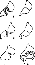
Figure 31. Operations for bilocular stomach: 1-transverse resection; 2-wedge resection; 3-gastro-gastroanastomosis; 4-double gastroenterostomy.
the shock is not great; otherwise, stagnation may occur in it; therefore, it is necessary to create an anastomosis on the pyloric sac as well, i.e., to perform a double gastroenterostomy (fig. 31). One must completely reject plastic surgery of a narrowed Stomach according to the pyloroplasty type. In the presence of a trilocular Stomach, the relationships are of course even more complicated, and a way out in each individual case can be found in various combinations of the techniques just mentioned. Phlegmonous inflammation of the stomach develops either as an independent disease or complicates general infections - typhoid, smallpox, pyemia, thus being a metastatic process; it more often develops after burns and injuries to the Stomach, including operative injuries - gastroenterostomy, dilation of the pylorus; finally, it complicates other stomach diseases, such as stomach cancer, ulcer, and even gastritis, especially in alcoholics. Usually, phlegmonous inflammation takes a diffuse character, and more rarely the case ends with the formation of an delimited abscess, which opens into the cavity of the Stomach, into neighboring organs, or outward. The disease begins and proceeds with severe general symptoms: high temperature, chills, general prostration, frequent, small pulse, shallow breathing, severe thirst, and general restlessness; from local symptoms, pain and swelling in the epigastric region, tenderness on palpation, vomiting, meteorism are usually observed; to these initial symptoms are added signs of local and then general peritonitis, or from the very beginning the picture of the disease proceeds as acute general peritonitis. The disease always has a very poor prognosis, and only in exceptional cases does it end with spontaneous emptying of the abscess into the Stomach or by other means. In a few cases, it has been possible to save patients by timely operation - resection of the Stomach or delimitation of the Stomach by tampons introduced into the abdominal cavity. Bleeding. When indications for surgical intervention for bleeding from stomach ulcers are considered, two forms should be distinguished: 1) profuse bleeding, usually in the form of abundant bloody vomiting, and 2) recurrent, chronically proceeding, not abundant, but leading to general anemia and general exhaustion from blood loss. In acute cases, the source of bleeding is arterial branches of the wall or one of the main arterial trunks of the stomach (a. coronar., a. gastro-duodenal., a. splenica). In chronic, recurrent cases, bleeding may indeed, in exceptional cases, have its source in the veins of the Stomach. It should also be noted that sometimes the source of bleeding is not even established at autopsy, despite the most careful searches. Surgical intervention for profuse bleeding does not, generally speaking, have many supporters, and most modern surgeons prefer in such cases a wait-and-see attitude and conservative treatment. The reason for this attitude is that in the vast majority of cases, abundant bleeding is single and not prone to repetition, at least in the nearest days. Haberer, one of the most active and radical surgeons in the field of the Stomach, who obtained very good results from surgical intervention in acute bleeding on his own material, nevertheless stated that of those cases in which the operation was rejected because of its apparent hopelessness, not a single patient died from bleeding. The second reason for the restrained attitude toward such bleeding is the danger of the intervention itself in weakened, bloodless patients; moreover, the operation itself appears rather uncertain: what exactly should be done? Practically, two cases may arise: a callous ulcer, easily opened during operation, or a 'soft' ulcer, not found on external examination of the Stomach. In the first case, to stop bleeding directly at its source, three possibilities present themselves: to excise the ulcer itself, to ligate the supplying vessels, as recommended by Witzel, or to open the Stomach and inject the bleeding vessel from the inside at the bottom of the ulcer. Resection in severely anemic patients represents a very serious intervention, injecting the supplying vessels from the outside is also not an easy task and moreover a less reliable method. Stopping bleeding by injection may fail completely due to the sutures being cut through; cauterization with a pacelon may also not achieve the goal. In the second case, i.e., with ulcers not immediately found, it is necessary to perform a wide gastrotomy to find the ulcer. With such ulcers, it is of course easier to cope with bleeding by injection from the inside, cauterization, and sequential suturing of the ulcer, but to find the ulcer from the stomach cavity is also not an easy matter; moreover, ulcers may be multiple. All these circumstances explain the very high mortality rate, reaching 25-37-65 (Hartmann, Mayo-Robson, Deaver), and the restrained attitude of modern surgery toward activity in acute abundant bleeding. It should also be noted that the widespread introduction into the clinic of blood transfusion as a hemostatic method and of hypertonic solutions is a new argument in favor of a wait-and-see attitude.-As for palliative operations for bleeding from ulcers, gastroenterostomy should be placed first. Its role in this respect is that the Stomach is unloaded from distension by blood, its cavity collapses and thus conditions are created for spontaneous cessation of bleeding, but the main thing is that reflexively the movements of the Stomach are calmed down - irritation of the small intestine inhibits the motor activity of the Stomach. However, gastroenterostomy does not always, contrary to expectations, lead to the goal - according to Kenue's statistics, in 25% of patients died from continued bleeding.-The second group of cases - chronic recurrent bleeding not responsive to therapeutic measures - unquestionably requires surgical treatment, which consists in excising the ulcer, resection of the stomach, or gastroenterostomy; the latter has chances of success with parapyloric ulcers, with duodenal ulcers, and with accompanying pylorospasm. Perforations. In ulcers that have perforated the Stomach, surgical intervention has an absolute indication and moreover should be done as early as possible. Cases ending sometimes in spontaneous recovery constitute such an insignificant exception that they should not be taken into account. The question practically reduces only to timely diagnosis and as rapid as possible organization of help. In most cases, perforation occurs suddenly and unexpectedly for the patient and physician, sometimes at the height of exacerbation of ulcer pains. The main and first symptom of perforation is severe pain in the epigastric region, accompanied by signs of shock. Very quickly follows then tension of the abdominal walls and local tenderness on palpation of the boat-like drawn-in abdomen. Vomiting is not an obligatory sign at the beginning. One should always remember that sometimes very quickly severe pain appears in the right iliac fossa, which is explained by the rapid descent of stomach contents downward along the lateral, right canal. This sign is practically important because on its basis a false diagnosis of perforating appendicitis is often made and the operation is begun not in the proper place. Following the initial, already very characteristic signs of perforation, the signs of general peritonitis increase. In the presence of old adhesions, the process may become delimited and lead to the formation of a local abscess. The operation, as already said above, must be performed as early as possible, and its outcome is determined precisely by this circumstance. General anesthesia is almost always necessary. The incision must be sufficiently large to allow freedom in examining and suturing the site of perforation. It is usually made along the median line upward to the navel, but if necessary it must be extended to the right or left by cutting through the rectus muscle. Finding the site of perforation usually does not present great difficulty if the perforation is on the anterior wall or on one or the other curvature of the Stomach; it is more difficult to orient oneself if the perforation occurred on the posterior wall toward the omental bursa. After finding the site of perforation and toilet of the nearest surroundings, they proceed to close the perforation by applying sutures; the difficulty of this moment lies in the fact that the edges of the ulcer are infiltrated and the sutures are easily cut through, therefore one has to take the nearest non-infiltrated areas of the walls into the suture. With extensive callous ulcers, direct suturing may not succeed, then it remains to apply plastic surgery with an omental flap on a pedicle. The impossibility of applying a firm suture to the infiltrated edges of the ulcer forces many surgeons to resort to some other techniques: freshening the edges of the ulcer, up to its complete excision (Finsterer), or conversely - refusal to suture the perforation, introduction of a drain into the perforation hole, fixation of the site of perforation to the abdominal wall. In extreme cases, the introduction of delimiting tampons (Petrov) is used.
In cases where the ulcer occupied the pyloric part of the Stomach or when suturing resulted in narrowing of the outlet, gastroenterostomy is performed as an additional operation; some authors consider gastroenterostomy never superfluous, while others never resort to it. Finally, some surgeons in operations at early stages resort to resection. After all manipulations with closure of the perforation site, the abdominal cavity is cleansed. If perforation occurred with an empty Stomach and there are no food masses in the abdominal cavity, it is simplest to limit oneself to wiping with moist gauze sponges and tightly suture the abdominal cavity. In the presence of large amounts of food masses, either the same method is used or the abdominal cavity is irrigated with large quantities of hot saline solution. If operating already in the stage of purulent peritonitis, drainage of the abdominal cavity is necessary both through the operative wound and through additional incisions above the pubis, in the iliac and lumbar regions. The outcomes of surgical intervention depend, as already indicated, on the time; mortality according to large statistics averages 16-30%, but if we take cases of early intervention, operations in the first hours give 0-4% mortality, after 10 hours-25%, after a day the mortality rate increases to 75%; later recovery is noted only in individual cases. Syphilis and tuberculosis. Surgical intervention in stomach syphilis is indicated in cases that have resulted in narrowing of the pylorus or deformation of the stomach of a persistent nature, with subsequent violation of motor activity; in such cases there is no significant difference between similar cases of other etiology. In open stomach ulcers of syphilitic origin, specific therapy is indicated if the diagnosis is established at least only presumptively; surgical intervention here may also prove inevitable if severe complications occur in the form of bleeding and subsequent cachexia or if fistulas or perforations form. In some cases, a palpable tumor in the epigastrium was mistaken for cancer of the pylorus and was removed by resection, while the diagnosis was established only by microscopic examination. Surgical treatment for tuberculous stomach ulcers in the form of radical excision is possible only in rare cases due to the extensive nature of adhesions or multiple involvement of the glands or dissemination in the peritoneum. In Bobrov's case, due to an extensive tuberculous ulcer, it was necessary to extirpate 3/4 of the Stomach with recovery. More often in tbc of the Stomach, palliative operations are performed in the form of gastroenterostomy in the presence of pyloric narrowing or jejunostomy if it is necessary to completely exclude the Stomach from the act of digestion. Furthermore, in cases of other complications in the form of formation of internal fistulas or external fistulas, various palliative operative measures are undertaken with the aim of excluding these fistulas. Finally, in any other complications of tuberculous ulcers, the operation is indicated for the same reasons as in simple stomach ulcers. Operations on the stomach. Stomach resection. By stomach resection is meant the excision of a larger or smaller portion of the stomach walls. Three types of resections are distinguished by shape: wedge-shaped, segmental, and circular resection. The volume of resection in individual cases varies widely, up to removal of the entire Stomach (total resection, extirpation of the Stomach); along with these terms, other equally common terms are used-gastrectomy' partial or total. The prevailing methods of circular resection remain two methods proposed by Billroth (see Billroth's operations on the stomach). The main modifications introduced by other surgeons are as follows: Kocher in 1890 modified Billroth's first method in that he sutured the end of the duodenum not into the incision in the Stomach, but into its posterior wall, retreating 1-2 cm from the tightly sutured end (figure 32). The purpose of this modification is to avoid the junction of 3kocher?yKT0МИЯ two lines of sutures as the most dangerous place in terms of rupture. In order to reduce tension in the sutures, Kocher preliminarily mobilizes the duodenum by making an arcuate incision at its upper angle. Hacker and Mikulich introduced into Billroth's second method a modification consisting in that instead of typical gastroenterostomy, they sutured the lower end of the Stomach into a loop of the jejunum, with the upper half of the gastric incision being preliminarily sutured tightly. Polya (1911) and Balfour (1912) modified this last modification (fig. 33), suturing the entire length of the gastric incision into the jejunum. The purpose of this last proposal: 1) to avoid narrowing of the anastomosis, 2) to avoid the development of perverted movement of food masses, 3) to shorten the duration of the operation by eliminating the occlusive suture of the Stomach. All these advantages are actually illus


Figure 33. Resection according to Polya-Balfour.
zeks,

£3<
zorny, and none of the modifications of Billroth's method finds a large circle of followers. Riedel's method-circular resection of the middle part of the Stomach and suturing the ends together-is in essence no different from Billroth's method. Saddle-shaped resection according to Schmiden's method (fig. 34) is a combination of Billroth's first method or Riedel's method with excision of the lesser curvature of the stomach in order to remove the entire length of Waldeyer's pathway as the area of predominant development of stomach ulcers. - Partial resections of the Stomach walls, not counting atypical operations sometimes used in the removal of benign stomach tumors, consist in excision of a part of the lesser curvature in the form of a wedge or in the form of a segment, depending on the shape and extent of the process (fig. 35). In the first case, the defect is sutured in the transverse direction, in the second-in the longitudinal direction. In these forms, re-proposed by Mikulich and Kaiser, after suturing the Stomach is somewhat deformed, taking the form of a sac or a long narrow elongated canal. But in severe form these deformations occur only after more extensive resections; in such cases the operation should be supplemented by the creation of a gastroenteroanastomosis to prevent stagnation in the stomach, since in addition to deformation the function of the antral part of the stomach suffers due to the cutting of the vagus nerve. Gastrotomy was performed three centuries ago for the removal of swallowed foreign bodies (Schrwabe; 1635). At present, gastrotomy is performed either for the removal of foreign bodies from the stomach, or as a preliminary act of other operations on the Stomach (dilation of the cardia, retrograde bougienage of the esophagus), or to stop bleeding from ulcers, or simply for examination of the Stomach from within. Depending on the purposes, the size of the opening in the Stomach is made from 1-2 cm to a size that allows four fingers or even the entire hand to be introduced into its cavity. The incision is made as close as possible to the place where further manipulations are planned, and in order to avoid excessive bleeding, it is conducted either in the direction of the vessels, from the greater curvature to the lesser, or along the anterior wall of the Stomach, along the axis of the Stomach, at an equal distance from both curvatures. Before opening the Stomach, the abdominal cavity must be well Figure 34. Saddle-shaped resection according to Schmiden's.


Figure 35. Segmental and wedge-shaped resections of the lesser curvature.
protected by gauze sponges, and to hold the Stomach, ligatures are passed through its wall or various clamps are applied. When suturing the incision in the Stomach, attention should be paid to the thorough stopping of bleeding from the mucous membrane. The incision itself is sutured with a two-layer suture.-The gastrotomy method has again been widely used in recent years for dilation of the pylorus in cardiospasm. The operation in this form was proposed in 1900 by Mikulich and consists in that after wide opening of the Stomach, four fingers or even the entire hand are introduced into it, the cardia is located and gradually dilated, first introducing one finger, then two, three, and even four; thus the diameter of the cardia can be dilated to 10-12 cm. The results of such an operation are usually brilliant and persistent, but of course the possibility of recurrence is not excluded. Of the 60 operations described in the literature, fatal outcomes were observed only in individual cases. Pyloroplasty. Plastic dilation of the pylorus in its cicatricial narrowing was proposed by Heinicke-Mikulich in 1887. The operation consists in that through the narrowed pylorus, a longitudinal incision 5-6 cm long is made through the entire thickness of the wall, the edges of the incision are retracted to the sides with hooks and the wound is sutured in the transverse direction, with the first suture being applied . L-ч in the middle as a guiding ^JLr ) (fig. 36). Practice has shown ma ??г\_^/ little applicability of this operation v>»
in the case of scar strictures, and in any case, it is permissible to resort to it only on the condition of mobility of the pylorus and the absence of extensive scars; otherwise the operation is either not feasible, leads to relapses, or from the very beginning proves to be useless. If the indication for it is not scar narrowing, but pylorospasm or congenital narrowing of the pylorus (spasm and hypertrophy of the pylorus), then technically the operation becomes easily feasible, but here it competes with another of its modifications - extramucosal pyloroplasty: the incision is made in the same way as in the first, i.e., longitudinal, 2 cm long; the pyloric muscle is mobilized and cut transversely or a piece 1 cm long is excised from it, after which the incision is sutured in the transverse direction (Weber, Payr). It should be noted that during this operation the gastric mucosa can easily be opened, but this is of no great importance. As an operation standing on the border of pyloroplasty and gastroduodenostomy, the Finney method must be mentioned. Operations on the cardiac part of the Stomach. Some of them are of a plastic nature and aim to correct mechanical obstacles on the basis of spasm, scars from burns, etc. Others are undertaken for the removal of neoplasms and therefore must be radical and are reduced to resection. In addition to the Mikulich operation (digital dilatation of the cardia) described above for cardiospasms, other methods serve the same purpose: cardiotomy, cardioplasty, esophago-gastrostomy, esophago-jejunostomy. Transverse cardiotomy according to Marwedel with sequential suture of the incision in the transverse direction appears to be a difficult operation due to the poor accessibility of the operative field. Out of 14 cases of operations by this method, 1 ended in death, in 2 a relapse occurred, in 9 there was recovery. The Heller operation is simpler and safer - extramucosal cardiotomy on the anterior and posterior wall with subsequent wrapping with omentum. Out of 49 cases, 28 resulted in cure, 10 in improvement. Esophago-gastrostomy according to Heyrovsky between the bottom of the Stomach and the esophagus is possible if the lower end of the esophagus is dilated and descends into the abdominal cavity, sometimes in the form of a sac. In the case of scar narrowing of the cardia with scar wrinkling of the entire stomach after a burn, Hesse made an anastomosis between the mobilized abdominal part of the esophagus and the preserved part of the bottom of the stomach on one side and the jejunum on the other. The results of the operations were excellent. In the literature, 15 similar cases are described with 12 good outcomes. - Resection of the cardiac part of the stomach by the abdominal route is one of the difficult operations. Indications for it are cancers of the cardia; the first attempt to excise such a tumor belongs to Mikulich, but the case ended in failure. The operation is possible and makes sense only in the case when the tumor does not extend to the diaphragm and adjacent organs, therefore the question of resectability can finally be decided only after laparotomy. The operation requires wide access, which is provided either by Marwedel's incision or by a midline incision with an additional transverse incision to the left. The excision should begin with the mobilization and elongation of the abdominal part of the esophagus, for which the peritoneum is incised, both vagus nerves are cut, and the esophagus is freed from the cellular tissue with a blunt method, with a finger, special attention must be paid to not opening the pleura. Then the upper part of the Stomach is mobilized together with the tumor and the left gastric artery is ligated. Clairmont recommends starting the entire operation precisely with the ligation of this artery, after which the further manipulations are facilitated. The freed and elongated esophagus is fixed with sutures to the edges of the esophageal hiatus. After the tumor is removed, the connection of the end of the esophagus with the Stomach is performed by various methods: the end of the esophagus can be sutured end-to-end, but it is more advantageous to suture the Stomach tightly and suture the esophagus into a new opening made in the stomach. Bircher immersed the end of the esophagus into the incision in the Stomach and sutured it above. Gerhammer suggests suturing the end of the esophagus with a seromuscular cuff from the walls of the Stomach. The main dangers associated with this operation consist in the possibility of divergence of the sutures due to tension or due to marginal necrosis of the end of the esophagus, finally due to infection of the peritoneum and especially of the pleura and mediastinal cellular tissue. To date, 33 cases of abdominal resection of the cardia have been described (Borchers) with 9 recoveries and 70% mortality. In conclusion, it is necessary to mention repeated operations on the Stomach after gastroenterostomies and after resections undertaken for so-called peptic ulcers, requiring excisions, anastomoses, and resections of the Stomach and intestines - small and large.
V. Dobrotvorsky. IX. Stomach Neuroses. By neuroses of the St. should be understood those pathological nervous reactions that arise in it as manifestations of the general neurasthenic constitution and are caused by psychic affects. From a symptomatic point of view, the neuroses of the St. include on the one hand such diseases of it, when in the presence of a number of subjective disorders, with the most thorough examination, it is impossible to discover any objective signs of organic disease of the stomach, and on the other hand, those in which the intensity of subjective sensations sharply does not correspond to objectively ascertainable changes. The main contingent of patients with neuroses of the St. are constitutional psychoneurotics; but neuroses of the St. can also arise on the basis of organic diseases of the St., with a certain predisposition of the nervous system. -The mechanism of the occurrence of neuroses of the St. is as follows. A psychic trauma caused a disturbance of psychic balance-affect. The central nervous system seeks to "unload itself", restore the lost balance-"react to the affect". This reaction goes along the autonomic nervous system and due to this or other secondary reasons can go precisely in the direction of the St. and cause in it this or that pathological reaction. The main condition for the occurrence of a neurosis of the St. is a psychically and vegetatively stigmatized personality. It has a number of peculiarities, a number of deviations in the psychic and emotional structure, namely: overestimation of oneself with insufficient faith in one's own strength, weakness of will and easy distractibility, irritability and hence an unjust attitude toward others, and as a consequence, self-absorption, hypochondria-fear of possible disturbances from one's own organs. Such a personality easily enters into conflict with the surrounding world and cannot withstand the demands made on it by the latter. Hence arises a psychic trauma, i.e., a disturbance of equilibrium in the central nervous system. Such persons are generally characterized by instability of equilibrium in the sphere of the autonomic nervous system (G. Bergmann). If at the same time its department, which manages the functions of the digestive apparatus, is distinguished by increased excitability, then the autonomic-nerve pathways going from the central nervous system to the St. offer less resistance to impulses coming from the central nervous system, and in particular to the "reaction" of affects arising in it. A secondary cause of the development of stomach neurosis can also be the inadequacy of the St. itself, the threshold of irritation of which is significantly lower than that of other organs. Thus, neuroses of the St. can arise if in a psychasthenic the affect of displeasure repeatedly coincides in time with some accidental stomach disorder: the resulting engram sensitizes the nervous system of the St. so much that the repetition of the affect of displeasure will cause the same picture of stomach suffering (according to the type of formation of conditioned reflexes of Pavlov). Neuroses of the St. also arise in such a way that in a nervously stigmatized person an acute stomach disease appears, after which there remains a fear of the recurrence of the disease, and therefore attention is fixed on the St. This is sufficient for a picture of stomach neurosis to develop. In addition, with increased excitability of the autonomic nervous system and in particular its stomach department, neuroses of the St. easily arise due to reflex transmission of irritation from other organs, especially from organs of the abdominal cavity when the latter are affected by this or that pathological process (diseases of the liver, intestines, uterus, peritoneum, etc.). The so-called stomach dyspepsia is classified among neuroses of the St. (see below). Other neuroses of the St., namely-sensory neuroses: bulimia, anorexia, gastralgia, pyrosis nervosa, gastralgokenosis (see); motor neuroses: nervous eructation, nervous rumination, peristaltic restlessness of the stomach, nervous vomiting (see below)-are distinguished from a purely symptomatic point of view by this or other outstanding manifestations. Stomach dyspepsia is predominantly a polysymptomatic disease, while other stomach neuroses are monosymptomatic. Neuroses of the St. undoubtedly also include many of those diseases that are considered violations of the secretory and motor functions of the St.: independent (primary) forms of hypersecretion, hypo- and achylia, pylorospasms, atony, etc. However, in view of the fact that with these forms a picture of the disease develops that is characteristic of a certain form of violation of one or another function of the stomach, they are singled out in a special section-"violations of the secretory and motor function of the St.", where they are described in parallel with violations of the same functions of the stomach that develop on the basis of organic diseases and manifest themselves with the same seemingly independent picture of the disease. Nervous stomach dyspepsia. By stomach dyspepsia most authors understand those forms of stomach neuroses in which a number of subjective disorders from the stomach are observed in the absence of signs of this or that more persistent violation of the function of the secretory and motor apparatus of the stomach. In fact, there is a fundamental difference between the concepts of intestinal and stomach dyspepsia: while intestinal dyspepsia is defined as a violation of the processes of digestion in the intestine, for example, violation of the processes of fermentation or putrefaction in the absence of not only organic changes of the intestine itself, but also of its neurosis, under stomach dyspepsia, as indicated, most authors at present understand only a special and moreover the most important group of neuroses of the St. Many do not consider all cases of stomach dyspepsia as neuroses. Thus, Cohnheim divides stomach dyspepsias into different types, among which nervous stomach dyspepsia is considered only one type, and in addition to it, dyspepsia in anemic subjects, in those with gastoptosis (enteroptotics), and in those suffering from tbc is also distinguished. Most authors, however, recognize that in fact, even with these latter types of stomach dyspepsia, the basis lies in violations of the function of the nervous system, with the only difference that in addition to the general neurasthenic constitution there is also general asthenia with this or other or already developed or developing on this basis suffering: enteroptosis, tbc of the lungs, etc., which in turn further weaken, resp. irritate the nervous system and thereby contribute to the development of stomach neurosis. Dreyfuss, who considers all stomach dyspepsias as psychogenic diseases, divides nervous stomach dyspepsia into purely psychopathic, congenital hysterical, resp. neurasthenic forms, and into psychogenic and cyclothymic-circular forms. In any case, the most accepted opinion is that which considers all dyspepsias as arising on a nervous basis. History of the doctrine of stomach dyspepsia. The doctrine of nervous stomach dyspepsia in the last 50 years has been most closely connected with the names of Leube, Ewald, Stiller, Glenard, Strümpell, and Dejerine. In 1879 Leube for the first time described a strictly delimited symptom complex, which he named nervous dyspepsia, considering it a pure sensory neurosis of the St., assuming that the cause and essence of this disease is the increased irritability of the peripheral nerves of the St., to which the entire nervous system reacts secondarily. Ewald held diametrically opposite views, considering that dyspepsia in general is not a clinically delimited disease, but only a symptom complex that is a partial manifestation (and often the only manifestation) of general neurasthenia or hysteria. At the same time, he expressed the assumption that in dyspepsia, a violation of the secretory and motor work of the St. and intestines is also often observed without organic disease of the St. The thought of clinicians, however, was directed to the search for organic bases for nervous dyspepsia. In this respect, the views of Glenard (1885) played a major role, who in a number of cases of nervous dyspepsia found anatomical changes not in the St., but in the position of the viscera of the abdominal cavity, and therefore considered responsible for the occurrence of this suffering general enteroptosis. According to his observations, all subjective and objective disorders of these patients disappeared after eliminating the indicated anomalies in the position of the abdominal viscera by means of this or other therapeutic measures. The neurasthenia and neurasthenic symptoms observed in such patients, Glenard considered a consequence of their dyspeptic disorders. In 1907 Stiller drew attention to the constitutional factor in the pathogenesis of stomach dyspepsia. He believed that nervous dyspepsia is only a partial manifestation of congenital general asthenia (asthenia universalis congenita), and not a morbus sui generis or a symptom of general neurasthenia, and that dyspepsia represents a phenomenon coordinated with the latter and is based on a congenital constitutional predisposition.
Painful sensations in neuro-dyspeptic patients, according to Stiller, are based on nervous disorders, which depend partially on hyperesthesia of the abdominal sympathetic nerve (resp. solar plexus), partially on hyperesthesia of central origin, as well as on the general psychological attitude of the patient. This psychological moment in the pathogenesis of nervous dyspepsia is particularly emphasized by Strümpel, who even introduces a new designation for so-called psychogenic dyspepsias, believing that the overwhelming majority of nervous dyspepsias depend on primary changes in the emotional life of the patient, among which he places fear in the first place. Strümpel's doctrine of the primary role of psychological factors in the pathogenesis of gastric dyspepsia is further developed by Dubois and Déjerine, who give such patients a corresponding name: pseudogastrapathies. In more recent times, the French school, without denying the role of the psychogenic factor, returns to the opinion that the cause of dyspepsias is hyperesthesia of the solar plexus. Finally, in the most recent times, Strauss and Kraus attach particular importance in the etiology and pathogenesis of dyspepsias to constitutional inadequacy, heredity, and disruption of endocrine balance. Etiology. Gastric dyspepsias can be of purely psychogenic origin as a result of so-called reacting of psychological affects in the direction of the autonomic nervous system; therefore they are observed in psychoneuroses; but they can also be of reflex origin (in the Pavlovian sense) in diseases of the most diverse organs of the abdominal and even non-abdominal region. The characteristic disorders of gastric dyspepsia have as their cause increased irritability, apparently predominantly of the sympathetic plexus of the Stomach, and generally a disturbance of equilibrium in the autonomic nervous system. In the presence of such conditions, even the slightest irritants, e.g., normal contractions of the Stomach, are sufficient to cause unpleasant subjective sensations. Along with this increased irritability of the gastric nerves, the increased irritability of the central nervous system also plays a major role. This is why nervous dyspepsias are often encountered as a partial phenomenon in general neurasthenia, resp. hysteria, although one cannot deny that sometimes dyspepsia occurs as an independent neurosis (monosymptomatic hysteria-Levin). Often the cause of the disease can be psychological excitations, grief, worry, fear, etc. Fear of infection, fear of getting cancer, unhappy love, grief, family and business worries and anxieties, and generally speaking, all psychological traumas are quite often an etiological factor in gastric dyspepsia. Violation of normal sexual life, excesses in Baccho et in Venere, masturbation, coitus interruptus, etc., can also serve as a cause of the occurrence of gastric dyspepsia; but also physical trauma with subsequent development of traumatic neurosis can play a certain role. In a large number of cases, gastric dyspepsia, as already stated, arises reflexively, and the excitation can come from the most diverse, even very distant organs. Thus, in diseases of the liver, intestines, dyspeptic phenomena can appear from the side of the Stomach. Often in persistent constipation, in worms (K. Faber), gastric dyspepsia can arise. It is often observed in diseases of the female genital sphere (Kehrer) and even in physiological changes in the genital sphere, during menstruation and especially during pregnancy, in the form of disturbance of appetite, paresthesia of appetite (Stiller), disturbance of motor function of the Stomach, vomiting, etc. For these disorders, a special name has even been proposed-dyspepsia uterina (Kisch). Similar changes can be observed in men in prostatitis, prostatorea, spermatorrhea, phosphaturia, sexual neurasthenia. All these disorders cannot of course be considered the main causes of the occurrence of neuro-dyspeptic phenomena from the side of the Stomach. For the most part they are coordinated, and a primum movens for the occurrence of the typical phenomena of the disease in question is the general neuropathic constitution (status enteroptoticus, asthenia universalis). One cannot further deny that the diseases already mentioned above, to which French and partly German authors point (syphilis, tbc, anemia, reduced nutrition, etc.), quite often play a certain role in the etiology of neuro-dyspeptic disorders, creating a predisposition to the occurrence of dyspepsia. The mechanism of development of gastric dyspepsia in the mentioned diseases may consist in the fact that in constitutional enteroptosis, the vertically located Stomach, in emaciation, descends even further, which entails a series of nervous disorders. In turn, subjective disorders in nervous dyspepsia often lead to reduced nutrition due to lack of appetite or fear of eating, and this causes even greater emaciation. Thus, a vicious circle is often created, which is not always easy to break. Symptomatology. Among the subjective signs of gastric dyspepsia, the symptoms of general neurasthenia play the main role. The latter gives the patients' complaints a characteristic coloration. Sometimes local symptoms from the Stomach play a dominant role, sometimes general nervous phenomena prevail. But in both cases, subjective symptoms are characterized by three moments. First, the complete irregularity and capriciousness of the onset of these symptoms and the variability of their intensity. Almost always in these patients there are intervals during which they do not experience any disorders at all; but even in periods of poor health, the intensity and character of the symptoms change. The second moment is the dependence of subjective disorders on mental disturbances, excitations, depressions, dissatisfaction, which either themselves cause subjective disorders or worsen existing ones. Finally, the third moment is the absence of any other factors that could cause a disturbance in the sensory equilibrium of the Stomach. Thus, the intake of food, its quality and quantity, bed rest, warm procedures, etc., have no significance in the appearance or change in intensity of neuro-dyspeptic disorders. Often heavy food is well tolerated, while there are periods of psychological excitement when even light food causes severe pain.-As for the character of these subjective disorders, their picture is so diverse that it is difficult to fit it into definite frameworks. Among the general nervous disorders, on the one hand general irritability and hypochondriac-depressive state stand out, and on the other-hand quick fatigability, disinclination to work, tendency to pessimistic ideas about the curability of the disease, heaviness in the head, constant or in attacks; often headache is observed, occurring soon after food intake, and sometimes on an empty Stomach, polyuria and pollakiuria, pains in the back, especially between the shoulder blades, dizziness and palpitations, which are associated with constipation or generally insufficient excretion of feces. Often insomnia or restless sleep disturb these patients. The local phenomena from the Stomach are equally diverse. These include salivation, belching, pains, nausea, vomiting, attacks of shortness of breath, sometimes even asthmatic attacks after food intake. These symptoms are observed either separately or combine in a motley picture of the disease, replacing one another or occurring simultaneously. It is especially necessary to note heartburn in dyspeptics in contrast to the burning gastric pains in hypersecretions and gastritis. Heartburn in gastric dyspepsia is completely independent of food intake. It is often observed on an empty stomach (Gastralgokenose Boas's). It does not pass either from the horizontal position of the patients or from therapeutic procedures in the form of heat or hot fomentations. At the same time, psychological irritation (even the minimal) can undoubtedly to a significant degree worsen the painful sensations in the area of the Stomach. Often complete loss of appetite is observed, sometimes reaching complete aversion to food; however, it should be noted that sometimes there is no real absence of appetite, but sitophobia is observed, i.e., fear of eating due to disorders that may arise after food intake. The function of the intestine is also extremely changeable. Periods of constipation can be replaced by periods of normal stool or periods of diarrhea. On objective examination, the general habitus asthenicus, s. enteroptoticus and a series of general nervous phenomena are most often noted-decrease or increase in skin reflexes and reflexes from mucous membranes, often a sharp increase in knee reflexes and reflexes from the Achilles tendon, dilation of the pupils and vasomotor disorders in various areas of the body, mild excitability, trembling of hands and tongue, phenomena of dysfunction in the area of the autonomic nervous system. Nutrition is often impaired, as patients, due to fear of eating, try to limit food intake, and due to reduced nutrition they become even more nervous and irritable, which in turn leads to worsening of subjective disorders. Reduced nutrition also leads to insomnia (Mathieu, Roux).
When examining the abdomen, patients with gastric dyspepsia are often found to have flabby abdominal walls with reduced fat content, low position of all organs, and a lowered lower border of the Stomach. The splashing sound in the Stomach is extremely easy to produce, which indicates hypotonia of the stomach walls. Painful points on pressure are frequently observed; hyperesthesia of the solar plexus can often be found. The sensitivity in this case can be so great that it creates difficulty in the differential diagnosis between nervous dyspepsia and ulcer of the Stomach. In addition to hyperesthesia of the specified area, painful points corresponding to the upper and lower mesenteric plexuses are often observed. Sometimes the entire white line from the navel to the xiphoid process and even McBurney's point becomes sensitive, which can lead to confusion with appendicitis; sometimes increased sensitivity on pressure on the spinous processes of individual vertebrae is noted.- As for the function of the Stomach, according to Boas, three types of gastric dyspepsia can be observed. In the first type, both motor and secretory functions are completely normal; in the second type, there is a disturbance of one or the other functions, continuing for a more or less long period; and finally the third type, when sharp fluctuations are observed in both functions. Sharp fluctuations are particularly characteristic for the secretory function, when after several days of hypochilia, hypersecretion can be observed and vice versa, i.e., there is so-called heterochylia (Hemmeter). The fluctuations observed in the third type can be capricious, sometimes even contradictory. Thus, sometimes atony is observed simultaneously with increased acidity or decreased acidity simultaneously with increased motor activity. X-ray examination sometimes gives an equally capricious picture: sometimes there is gastoptosis and atony, sometimes strengthening of peristalsis and peristalsis, and finally normal relations. In any case, a decrease in motor activity in the sense of longer retention of food in the Stomach in this disease has not been established. In the large intestine, spastic and atonic states are observed. Diagnosis. To establish the correct diagnosis, one must first take into account the three characteristic features mentioned above; secondly, the complete ineffectiveness of a strict diet and other usual therapeutic measures in terms of their effect on subjective disorders; finally, the discrepancy between subjective disorders and objective examination in the sense that the severity of subjective sensations does not correspond at all to the insignificance of objective symptoms found in the examination of abdominal organs. In differential diagnosis, it is necessary to distinguish nervous dyspepsia from chronic gastritis, stomach ulcer, gallstone disease, and hernia of the white line. The differential diagnosis must be made with extreme caution so that in the presence of even general neuropathy, some organic disease is not overlooked, which can extremely resemble gastric dyspepsia in its picture, since even in cancer of the Stomach, nervous dyspeptic symptoms can be observed, and according to modern views, ulcers of the Stomach are observed particularly often in people with an unbalanced nervous system. Finally, in making a diagnosis, in addition to a detailed examination of the gastrointestinal tract, the general condition of the nervous system must be investigated, and what is no less important, the condition of various organs that can serve, as mentioned above, as a starting point for the development of the neuro-reflex form of dyspepsia. In any case, in differential diagnosis, all organic diseases of the Stomach must first be excluded, and only in their absence can the diagnosis of gastric dyspepsia be made. -The prognosis of nervous dyspepsia is rather doubtful; as for recovery, a long disappearance of nervous dyspeptic symptoms can be expected only where the exclusive cause of them is a primary disease in some other organ. Elimination of the latter can lead to the disappearance of dyspeptic phenomena from the Stomach. But such cases are relatively rare. As a rule, recovery from nervous dyspepsia is only temporary. However, with the help of appropriate therapeutic measures, it is possible to make all subjective disorders disappear for a more or less long time. Therapy. Since gastric dyspepsia most often arises on the basis of general instability of the nervous system, which is already manifested in childhood, prevention is of great importance in treatment, therefore in such cases from the earliest childhood, all measures should be taken to eliminate or reduce the predisposition to general neurosis. Proper nutrition, hardening, strengthening of muscles, elimination of all harmful factors that can lead to general neurosis, regulation of sexual and mental life - these are the main points in preventing this disease. As for the treatment of nervous dyspepsia itself, here, due to the extraordinary diversity of the picture of this disease, individualization plays a greater role than anywhere else. First of all, it is necessary to clarify the basic suffering that serves as a starting point for the development of neuroses - disease of the liver, female sexual sphere, etc. - and direct the treatment in this direction. Then causal therapy also presupposes strengthening of the nervous system. Here, treatment with rest should be placed first. It is especially indicated where there is a disorder of nutrition and especially its decrease. Removal from the usual home environment to a sanatorium or to favorable climatic conditions, prohibition of professional work for a more or less long period, calm the nervous system and reduce dyspeptic disorders.--As for dietary treatment, here the first place is occupied by increased nutrition (Weir-Mitchell). Often with an increase in weight, the subjective disorders of patients also disappear. Heavily digestible dishes often lead to a faster disappearance of subjective disorders than easily digestible ones. A vegetarian diet is recommended, and simultaneously with it, restriction of protein and especially animal food. Light massage, electro-hydrotherapy, especially gentle hydrotherapeutic procedures not associated with sharp temperature changes, are extremely beneficial.--Drug therapy should be reduced to a minimum. For insomnia, hypnotics-veronal, trional, hedonal, luminal sometimes have to be used. For severe excitement of the nervous system, valerian and bromides are given. For nervous diarrhea, sometimes simultaneously with a change in diet, calcium (Calcium carbonicum, Calcium phosphoric) and sometimes bismuth have to be used. Narcotics (opium and morphine) are not recommended. Almost never does it become necessary to drink any mineral waters, except those used for anemia and containing iron (waters of Zheleznovodsk, Levico, Ronceno, etc.). M. Gubergritz. Gastralgia (gastralgia nervosa), a term used only in relation to those pains arising in the stomach and having a paroxysmal character, which are not caused by organic diseases of the Stomach. These are usually periodically or irregularly occurring paroxysms of severe pain in the epigastric region, independent of food intake, accompanied by nausea, and sometimes vomiting and often radiating to the hypochondria, chest, and back. These paroxysms last from several minutes to many hours and are most often observed in neurasthenics and hysterical individuals, appearing in them mostly under the influence of excitement, grief, etc. The diagnosis of gastralgia should be made with great caution, since with the development of diagnostic methods, a larger and larger part of so-called nervous gastralgias turns out to be pains caused either by organic diseases of the Stomach (ulcer, cancer, perigastritis) or diseases of neighboring organs (liver, kidney, and pancreatic stones, wandering kidney, angina pectoris and abdominal, hernias of the white line, etc.). Gastralgia is probably caused by spastic contraction of the Stomach, most often of the pylorus (pylorospasmus). Gastralgia can also be of reflex origin, with the source of the reflex being chronic appendicitis, inflammation of the peritoneum, and diseases of the sexual sphere in women (with irregular menstruation, diseases of the ovaries and tubes, with incorrect positions of the uterus) and in men (with hypertrophy of the prostate gland, spermatorrhea, etc.). Gastralgia can also occur in diseases of the central nervous system (tumors of the brain, myelitis, tabes dorsalis, etc.), in chronic poisoning with nicotine, etc. But it should be remembered that pylorospasm - this main source of stomach pains in general and of nervous gastralgia in particular - in the vast majority of cases is caused by organic diseases. The paroxysm of nervous gastralgia itself is not typical, and its neurotic nature is established only by excluding the organic diseases listed above on the one hand and establishing the presence of neuropathic constitutional factors on the other. Nervous pyrosis (pyrosis nervosa).
Under nervous heartburn, in contrast to pyrosis hydrochlorica, is understood a type of heartburn that is caused not by increased acidity of the stomach contents, but by other, not always clear factors (see Heartburn). Nervous rumination (ruminatio, merycismus) differs from simple regurgitation (regurgitatio) in that food that has entered the mouth from the esophagus is chewed again and swallowed. (For the pathogenesis and clinical picture of this phenomenon, see Rumination.) Special treatment of nervous rumination consists in making the patient by force of will suppress the act of regurgitation. It is very advisable to make such patients eat in the presence of others, so that they carefully watch themselves. One should insist that the patient eats slowly and chews food thoroughly. Based on the fact that liquids or a combination of solid and liquid food more often cause regurgitation, Boas recommends a 'dry diet,' which in a number of cases proved to be very advisable. Nervous belching (eructatio nervosa, aerophagia, morbus ructuosus). Nervous belching is the belching of air in the form of separate attacks, quickly following one another, not depending on food intake. At night, these attacks usually cease. Patients, without noticing it themselves, swallow a significant amount of air (aerophagia), which accumulates in the esophagus or in the Stomach and is then expelled outward (eructatio). In rarer cases, almost exclusively in hysterical women, belching is accompanied by loud convulsive screaming (eructatio tonans). It most often arises on the basis of hysteria under the influence of such affects as fear, grief, etc., but there is a significant contingent of patients in whom the initial factor of this functional disorder is other stomach diseases, accompanied by a feeling of tension in the area of the Stomach. This sensation in some such patients causes a desire to free themselves from it by means of peculiar movements of the pharynx and esophagus, which contribute to the gradual swallowing of atmospheric air. Having accumulated in a significant amount, it is then expelled outward by loud prolonged belching. At the same time, the patient gets instant relief and, having experienced this, then begins to often resort to this method, imagining that in this way they are freeing themselves from gases formed in the stomach. Aerophagia in cardiac patients deserves special attention, the condition of which, as is known, sharply worsens under the influence of the Stomach and intestines being filled with gas, resp. the high position of the diaphragm. Patients with aerophagia sometimes accumulate so much air in the Stomach that their condition takes on a threatening character. In such cases, the best remedy is the rapid introduction of a stomach tube, which immediately frees the patient from the distressing condition.-Special treatment of nervous belching consists first of all in the application of appropriate psychotherapy. It is necessary to explain in detail to the patient that their illness depends on their swallowing of air. Suggestion and hypnosis often also lead to good results here. Systematic upbringing of such patients also plays a big role. To help psychotherapy, it is recommended (Bouveret, j. Ch. Roux and Mathieu) to keep the mouth open for a long time, making such patients hold a cork or a wide cigar holder between their teeth. The results of this method are quite quickly apparent. Boas advises introducing a stomach tube daily to such patients for 5-7 days. All these methods are nothing other than 'instrumental psychotherapy.' Peristaltic restlessness of the Stomach (Kussmaul) (tormina ventriculi nervosa). The basis of this motor neurosis of the Stomach is hyperperistalsis of the Stomach ['segmental' peristalsis (see above - X-ray examination)]. In the vast majority of cases, hyperperistalsis is caused either by organic stenosis of the pylorus or by an ulcer of the duodenum. Only in a negligible number of cases are we dealing not with a reflex act, but with a pure motor neurosis of the Stomach on the basis of pathological function of the autonomic nervous system. Such conditions are most often observed in exhausted individuals with very flabby abdominal walls, mainly in women, and at the same time, along with the Stomach, the entire intestine participates in the peristaltic restlessness (tormina ventriculi et intestinorum). During such visible peristaltic agitations, rumbling and gurgling noises are heard at a distance throughout the abdomen, and patients complain of painful sensations and a feeling of tension in the abdomen. Only gradually, sometimes after many hours, does calmness set in. Only the most careful clinical and especially X-ray examination can save from erroneous diagnoses, especially in the sense of missing the recognition of stenosis in the area of the Stomach or intestine. As with all neuroses, treatment should first of all be directed at the general neurosis. The use of bromides and atropine proves to be very effective in such cases as symptomatic treatment. Nervous vomiting (vomitus nervosus). By this term, applying it to denote a neurosis of the Stomach, is meant not vomiting caused reflexively, and not vomiting caused by pathological conditions of the central nervous system, but vomiting of a purely psychogenic nature, arising under the influence of heavy experiences, excitement, grief, worry, mental fatigue, feeling of fear, disgust, etc. This type of vomiting is especially often encountered in young girls and women on the basis of hysteria and neurasthenia. Nervous vomiting is characterized by independence from the quality and quantity of food, the extraordinary ease of onset, frequent absence of preliminary symptoms (nausea, heaviness and pains in the area of the Stomach), absence of secretory and motor disorders from the stomach, and a sharp dependence on factors affecting the patient's mood.-The treatment of all the neuroses described here coincides with the treatment of nervous dyspepsia.
Vichunevii. X. Tuberculosis of the stomach. Frerichs found tuberculosis of the stomach in 2.4% of all autopsies of tuberculosis patients; Simmonds and Glaubitt - only in 0.4-0.76%, and in Simmonds' material in 92% of cases of stomach tuberculosis there was accompanying tuberculosis of the intestines. Consequently, the stomach is affected by tuberculosis much less frequently than the intestines. This is explained by a number of reasons causing the low susceptibility of the stomach to tuberculosis infection, namely: 1) the scarcity of lymphatic follicles in the stomach (lymphoglandulae), - the latter are found in somewhat larger numbers only in the pyloric part of the stomach, which is indeed the favorite site of localization of tuberculosis of the follicles; 2) the great resistance of the gastric mucosa to infections and 3) the layer of mucus covering the mucosa (Arloing); 4) the bactericidal influence of HCl (according to Weichselbaum's opinion; at present this influence is disputed). Due to all these reasons, tuberculous sputum usually passes through the stomach without infecting it. Nevertheless, contact infection of the stomach by sputum is apparently one of the frequent ways of infection with stomach tuberculosis. The question of whether swallowed sputum containing tubercle bacilli can infect a simple stomach ulcer remains open. Another route of infection of the stomach, the lymphogenous, occurs in tuberculosis of the peritoneum or lymphatic glands. The third route, the hematogenous, according to Arloing - is the only way of infection with stomach tuberculosis. Taking into account the importance which the hematogenous route has for the spread of tuberculosis in general, it is necessary to recognize this route as frequent; in miliary tuberculosis it is beyond doubt. Tuberculosis of the stomach manifests itself in 4 main forms. 1. Miliary tuberculosis in general acute and subacute miliary tuberculosis. 2. Solitary caseous tubercles, sometimes in the form of conglomerates (e.g. in the pyloric region). 3. Tuberculous ulcers of various kinds: a) small crater-shaped ulcers, deep, reaching the submucosa, having an undermined, overhanging, infiltrated edge; the base of the ulcer consists of granulation tissue; b) flat ulcers of various sizes (from a pea to the palm of the hand), often multiple; they either do not reach the submucosa or involve only its superficial layer. 4. The hyperplastic diffuse form, so-called tuberculose inflammatoire Poncet, in which there are no specific tuberculous tissue elements, but there is development of diffuse granulation tissue and then sclerosing tissue. The favorite site for tuberculous processes in the stomach, as already mentioned, are the pylorus and the prepyloric region. The clinical manifestations of stomach tuberculosis present nothing characteristic. General dyspeptic phenomena and pains of gastros-pastic character cannot serve as an indication of tuberculous affection of the stomach. In tuberculous ulcer the pains are less sharply expressed than in simple ulcer, and bloody vomiting occurs less frequently due to obliterative processes in the arteries in tuberculosis. Organic disease of the stomach becomes evident when signs of pyloric stenosis appear, which forms either as a result of ulcer cicatrization, or as a result of the hyperplastic process, or as a result of pyloric abscess, or as a result of the spread to the pylorus of the process from caseous glands adherent to it. In tuberculous ulcer of the stomach and in tuberculous narrowing of the pylorus, either a decrease in the acidity of the gastric contents or the absence of free HCl is usually observed. To determine the tuberculous nature of the process, valuable service can be rendered by the injection of alt-tuberculin: if during the general reaction a marked exacerbation of local gastric phenomena (focal reaction) is also noted, this indicates tuberculous affection of the organ located in the epigastric region (liver, stomach, pancreas, etc.). In diseases of the stomach suggesting its tuberculous nature, the chest cavity should always be investigated with special care. On the other hand, in the presence of pulmonary tuberculosis, dyspeptic phenomena from the stomach and even signs of stomach ulcer do not yet give the right to make a diagnosis of stomach tuberculosis, since in pulmonary tuberculosis, especially in later stages, dyspeptic phenomena due to accompanying gastritis are observed very frequently; the combination of pulmonary tuberculosis and simple ulcer is also not a rare phenomenon. - Since tuberculous processes in the stomach can heal spontaneously, the prognosis depends less on the local process in the stomach than on the nature of the pulmonary process and on the general condition of the patient. Tuberculin therapy is indicated provided there are also indications from the lungs. If there is a fibrous process in the lungs, such therapy gives good results both for the lungs and for the stomach. In pyloric stenosis, surgical intervention is indicated. Climatotherapy and other measures recommended for tuberculosis in general are also beneficial in stomach tuberculosis, but it should not be forgotten that dyspeptic phenomena make intensive feeding difficult. XI. Syphilis of the stomach. Syphilis of the stomach compared with syphilitic affection of other internal organs (aorta, liver, etc.) occurs rarely. Chiari in the autopsy material of 343 syphilitics found syphilis of the stomach 2 times, Stolper in 61 autopsies of syphilitics - 1 time, and Gmelin in the general autopsy material in 10,000 cases - 2 times. In the clinic of internal diseases the diagnosis of syphilis of the stomach is made significantly more often than by pathologists at autopsies, because clinicians often attribute to syphilis of the stomach also various functional disorders of the stomach's activity in syphilitics, which have no specific patho-anatomical basis; such non-specific phenomena from the stomach in syphilitics may respond to specific therapy; they find explanation in the histological research of Rudnev, who discovered specific changes in the stomach in the early period of syphilis. Already during the early secondary period of syphilis pathological phenomena from the stomach appear, and in most cases the examination of the gastric juice reveals more or less diminished secretory activity of the stomach (Neugebauer, Luria). Under specific treatment this specific early process passes away just as all other phenomena do. During the tertiary period more definite changes from the stomach are already encountered, and Gaumann patho-anatomically distinguishes: 1) chronic syphilitic gastritis; 2) gummas in the form of flat plaques of various sizes (from a pea to the size of the palm) consisting of gummatous infiltrate of the mucosa, which later extends to the submucosa; sometimes in cases of old syphilis the infiltration begins in the submucosa and extends to the mucosa; 3) a syphilitic ulcer on the basis of a disintegrated gummatous plaque (Fig. 37); the base of the ulcer consists of sclerosed tissue of the submucosa; a syphilitic ulcer can heal or perforate; in case of adhesion, the calloused ulcer can penetrate into a neighboring organ with which the stomach is adherent (Gaumann), -ulcus callosum penetrans; 4) scars, usually star-shaped, forming either on the basis of an ulcer or by direct sclerosing of the gummatous plaque; 5) diffuse gummatous infiltrate, or hyperplasia, "Figure 37. Multiple gummatous ulcers of the stomach. (After Frankel.)"
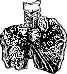
XI. Syphilis of the stomach. Syphilis of the stomach compared with syphilitic affection of other internal organs (aorta, liver, etc.) occurs rarely. Chiari in the autopsy material of 343 syphilitics found syphilis of the stomach 2 times, Stolper in 61 autopsies of syphilitics - 1 time, and Gmelin in the general autopsy material in 10,000 cases - 2 times. In the clinic of internal diseases the diagnosis of syphilis of the stomach is made significantly more often than by pathologists at autopsies, because clinicians often attribute to syphilis of the stomach also various functional disorders of the stomach's activity in syphilitics, which have no specific patho-anatomical basis; such non-specific phenomena from the stomach in syphilitics may respond to specific therapy; they find explanation in the histological research of Rudnev, who discovered specific changes in the stomach in the early period of syphilis. Already during the early secondary period of syphilis pathological phenomena from the stomach appear, and in most cases the examination of the gastric juice reveals more or less diminished secretory activity of the stomach (Neugebauer, Luria). Under specific treatment this specific early process passes away just as all other phenomena do. During the tertiary period more definite changes from the stomach are already encountered, and Gaumann patho-anatomically distinguishes: 1) chronic syphilitic gastritis; 2) gummas in the form of flat plaques of various sizes (from a pea to the size of the palm) consisting of gummatous infiltrate of the mucosa, which later extends to the submucosa; sometimes in cases of old syphilis the infiltration begins in the submucosa and extends to the mucosa; 3) a syphilitic ulcer on the basis of a disintegrated gummatous plaque (Fig. 37); the base of the ulcer consists of sclerosed tissue of the submucosa; a syphilitic ulcer can heal or perforate; in case of adhesion, the calloused ulcer can penetrate into a neighboring organ with which the stomach is adherent (Gaumann), -ulcus callosum penetrans; 4) scars, usually star-shaped, forming either on the basis of an ulcer or by direct sclerosing of the gummatous plaque; 5) diffuse gummatous infiltrate, or hyperplasia, "Figure 37. Multiple gummatous ulcers of the stomach. (After Frankel.)"
the specific form of syphilis of the Stomach with involvement of the mucous membrane, muscular layer, and especially the submucous layer, which sometimes reaches a thickness of one or more centimeters (linitis plastica). In this process, either the entire Stomach or only the prepyloric part and the pylorus is involved. Subsequently, cirrhotic shrinkage of the stomach (microgastria) occurs. In the stage of scarring and complete shrinkage, the syphilitic nature is histologically difficult to prove. Endophlebitis and endarteritis are also observed in non-specific inflammation and are by no means characteristic of the specific form, as was previously thought. The symptomatology of syphilis of the Stomach has nothing characteristic of syphilis and depends 1) on the form of syphilis, 2) on the localization of the process. Sometimes there are signs of ulcer of the Stomach - either of the body or the pylorus; in the latter case, the signs of stenosis come to the forefront. There are also bloody vomitings, but they are not yet proof of a syphilitic ulcer of the Stomach, as they can also occur in syphilitic cirrhosis of the liver and in syphilitic lesions of the mediastinum. The pains are not constant. Their cause is most often syphilitic stenosis and perigastritis. The pains are sometimes prolonged, sometimes appear only after eating. Characteristic of all forms of syphilis of the Stomach is a decrease in secretion of the Stomach up to achylia (Gausmann). As for palpation, a gumma of the Stomach is usually not palpable. Syphilitic lesions of the Stomach create the impression of a tumor in the following cases: 1) when dense adhesions have formed at the site of a syphilitic ulcer with neighboring parts, especially when a callous syphilitic ulcer penetrates into a neighboring organ; 2) when there is the hyperplastic form of syphilis of the Stomach - sometimes the entire Stomach is palpated as a large elastic balloon resembling a pregnant uterus. The shrunken Stomach, lying high in the epigastric region, is palpated by deep palpation as a dense cord lying closer to the xiphoid process. In palpatory diagnosis, it must be borne in mind that a retroperitoneal gummatous tumor can easily be confused with a tumor of the Stomach. "If the retroperitoneal tumor lies behind the Stomach, then the greater curvature, mobile during respiration, is palpated on the tumor; if the retroperitoneal tumor lies below the Stomach, then the curvature is palpated above the tumor; if the tumor lies above the Stomach, then the curvature is palpated below the tumor. Distension of the Stomach in doubtful cases also sometimes gives an answer to the question of whether the tumor belongs to the stomach or not. In the diagnosis of syphilis of the stomach, one proceeds from the position that with normal or increased acidity, it is hardly a case of syphilis of the Stomach. First, an attempt is made to establish the presence of stenosis, palpate the tumor and localize it (gastric or extragastric tumor, tumor of the body of the Stomach or the pylorus), and only then is the question of the possible etiological role of syphilis raised. Personal and family history, W.R. may serve as an auxiliary element of diagnosis, while it must be remembered that both history and W.R. may be absent, as in visceral lues generally. Syphilis of the Stomach in the strict sense of the word must be distinguished from all kinds of gastric phenomena caused by neurosyphilis of the autonomic nerves (Mogilnitsky, Luria), which can cause either hypersecretion or hyposecretion, or gastrospastic or gastratonic states, or, by disturbing trophism, - erosions and simple ulcers and hemorrhages (Lenzmann). The therapy of syphilis of the Stomach should be combined. Along with iodine, treatment with arsenic preparations, mercury, and BISMUTH should be prescribed. F. Gausmann. XII. Parasites of the stomach. The human Stomach is a stage through which various parasites penetrate into the digestive canal. Through the stomach pass, without opening, cysts of the dysentery amoeba and eggs of ascarids. On the contrary, capsules of trichinae, the shell of encysted cercariae of flukes, and eggs of some worms (e.g., the pork tapeworm Taenia solium) are opened under the action of gastric juice. Parasites are found in the Stomach itself as an exception. Lambliae (Giardia intestinalis) were found in the Stomach in the presence of a cancerous tumor in it. It is possible for an amoebic abscess of the liver to be opened in the Stomach. Among parasitic worms, there are cases of atypical location of ascarids in the stomach. The fluke Fasciolopsis can live in the human stomach, which has so far been established experimentally. As pseudoparasites in the human stomach, larvae of various flies can live. Cheese mites (Tyroglyphus siro) can cause catarrh of the stomach. It is claimed that there have been cases of slugs being vomited up.
Gastric juice
166 logie innerer Krankheiten, heransgegeben v. F. Kraus u. Th. Brugsch, B. V, T. 1, B.-Wien, 1921 (lit.). Anatomy and physiology.-Babkin B., External secretion of the digestive glands, M.-L., 1927; Batuev N., Varieties of the form of the human stomach in connection with its development, structure, function and position, Russ. physician, 1913, № 27-28; Deshin A., On the question of the diversity of forms of the human stomach, Bull. Moscow Soc. Nat. Sci., New series, vol. XXXII, issue 1-2, 1923-24; London E., Physiology and pathology of digestion, M.-P., 1924; Pavlov I., Lectures on the work of the main digestive glands, L., 1924; Alvarez W., The mechanics of the digestive tract, N. Y., 1928; Cannon W., The mechanical factors of digestion, L., 1911; Handbook of normal and pathological physiology, ed. by A. Bethe, G. Bergmann et al., Vol. III, B., 1927; Heidenhain R., Secretion processes in the stomach (Handbook of Physiology, ed. by L. Hermann, Vol. V, Lpz., 1880; Russian ed.- St. Petersburg, 1886). Methods of investigation.-Gorshkov M., The constant thin probe as a method for studying the activity of the human stomach, Arch. clin. and exp. med., 1922, № 1; Gurevich G., A new method for obtaining gastric juice in humans, diss., St. Petersburg, 1903; Den O., Fundamentals of radiology, L., 1928; Zimnitsky S., On disorders of the secretory activity of gastric glands from the point of view of functional diagnosis, M., 1926; Levin A., On the methodology of functional investigation of the stomach. Therapeutic archive, vol. V, issue 2, 1927; Leporsky N., On a new test breakfast, Sib. med. journal, 1922, № 7-8; Pletnev D., Radiodiagnosis of the respiratory organs, circulatory system and digestive system, M.-L., 1926; Strazhesko N., Fundamentals of physical diagnosis of diseases of the abdominal cavity, Odessa, 1924; Lugia R. and Mogilewsky E., New contributions to gastric chromoscopy, Arch. f. Verdauungs-krankheiten, Vol. XLIV, 1928; Schlesinger E., The X-ray diagnosis of stomach and intestinal diseases, B., 1927; Stierlin E., Clinical X-ray diagnosis of the digestive tract, B., 1928; Rezlaff K., Methodology of functional diagnosis of abdominal organs (Clinical Laboratory Technique, ed. by Th. Brugsch and Schittenhelm, Vol. III, B.-Wien, 1928). Prolapse of the stomach.-Braitsiev V., Experience in the surgical treatment of gastoptosis, Vestnik khirurgii, vol. VIII, book 1, 23, 1926; Volkov M. and Delitsyn S., Pathogenesis of movable kidney, St. Petersburg, 1897; Vvedensky K., Results of surgical treatment of gastoptosis, Vestn. khir., vol. V, book 14, 1925; Golubinin L., Enteroptosis, its pathogenesis, symptomatology and treatment, M., 1912; Morozova A., Gastropexy for gastoptosis (Jubilee collection of Prof. I. Grekov, P., 1921); Fiber K., The gastoptosis question, Klin. Wochenschr., 1923, № 18; Glionard F., Visceral ptoses, P., 1899; Liek E., Should we operate on the sinking stomach? Arch. f. klin. Chir., Vol. CXXXVII, 1925; Maluschew D., On the various modifications of Biers' gastoptosis operation, Zentralbl. f. Chir., 1923, № 2; Martin B., Treatment of gastoptosis by resection, ibid., 1925, № 40. Ulcers and tumors of the stomach.-Blumenthal N., On the question of perforation of ulcers of the stomach and duodenum, New surg. archive, vol. II, book 2, № 6, 1922; Bogoraz N., A new principle in the surgical treatment of stomach ulcers, ibid., vol. III, book 2, № 10, 1923; Braitsiev V., On the etiology and treatment of round ulcers of the stomach and duodenum, ibid., vol. III, book 4, № 12, 1923; Galperin Ya., 548 operations on the stomach, ibid., vol. III, book 1, № 9, 1923; ibid., Causes of recurrences after operations for stomach ulcers, ibid., vol. III, book 2, № 10, 1923; Hesse E., Resection of the stomach for ulcers, XV Congress Russ. surgeons, M., 1922; Dobrotvorsky V., On the question of gastroenterostomy, diss., St. Petersburg, 1909; Martynov A., Ventriculus, XV Congress Russ. surgeons, M., 1922; Petranovsky G., Perforating ulcers of the stomach and duodenum, Vestnik khirurgii, vol. XIV, book 42, 1928; Spasokukotsky S., Repeated operations on the stomach, New surg. arch., vol. III, book 1, № 9, 1923; Yarotsky A., Dietetic treatment of round ulcers of the stomach and duodenum, L., 1928; Veil H., The perforated stomach and duodenal ulcer, Erg. d. Chir., Vol. XVI, 1923; Konjetzny G., The stomach cancer, Results of Surgery and Orthopedics, Vol. XIV, 1921; Pauchet V. et Hirchberg A., Cancer of the stomach, P., 1928; Starlinger F., Results of 25 years of operative therapy of ulcer disease of the stomach and duodenum, Archiv f. klin. Chir., Vol. CXLVII, 1927. Syphilis and tuberculosis of the stomach.-Gausman F., On syphilis of the stomach, Prakt. physician, 1912, № 27-28; Luriya R., Syphilis and the stomach, M., 1928; Mogilnitsky B., Syphilis of the autonomic nervous system and the endocrine apparatus (Syphilis of the nervous system, ed. by A. Abricosov, P. Gannushkin and M. Margulis, M.-L., 1927); Svetukhin K., On syphilis of internal organs, Kharkov, 1904; Voas K., Syphilogenic diseases of the stomach in the light of modern research results, Zentralbl. f. Haut-u. Geschlechtskrankheiten, Vol. XIII, 1924.
Related articles
Cite this page
“Stomach.” Soviet Medical Encyclopedia. English translation of Bolshaya Meditsinskaya Entsiklopediya, 1st ed. (Moscow, 1928–1936), ed. N. A. Semashko. https://sovietmedicalencyclopedia.pages.dev/article/stomach/