Glands
Historical document, translated for reference. It reflects medical knowledge of the 1920s–30s and is not medical advice.
Summary
This article provides a comprehensive overview of the classification, structure, and development of glands in the human body as understood in the early 20th century. It details the distinction between unicellular, multicellular, and epithelial surface glands, as well as the functional differences between exocrine and endocrine systems.
Encyclopedia article (1928–1936)
GLANDS (Latin: glandulae), formations that secrete various kinds of substances from the body, which either may find some physiological use later (secretions and incretions) or must simply be removed from the organism as unnecessary and harmful (excretions). An essential part of glands is a special modification of epithelial cells—glandular epithelium. Glandular cells can be located singly among the cells of the covering epithelium, forming so-called unicellular glands, or they can cover the surface of an organ in a continuous layer (secreting epithelial surfaces), or finally, they can form multicellular glands, which are organs lying beneath the epithelium.
Types of Glands. A. Unicellular glands include mucus-secreting goblet cells (see), which are located in higher vertebrates among the cells of the cylindrical epithelium in the digestive, respiratory, and urogenital organs, and in lower vertebrates, also in the stratified epithelium of the skin and mucous membranes. Sometimes mucous cells cluster in the epithelium, forming glandular buds that resemble taste buds in appearance (gill cavity of the lamprey), or forming sac-like structures: the mucous membrane of the nose, larynx, and urinary tract in humans (see Brunn's epithelial nests), and the esophagus of birds (in humans, these are apparently pathological formations); the mucous cells of invertebrates (mollusks, worms) can be located under the epithelium, connecting to the surface by a thin neck (exoepithelial unicellular glands).
B. Secreting epithelial surfaces (absondernde Epithelflächen, Schaffer; glandes en nappes, Prenant) are present in organs where the covering epithelium can participate in the secretion process throughout its entire extent. These include: the epithelium of the choroid plexus (plexus chorioideus), which, along with a striated border and a bundle of hairs passing through it, contains granules and vacuoles in its body and participates in the formation of cerebrospinal fluid; the epithelium of the stomach surface, which contains mucigen granules in its upper part and secretes mucus in small portions; some authors also include here the epithelium of the gallbladder, the epididymis, the amnion, and even the uterus and fallopian tubes (during a certain period). In some reptiles, the stratified epithelium of the tongue and esophagus is entirely covered with goblet cells; the same is observed in the epidermis of cyclostomes (hagfish).
C. Multicellular glands (exoepithelial) represent sections of glandular epithelium in the form of tubes, sacs, or vesicles, embedded deep into the underlying connective tissue, receiving a sheath from it, having their own vessels and nerves, and thus forming special organs. The indicated three groups of glands, however, do not exhaust all possible secretory epithelial surfaces; a whole series of the latter, not having a typical glandular structure (for example, the stratified squamous epithelium of the skin and mucous membranes), can carry out a secretory function characteristic of typical glands, especially under certain pathological conditions (see below).
Development of Glands. Multicellular glands originate from the covering epithelium of the outer (skin glands), inner (glands of the digestive and respiratory tract), or middle (glands of the urogenital tract) germ layer by growing deep into the tissue in the form of solid cords, which are bulbously expanded at the ends; the cords can remain simple or branch dichotomously at the ends. The primordium of glands occurs during fetal life, at different times for different glands; the large glands of the digestive apparatus are the first to be laid down (at the end of the 1st month). The lumen of the gland is formed much later; in rare cases (stomach), glands arise immediately in the form of a tubular depression. The indifferent epithelium of the primordium turns into glandular epithelium only in the distal, terminal parts, which can expand in the process; the proximal sections retain the shape of narrow tubes, lined with covering epithelium, and form the excretory duct (see), through which the secretion exits to the surface [see Vol. VIII (art. 159–160), Fig. 9–11]. Thus arise glands of external secretion, or exocrine glands. In another series of cases, the excretory duct disintegrates into separate groups of cells at early stages and disappears; only the glandular sections remain, lying in the depths, without connection to the epithelium, and the removal of the secretion is taken over by blood and lymph vessels. These are glands of internal secretion, or endocrine glands (blood glands, according to old terminology).
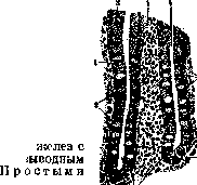
Classification. A completely satisfactory classification of multicellular glands has not yet been developed. The classification usually cited in textbooks is based on the shape of the glandular (terminal, main) sections, otherwise known as adenomeres (M. Heidenhain), and their number. First of all, a distinction is made between tubular and vesicular (alveolar) glands, depending on whether the glandular section has the shape of a tube with a lumen of uniform width or swells into a rounded or oval sac (alveolus, acinus) with a lumen of various shapes and widths, sometimes even without a lumen at all. Both are divided into simple glands (a single glandular section) and complex glands (several glandular sections). From the number of complex glands, some are sometimes distinguished under the name of branched glands, which, with several glandular sections, have a common excretory duct in the form of a simple tube; they correspond to one lobule of other complex glands with a branched duct. Tubular glands include the Lieberkühn glands of the intestine (Fig. 1), as well as the sweat glands of the skin, the lower end of which curls into a glomerulus; branched tubular glands include the glands of the stomach floor, the pyloric glands, and Brunner's glands. Complex tubular glands include the kidneys and the liver, the tubular nature of which is clearly expressed in lower vertebrates (for example, snakes), while in higher vertebrates it is masked by the distribution of connective tissue and vessels, which divide the liver into lobules. Simple alveolar glands are present in the skin of amphibians; in humans, small sebaceous glands can have such a form; ordinary sebaceous glands of the skin belong to branched alveolar, or saccular, glands; the sebaceous glands of the eyelids (Meibomian glands) have a more complex structure.
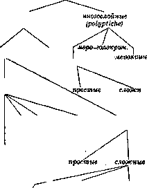
Complex alveolar glands with a branched excretory duct (racemose) are the most common type of gland; this includes the small glands of the oral cavity, esophagus, respiratory tract, salivary glands, pancreas, glands of the urogenital apparatus, and the mammary gland. The shape of the glandular sections of this type varies quite significantly—from a rounded vesicle to an elongated sac, sometimes branched, which is why it has become customary to call these glands tubulo-acinar. Difficulties in determining the shape of the terminal sections and disagreements regarding individual cases force some authors to abandon the cited classification and base the division on other characteristics. Long ago, Ranvier divided glands into 2 types: holocrine, the cells of which entirely turn into secretion (sebaceous glands), and merocrine, in which a part of the cell turns into secretion, while the cell itself remains and continues to function (the majority of glands); subsequently (1917), Schifferdecker distinguished from the composition of merocrine glands a special type, approaching holocrine, the cells of which are partially destroyed during secretion—apocrine glands (see). The newest classification of glands, proposed by Schaffer, is based mainly on the nature of the glandular elements, the number of their layers, and the nature of the secretion. It takes into account glands described in various species of mammals, some of which are not yet sufficiently studied, and is quite complex. In the part relating to multicellular glands, it has the following form: Multicellular glands, single-layered (monoptyche), merocrine, apocrine, holocrine, homocrine (cells of one type), heterocrine (cells of different types), amphoteric (mucous-protein, secreting fat, protein-mucous).
Structure of Glands. An essential part of the glands consists of cells of the glandular epithelium, the diversity of which can be reduced to several types. 1. The cell of holocrine glands (Fig. 2), which secretes a fatty secretion, is a simple epithelial cell of the Malpighian layer, accumulating fat droplets in the protoplasm through fatty degeneration (as some assume) or through the condensation of fat molecules in protein granules. The accumulation of fat droplets gives the cell body a reticular appearance; simultaneously, regressive changes occur in the nucleus, which becomes pyknotic and shrivels. As a result, the cell disintegrates into a pile of fatty detritus. A merocrine cell of a protein (serous) character (Figs. 3 and 4) (salivary glands, stomach glands, pancreas, lacrimal glands, and others) is cited in textbooks as a typical example of a glandular cell.
[layer; 2-stratum lucidum; 3-stratum granulosum; 4-mucous or reticular layer]. It has the shape of a cylinder or truncated cone, with a nucleus in the middle of the polyhedral cells of the basal layer of the stratified squamous epithelium of the skin; 5-papillary layer. Secretion of a proteinaceous character in the form of droplets or granules of uniform size accumulates in the upper part of the cell, sometimes extending into its lateral parts (it is usually stained with acidic dyes and iron hematoxylin). In the basal part of the cell, rod-like striation is usually observed, which, according to the latest research (Zimmermann), represents the splitting of the body into thin rods or plates (lamelläre Streifung). The removal of the secretion occurs either by way of
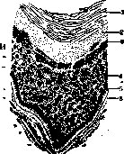
papillary layer of the dermis. (From Polyakov.)

Figure 3.
Figure 4. Figure 3. Part of a longitudinal section of the glands of the human stomach pylorus: 1-bottom of the gastric pit, into which two tubular glands of the outlet part of the stomach open-5; 2-submucosa; 3-lumen of the gland; 4-columnar cells. Figure 4. Longitudinal section of the tubular glands of the pylorus: 1-single-layer columnar epithelium; 2-canal of the gastric pit; 3-tubular glands of the stomach pylorus; 4-submucosa; 5-muscular layer. (From Polyakov.) the expulsion of individual granules through the free surface of the cell or through secretory intracellular capillaries, thin tubules that usually open on the lateral surface of the cell; in this case, the secretion is preliminarily liquefied. 3. Apocrine cell (Fig. 5) of the mammary and large sweat glands. At the base, rod-like striation is sometimes observed; the secretion accumulates above the nucleus in the form of fat droplets and protein granules; its release is accompanied by the detachment of the upper part of the cell (decapitation); sometimes, after preliminary liquefaction, the secretion flows out in the form of bubbles (vesicular secretion). After the removal of the secretion, the cell flattens. 4. Mucous cell—of the same character as the goblet cell. The secretion accumulates above the nucleus in the form of large granules (in vivo), which usually swell in fixed preparations; it stains with basic dyes, often metachromatically. As the secretion accumulates, the nucleus is pushed to the base of the cell, and the secretion is expelled through the free surface. There is never any rod-like striation of the base. 5. Excretory cell...
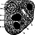
Figure 5. Section of a sweat gland: 1-cells of the sweat gland; 2-muscular elements; 3-nuclei of deeply lying tubules; 4-fat cells; 5-excretory duct; 6-basement membrane. (According to Szymonowicz.)
Besides the distinct striation at the base, on the free surface of the cell, a border of fine rods (Burstenbesatz, striated border) is noticeable. In the body of the cell, granules and vacuoles are noticeable in different places and in varying quantities; the actual process of secretion has not been clarified (see also Excretory processes). Besides secretion, the cell also possesses the ability of absorption (see) from the lumen (see Kidneys, Diuresis). In all types of glandular cells, necessary components are: mitochondria in the form of filaments and granules, accumulating mainly in the basal part; the microcenter, usually in the form of a diplosome, above the nucleus, often in the middle of the secretion accumulation; and finally, the Golgi apparatus in the form of a reticular basket, also located above the nucleus, but sometimes extending to its lateral surfaces. The accessory nuclei and ergastoplasm described previously are currently recognized as artifacts. Besides the glandular epithelium, in the wall of the glandular cell or tubule, there is a layer of special flat cells recognized as smooth muscle (myoepithelium), of spindle or stellate shape (basket cells); behind them lies a fine-fibrous proper membrane (membrana propria) of connective tissue origin. The terminal parts of the excretory duct can enter the lumen of the cell, forming centroacinar cells (pancreatic gland). Excretory ducts in simple and branched glands have the appearance of a short tube or neck, lined with a single-layered indifferent epithelium of cuboidal shape, but in sweat glands, the epithelium is double-layered. The excretory ducts of complex glands have a greater length and usually branch dichotomously, whereby the caliber of the tube gradually decreases; in the initial part, they are lined with multi-layered or double-layered epithelium, which is gradually replaced by single-layered columnar and finally low epithelium. In some glands, the continuation of these epithelium-lined ducts are intercellular secretory capillaries in the form of the finest tubes with homogeneous walls (liver, salivary glands), which can connect with intracellular capillaries. In serous and mixed salivary glands, the interlobular excretory ducts are lined for a certain distance with high columnar epithelium with rod-like striation of a glandular character (secretory segments or salivary tubules); immediately after them begin thin segments with low epithelium (intercalated ducts). In mucous glands, the terminal segments of the excretory duct have a larger caliber, and their cells can undergo mucification over a significant extent. Multicellular glands are structured with the help of fibrous connective tissue, which forms a capsule around the gland; in large glands, lamellar processes (septa) extend from it deep into the gland, dividing the gland into lobules of angular shape, which can be subdivided into secondary lobules; inside the lobules, there is looser tissue, enveloping the cells in a thin layer. In the connective tissue, blood vessels branch out, surrounding the glandular segments with a capillary network, as well as lymph vessels and nerves, which terminate partly on the vessels and partly on the glandular segments. Nerve endings on gland cells, discovered for the first time by Pflüger, were described in detail by Arnstein in salivary glands and by Retzius in many others. General physiology of glands. The functional activity of glands is divided into 2 periods: 1) the production (accumulation) of secretion or excretion in glandular cells and 2) its release. The latter period was previously described as the active state of the gland, in contrast to the period of accumulation, which was called rest. The production of secretion occurs at the expense of substances brought by the blood, whereby to explain the specificity of the secretion, it is necessary to acknowledge that the cell possesses a selective absorption capacity, depending on the properties of the cell membrane. The microphysiology of secretion production, despite a large number of studies, has been little clarified. It can be assumed that the rod-like striation of the basal end is connected with the process of absorption of substances, mainly water. It is further assumed that substances that have penetrated into the protoplasm undergo processing with the participation of the nucleus and are then released within it in the form of droplets or granules. According to Altmann's theory, the main participation in this process is taken by protoplasm granules, which condense certain substances within themselves and turn into secretion granules (granular synthesis); subsequently, the same role began to be attributed to the ergastoplasm (a convoluted bundle of fibrils at the base of the cell), and then to mitochondria (chondriosomes); the latter view still finds many defenders today. Recently, the accumulation and specific processing of substances began to be attributed to the reticular Golgi apparatus, with which, however, many scientists cannot agree. The participation of the nucleus in the process of secretion was usually proven by the change in its shape: during the period of secretion production, the nucleus in fixed preparations appears shriveled, but after release, it rounds out; however, intravital observations do not confirm this. Data regarding the exit of chromatin or the nucleolus from the nuclei of glandular cells are very doubtful. In a number of glands, the granules accumulating in the body of the cell do not represent the final secretion, but its preliminary phase—prosecretion (mucinogen, zymogen); the secretion is formed when the granules are pushed out and swell. The process of secretion release from cells during life has been observed repeatedly in the rabbit pancreas (Kühne and Lea, Langley). Upon nerve stimulation, a rounding of the cells and their clear demarcation were noted; the inner granular zone decreases significantly due to the expulsion of granules into the lumen and their dissolution. Old observations on the stimulation of cerebral and sympathetic nerves of salivary glands showed a difference between liquid "cerebral" and thick "sympathetic" saliva, on the basis of which R. Heidenhain considered the release of solid organic substances and water as two different processes. This theory is currently being developed by M. Heidenhain: he attributes the release of water to the intergranular substance of the cell and links it to the contraction of fibrils running along the length of the cell (tonofibrils); it proceeds in waves and squeezes water out of the cell. The removal of secretion granules is caused by the contraction of the myoepithelial cells of the glandular alveolus. Joint observations by physiologists and histologists on the pancreas (Babkin, Rubashkin, Savich) note a difference in the release of secretion under the influence of chemical stimulation (secretin) and nerve impulse (vagus nerve). In the first case, a stream of water passes through the cell, washing out the granules; in the second, the granules are almost not released, and structural changes occur in the cell (appearance of vacuoles) associated with a change in staining.
V. Karpov. Pathology of glands encompasses all those pathological processes which are observed in the suffering of other organs as well. Thus, in glands, one may observe developmental defects, for example, in the sense of excessive development, underdevelopment, incorrect positioning, etc. Inflammatory processes in glands are a very frequent phenomenon both in local and in general processes, for example, in infectious diseases, whereby the entry of infection can occur both from the side of the supplying blood and lymph vessels and from the side of the excretory ducts, especially in the presence of significant functional changes in the gland, for example, in the quantity and quality of the secretion, the delay of its excretion, etc. The very character of inflammatory processes, their morphological and clinical characteristics depend not only on the causative factor (tuberculosis, syphilis, etc.) but also on the structure and function of the gland itself, on its significance in the general balance of the organism, in metabolism, etc. Like other organs, glands can undergo atrophic, hypertrophic, and neoplastic processes; the main mass of cancers falls on glands, whereby some glands (stomach glands, mammary gland, liver, pancreas) play an especially important role in this regard, while others (for example, salivary, lacrimal), on the contrary, play a very modest one. The involvement of certain glands by a tumor (specifically cancer) is not always equivalent to the loss of their function; thus, in primary cancer of the entire pancreas or the entire thyroid gland, one may not observe any phenomena of the loss of the general physiological function of these glands; on the contrary, the removal of cancerously altered glands can produce such phenomena (in the indicated cases, phenomena of athyreosis, pancreatic diabetes, etc.). The excretory processes themselves in glands under pathological conditions can change significantly both in the sense of the quantitative and qualitative content of the secretion and in relation to their morphological expression. Thus, under certain conditions, the stomach and salivary glands can intensely excrete chlorides and nitrogen-containing compounds (in uremia); the very mucosa and contents of the stomach sometimes acquire a clear smell of alkaline urine. In the process of excreting pathological products, both those formed inside the organism (in infections, autointoxications) and those introduced from the outside, various acute and chronic processes can occur in glands, such as: inflammations (for example, "excretory" nephritis, colitis, etc.), necroses, degenerations, and in some cases, apparently, deeper changes, such as: cirrhosis of organs, cancerous neoplasms, etc. In general, it seems necessary to recognize that the main mass of pathological processes observed in glands has as the mechanism of its origin precisely the factor of the excretion of corresponding products of a pathological order, and in some cases also organized bodies, for example, certain bacteria. In a number of cases, such a function of excretion lies at the basis of the pathogenesis of known diseases or individual symptoms of a disease. Thus, in the origin of typhoid fever, dysentery, cholera (specifically the corresponding morphological pictures), it is important to consider the factor of the excretion by the liver (with bile) and the intestine of corresponding bacterial bodies, endotoxins, etc. The same factor should be considered in the mechanism of the development of bronchitis, pneumonia, nephritis, gastroenteritis, etc. The morphology of excretory processes in glands under pathological conditions may have nothing characteristic; more often, however, one observes certain deviations in secretion, predominantly of a quantitative character, such as: increased desquamation of epithelium, granular, lumpy, droplet disintegration of it—in general, phenomena bordering on or coinciding with those processes which are classified as degenerative and necrobiotic. Lymphatic glands do not represent glands in the proper sense of the word; it is more correct to designate them as lymph nodes. I. Davydovsky.
Related articles
Cite this page
“Glands.” Soviet Medical Encyclopedia. English translation of Bolshaya Meditsinskaya Entsiklopediya, 1st ed. (Moscow, 1928–1936), ed. N. A. Semashko. https://sovietmedicalencyclopedia.pages.dev/article/glands/