Cowper's Glands
Historical document, translated for reference. It reflects medical knowledge of the 1920s–30s and is not medical advice.
Summary
Cowper's glands are paired glandular organs located at the blind end of the bulb of the urethra. Their functions are not fully understood, but they may secrete mucus that mixes with semen and protects the urethra.
Encyclopedia article (1928–1936)
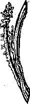
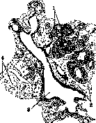
COWPER'S GLANDS, more correctly Cowper's (W. Cowper) (glandulae bulbo-urethrales Cowperi), were first described in 1684 by Cowper. Winslow called these glands the 'antiprostate' (Braus). They are paired glandular organs located at the blind end of the bulb of the urethra, near the membranous part (fig. 1), the size of a pea, yellowish-brown in color (Rauber-Kopsch). During dissection, it is not always possible to accurately determine the location and shape of these glands, as the muscle fibers of the deep transverse muscle of the perineum (m. transversus perinaei profundus; fig. 2) surround them and simultaneously compress the glandular tissue, so that the latter loses its shape and is macroscopically barely noticeable. Both glands lie quite close to each other and are sometimes connected by a bridge of muscle fibers forming an isthmus. The excretory duct of the gland, 5-6 cm long, ends with a slit-like opening in the fossa bulbi urethrae (fig. 3) (Braus). In addition to these Cowper's glands, additional glands (gl. Cowperi, glandulae accessoriae) with blindly ending ducts are also observed (fig. 4) (Lichtenberg). Microscopic examination of Cowper's glands (fig. 5) shows that the secretory epithelium lines not only the terminal branches of the gland but even the walls of the excretory duct up to its point of entry into the urethra. The cells lining the walls of both the main and terminal ducts consist of a single-layer cylindrical epithelium, resembling the epithelium of mucous glands and staining blue with hematoxylin. The mucus contained in them does not precipitate when treated with acetic acid. At the blind end of the gland, the terminal branches depart much more frequently; they are larger and dilate into ampullae, which serve as a reservoir for the gland's secretion (figs. 4 and 5) (Braus).
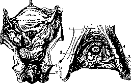
Figure 1.
Figure 2.
Figure 1. 1-Gl. bulbo-urethralis dextra; 2-bulbus urethrae. Figure 2. 1-symphysis os-pubis; 2-gl. bulbo-urethralis; 3-m. transvers. perinaei pro-fund.; 4-urethra (pars membranacea).
The functions of Cowper's glands have not yet been elucidated. Apparently, during ejaculation, the contents of Cowper's glands, under the influence of contraction of smooth and striated muscle, exit from the excretory duct and mix with the ejaculate. Some suggest that the contents of these glands cover the mucous membrane of the urethra with a protective layer, shielding the walls of the canal from irritation by urine residues.
Figure 3. Glandula bulbo-urethr. (1); 2-pars mem-bran. urethrae; 3-d. ex-cretor. gi. Cowperi.
Figure 4. Gl. bulbo-ure-thrales(i- gl. accessor.).
Cowperitis (inflammation of Cowper's glands), was first described by Gubler in 1849. Recently, cowperitis, especially gonorrheal, has received proper attention in Western and our literature. Cowper's glands, due to their anatomically close contact with the urethra, reflect the condition of the latter. Inflammation of Cowper's glands can occur through direct transmission of infection per continuitatem from the urethra, as occurs in gonorrheal and so-called non-gonorrheal, or catarrhal, urethritides. Hematogenous and lymphogenous routes of infection are observed much less frequently, for example in tuberculosis (isolated observations). If Ricord counted 6 cases of cowperitis per year in his extensive practice, it must be assumed that he meant only abscesses of this organ. Leszynsky, in a large series of autopsies of persons suffering from chronic gonorrhea, proved that cowperitis occurs as frequently as prostatitis. In acute gonorrhea, Muhlpfordt found cowperitis in 15% of cases, while Shishov and Smirnov found it in 12% of cases of chronic gonorrhea. Patho-anatomically, this is a catarrhal or purulent inflammation, sometimes with the formation of retention cysts. A special pathological form is independently developing cysts of Cowper's glands. Cowperitis is more often a unilateral disease. Usually the process goes unnoticed, and only later, when prolonged gonorrhea forces a careful search for the cause, systematic examination leads to the discovery of cowperitis. Diagnosis is made primarily on the basis of palpation. Examination of Cowper's glands according to Picker is performed either in the horizontal or knee-elbow position. The index finger is inserted into the rectum, bent into a hook, and then an attempt is made to connect its tip with the tip of the thumb of the same hand, bringing the thumb to the gland from the perineal side. Between the tips of both fingers, the inflamed gland is clearly palpable and appears painful. If during inflammation the long excretory duct remains open, it becomes possible by massage to express the gland's secretion and subject it to examination. Massage is performed in such a way as not to touch the prostate: the urethra is first washed and the bladder is filled with an indifferent solution. In the secretion during inflammation, pus and microorganisms are found. When edema closes the lumen of the excretory duct, acute cowperitis occurs, accompanied by a number of symptoms: stabbing pain in the perineum, formation of a tumor that interferes with urination, elevated temperature, and sometimes the rupture of the resulting abscess to the outside. Urinary infiltration can also have an abscess of Cowper's gland as its primary source. Chronic cowperitis usually proceeds without symptoms; less frequently, pain is observed when sitting on hard objects and the appearance of discharge from the urethra not in the morning but after walking, in the evening. In addition to palpation in chronic cases, diagnosis can be aided by urethroscopy and urethrography. In the first, dilation and inflammation of the opening of Cowper's gland are visible, while in the second, filling with contrast solution is visible.
Figure 5. Structure of Cowper's glands: 1-cylindrical epithelium; 2 and 4-smooth and striated muscle; 3-ampulla.
Treatment: in acute forms-rest, hot water bottles; in abscess-wide incision; in chronic cowperitis-massage, bougienage, various forms of heat, in stubborn cases-extirpation. Prevention of cowperitis is timely and proper treatment of urethritis.
Related articles
Mentioned in
Cite this page
“Cowper's Glands.” Soviet Medical Encyclopedia. English translation of Bolshaya Meditsinskaya Entsiklopediya, 1st ed. (Moscow, 1928–1936), ed. N. A. Semashko. https://sovietmedicalencyclopedia.pages.dev/article/cowpers-glands/