Bartholin's Glands
Historical document, translated for reference. It reflects medical knowledge of the 1920s–30s and is not medical advice.
Summary
This article describes the anatomy, development, and histology of the Bartholin's glands, which are the major vestibular glands in the female reproductive system. It also covers the pathology of bartholinitis, noting its frequent association with gonorrhea and discussing treatment methods such as excision.
Encyclopedia article (1928–1936)
BARTHOLIN'S GLANDS, or the greater vestibular glands (glandulae vestibulares majores), are accessory glands of the female reproductive apparatus. Bartholin's glands were first described by Duverney (17th century) in the cow, then by Bartholin (17th century) in women, and in the 19th century they were described in detail by Tiedemann and Méry, which is why the glands also bear the names of these authors. Bartholin's glands are located on both sides of the vaginal entrance, in its posterior section, between the m. constrictor cunni and the m. bulbo-cavernosus; they are often surrounded by muscle bundles; anteriorly, they border the posterior
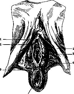
1-m. ischio-cavernosus; 2-orificium urethrae externum; 3-m. transversus perinei superficialis; 4-bulbus vestibuli; 5-orificium vaginale; 6-glandula vestibularis major (Bartholini); 7-anus (according to Spalteholz).
end of the vestibular bulb (bulbus vestibuli) and are in contact with the m. transversus perinei, being located between the two layers of the fasciae perinei. Bartholin's glands are rounded bodies the size of a pea (up to 1.3 cm), which can be palpated in the posterior part of the labia majora; their excretory ducts extend for a considerable distance (1.5 cm–2 cm) and open at the vaginal entrance on the inner side of the labia minora, in the so-called sulcus nympho-hymenalis. Upon compression of the gland, a transparent, odorless, neutral secretion is released, which does not become cloudy with acetic acid, i.e., it does not contain mucus; during reflex stimulation, the secretion is ejected in a stream; its purpose is to moisten the vaginal mucosa during sexual intercourse. Bartholin's glands arise in the 3rd month of fetal life in the dorsal wall of the urogenital sinus as a paired outgrowth that quickly begins to branch. By their site of development, their position in the developed organism, and their histological structure, Bartholin's glands correspond exactly to the bulbourethral, or Cowper's, glands of the male. Bartholin's glands break down into small lobules of 1–3 mm, enveloped by smooth and striated muscles; the lobules have a tubulo-acinar structure, with pear-shaped alveoli of 45–100 μ. The alveoli have a wide lumen with two types of cells: those with a cloudy body and light ones that stain weakly with hematoxylin. The latter resemble mucous cells, although, unlike them, they have terminal bars and secretory capillaries. Alveoli with low epithelium are also encountered. The excretory ducts are lined, depending on their caliber, with single-layered or multi-layered columnar epithelium, behind which lies a layer of smooth muscle; towards the end, they expand in the form of sinuses. Glandulae vestibulares minores are scattered throughout the vestibule, are smaller in size, and secrete mucus.
V. Karpov. Bartholinitis, an inflammatory disease of Bartholin's glands, usually caused by gonococci and occasionally by other bacteria. It is not the entire gland that is affected, but only its excretory duct. Inflammation begins with redness around the exit opening; upon pressure on the duct, a pus-like fluid containing gonococci is squeezed out; the duct itself can be felt as a spindle-shaped cord, sometimes painful upon pressure. When the exit opening is blocked, the purulent discharge accumulates and stretches the duct—it turns into a cyst-like, tense tumor, sometimes up to the size of a chicken egg. This results in a so-called false abscess (pseudo-abscessus), which can rupture on the inner surface of the labia. In treatment, attempts to introduce medicinal substances (silver nitrate, choleval, protargol) into the duct by means of various syringes do not yield satisfactory results; X-ray treatment is only just being developed. Reliable results are obtained with the easily performed excision of the duct during the acute period, when it is still palpable. The most radical approach is the complete excision of both the duct and the gland.
Related articles
Cite this page
“Bartholin's Glands.” Soviet Medical Encyclopedia. English translation of Bolshaya Meditsinskaya Entsiklopediya, 1st ed. (Moscow, 1928–1936), ed. N. A. Semashko. https://sovietmedicalencyclopedia.pages.dev/article/bartholin-s-glands/