Bartonella
Historical document, translated for reference. It reflects medical knowledge of the 1920s–30s and is not medical advice.
Summary
This article from the 1st edition of the Great Medical Encyclopedia (1928–1936) discusses Bartonella, covering bacterial microorganisms parasitic to erythrocytes and endothelial cells, specifically Bartonella bacilliformis (the causative agent of Carrion's disease, including Oroya fever and verruga peruviana) and Bartonella muris in rats.
Encyclopedia article (1928–1936)
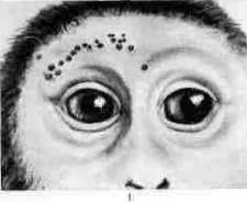
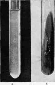
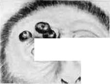
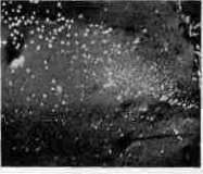
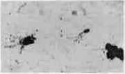
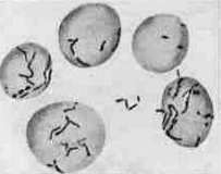
BARTONELLA, bartonella, a bacterial-type microorganism parasitizing the erythrocytes as well as the blood vessel endothelium in humans and rodents. A distinction is made between B. bacilliformis and B. muris. I. B. bacilliformis Strong, Tyzzer etc. 1915. Morphology: thin straight or curved rods, measuring 1.5–2.5 x 0.2–0.5 µ, sometimes with thickenings at the ends (see table, Fig. 7); motile, having a tuft of flagella up to 30 µ in length at one end (see Fig. 5). They also occur in the form of V- or Y-shaped rods and as rounded bodies with a diameter of 0.5–1 µ. They do not form spores. Stained with Giemsa, they color red; gram-negative. They do not pass through filters. Reproduction takes place within endothelial cells. They are best cultivated under aerobic conditions on Noguchi's medium, proposed for leptospires (0.9% NaCl - 800.0; rabbit serum - 100.0; 2% agar - 100.0; hemolyzed rabbit blood (1:3) - 10.0–20.0); optimum growth at 25–28°; pH = 7.8–8.0; grayish colonies reach a size of 1–2 mm on the 6th–8th day (see Figs. 4 and 6). They do not ferment carbohydrates and do not hemolyze erythrocytes. They are inoculable to monkeys (Macacus rhesus, Chimpanzee, Orangutan), guinea pigs, rabbits, and dogs (see Figs. 1 and 2). In humans, it is the causative agent of bartonellosis, known as Carrion's disease, which manifests as 1) the so-called Oroya fever—an acute febrile, frequently fatal septico-anemia, or 2) the so-called verruga (verruga peruviana, see)—a chronic local process lasting from 2 to 24 months and expressed by the formation of characteristic nodules (on the skin, conjunctiva, oral mucosa, and other sections of the digestive tract, etc.) (see Fig. 3). Verruga appears a few weeks or months after contracting Oroya fever or independently of it. Both diseases occur in Peru in valleys with a humid tropical climate. Cases of simultaneous or successive disease in the same person with both specified clinical forms, as well as Carrion's experiment (1885), who inoculated himself with material from a verruga patient and died as a result of Oroya fever, pointed to the possibility of a common origin for both diseases. However, due to the identical geographical distribution of both diseases, all these data did not exclude the possibility of the simultaneous presence of two different infections in one person. Only through Noguchi's experiments (1926, 1927) was the etiological identity of both diseases definitively established. Cultures of Bartonella isolated from the blood of patients with Oroya fever caused irregular intermittent fever in monkeys (Macacus rhesus) upon intravenous administration, and in particularly susceptible animals or in the case of particularly virulent cultures, also a typical picture of anemia. Administration of the same material deep into the skin of monkeys caused typical verruga in them (see Figs. 1 and 2), and cultures of Bartonella could be isolated from the nodules themselves, from lymph nodes, and from the blood. Administration of a suspension of human-origin verruga nodules to monkeys caused a picture of experimental verruga, and here too Bartonella were found not only in the nodules but also began to circulate in the peripheral blood. During experimental infection of monkeys with Bartonella isolated from patients, verruga-like nodules sometimes developed in their lungs and spleen as well. Serological examination of Bartonella bacilliformis isolated in cases of Oroya fever, as well as cross-immunization experiments, demonstrated their complete identity with Bartonella from cases of verruga.
Spread in the organism. Bacteriological examination in experimental Oroya fever has shown that Bartonella penetrate all organs and can be detected (microscopically and by cultures) in the blood, liver, spleen, lymph nodes, and bone marrow. In human peripheral blood, Bartonella affects up to 20–30% of all erythrocytes, and in some erythrocytes their number may reach up to 30 specimens.

Histological changes during Bartonella infection are very characteristic. In cases of natural or experimental Oroya fever, the following are observed: in the liver—necroses around the hepatic veins accompanied by macrophagocytosis, invasion of polymorphonuclear leukocytes into the necrotic zones, endothelial hyperplasia, and fatty infiltration of liver cells; in the spleen and lymph nodes—endothelial hyperplasia causing small infarcts accompanied by the disappearance of follicles and strong macrophagocytosis; in the lymph nodes—deposition of dark green pigment; in the bone marrow—macrophagocytosis, an increase in the number of normoblasts. In experimental verruga, the initial changes present a picture of angioendothelioma, which is then complicated by secondary connective tissue proliferation. The endothelial cells of blood vessels in various organs in Oroya fever and in the nodules in verruga are strongly hypertrophied and contain many Bartonella both in the form of rods and as small granular elements (see Fig. 3).
Diagnosis of both forms of the disease, as well as the onset of recovery, are ultimately established on the basis of cultures from blood and internal organs, especially from lymph nodes, where Bartonella persist for up to 5 months after clinical recovery. Positive cultures of Bartonella can be achieved at the peak of the disease even at a blood dilution of 1 million times. During the recovery period, Bartonella continues to circulate in the blood for up to 2 months, but their number steadily drops to a titer of 1:10 and lower.
The routes of entry of Bartonella into the organism have not yet been definitively clarified. In verruga, infection occurs, in all probability, through the skin, but under experimental conditions, penetration of Bartonella into the skin was possible only upon its scarification or intradermal administration. Apparently, the route of infection for Oroya fever also lies through the skin. Under experimental conditions, this infection was successfully induced by the bite of the tick Dermacentor andersoni. Under natural conditions, the role of Phlebotomus verrucarum is considered probable. The question has not yet been definitively clarified. Immunity. Contracting a Bartonella infection induces a state of complete immunity, which is also manifested in cross-immunization experiments. Contracting Oroya fever immunizes monkeys against the formation of verruga; however, in some cases of verruga infection, immunity develops so slowly that superinfection is still possible at the beginning of the process. Immunization of rabbits with Bartonella cultures imparts to their serum the ability to fix complement at a titer of 0.001.
II. Bartonella muris Mayer 1925 corresponds morphologically to Bartonella bacilliformis. In rats, Bartonella is the cause of a latent infection that under normal conditions is not detected in them and does not cause any disease phenomena. However, in rats with reduced resistance on the basis of splenectomy, Bartonella multiply strongly and cause their death with phenomena of severe anemia. Infection of rats with Bartonella muris is apparently very widespread; it has also been detected in Moscow, but in some geographical regions (Sicily) rats are free from this infection. Under natural conditions, the infection of rats with Bartonella is associated with the infectiousness of their excreta. This question has not been definitively clarified, just as the question of the relationship between Bartonella muris and Bartonella bacilliformis remains unresolved. Lit.: Odriozola E., La maladie de Carrion, P., 1898; Noguchi H., Journal of experimental medicine, 1926, № 43-44, 1927, № 45; Mayer M., Kirkuth W., Borchardt W., Klinische Wochenschrift, 1926, № 30.
Related articles
Mentioned in
Cite this page
“Bartonella.” Soviet Medical Encyclopedia. English translation of Bolshaya Meditsinskaya Entsiklopediya, 1st ed. (Moscow, 1928–1936), ed. N. A. Semashko. https://sovietmedicalencyclopedia.pages.dev/article/bartonella/