Scleroma
Historical document, translated for reference. It reflects medical knowledge of the 1920s–30s and is not medical advice.
Summary
An overview of scleroma (rhinoscleroma), detailing its history, geographical distribution, epidemiology, pathogenesis, clinical forms, and pathological anatomy as understood in 1930s medicine.
Encyclopedia article (1928–1936)
SCLEROMA. History. Scleroma was first described by Hebra and Kaposi (1870) under the name "rhinoscleroma", because the process in the nose was taken as the primary basis of the disease, while accompanying changes in the larynx and other organs seemed insignificant and accidental. But soon other authors (Gerhardt, 1873; Ganghofner, 1880) noted the unity of manifestations of the scleromatous process in various parts of the respiratory tract and the identity of scleroma with that disease which was described even earlier by Stork under the name of "chronic blennorrhea of the respiratory tracts." The elucidation of the histological structure of scleromatous infiltrates was especially facilitated by the works of Mikulicz (1876), while bacteriology had as its pioneers Frisch and Pelizzari (1882), who discovered a specific microorganism in the scleromatous tissue—Bact. rhinoscleromatis (see Microorganisms). Subsequent works by Cornil and Alvarez, Paltauf, Eiselsberg, and others clarified the details of the morphology and cultivation of the scleroma bacillus. Attempts to experimentally induce the scleromatous process in animals have been made since 1889 (E. M. Stepanov); positive results were obtained by Kraus, G. E. Zhukov, and Gerber. In 1909, Goldzieher and Neuber discovered a specific antigen in the blood of scleroma patients, and in 1930, Mantoux and Roux observed an allergic reaction, both local and focal, upon intradermal administration of the antigen. Social significance, geographical distribution, and epidemiology. The gradual spread of scleroma far beyond the borders of its endemic foci, a significant increase in the total number of patients, and the undoubted connection with living conditions determine the social character of this pathological form (Fig. 1). Statistics show that scleroma is distributed on the territory of our Union predominantly in the Ukrainian SSR and Belarusian SSR, where according to recent data up to 1,000 patients have been registered, while individual cases have been observed in other places, sometimes far removed from the western border, e.g., in Siberia (Pautov), Central Asia (Voyachek), etc. Abroad, scleroma has its main distribution in Poland, Czechoslovakia (several hundred cases each), more sporadically in Romania, Germany, Bulgaria, Switzerland, Italy; individual cases are encountered in France, the USA, the Sunda Islands (Sumatra), and other countries. There are a total of 2,631 cases (S. I. Belinov). Women predominate among the patients. The age from 15 to 35 accounts for half of all cases. The disease nests predominantly among the underprivileged strata of the population living in poor hygienic conditions. Spa
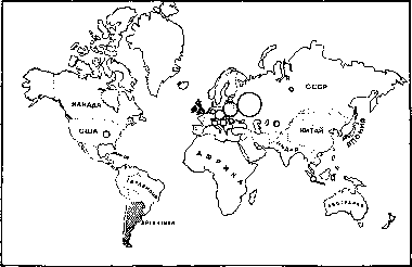
rno. 1. Geographical distribution of scleroma in various countries. Scleroma nests are indicated by circles; areas where scleroma does not occur at all are shaded; places of sporadic cases are marked with black dots (according to data of the II International Oto-Laryngological Congress).
mode of transmission is not precisely known; direct transmission from person to person is unlikely, although not completely excluded. From the "nests" of scleroma, the disease gradually spreads to neighboring areas or, through migration, is carried in leaps to distant countries. According to A. M. Puchkovsky, wars and the movements of human contingents associated with them also greatly contributed to the dissemination of scleroma. The relation to everyday life is expressed by the fact that nests of scleroma are more frequently found in swampy areas, with crowding of the population and other anti-hygienic conditions (shared quarters for people and domestic animals, poor nutrition, etc.). Pathogenesis and forms. Scleroma is a chronic infectious disease expressed by the development in various parts of the respiratory tract (hence the name scleroma respiratorium) and adjacent organs of granulomas, which over time turn into scar-transformed masses causing greater or lesser strictures of the respiratory tract. The incubation period is not precisely known. Even children in the first years of life can fall ill. The portal of entry is apparently the upper respiratory tract, but judging by the predominant localization of changes, one must think that the primary nest in different people may be located selectively either in the nose, or in the nasopharynx, or in the larynx, and there are three characteristic places where scleromatous granulomas are most often determined—this is the entrance to the nasal cavity, the choanae (rhino-pharyngoscleroma), and the subglottic space of the larynx (chorditis vocalis inferior hypertrophica); recently, frequent involvement of the tracheal bifurcation has also been indicated (all this corresponds to the sites of physiological narrowings). The disease is characterized by slow progression, but gradually the infiltrates can spread to other organs, capturing the walls of the oral cavity, pharynx, bronchi, external nose, lips, lacrimal pathways, Eustachian tube, tympanic cavity, and external auditory meatus. Each localization can be designated by a prefix to the main name of the organ designation, e.g., "bronchoscleroma" or "otoscleroma". In addition to typical scleroma, characterized by moderately developed granulomatous infiltrates and strictures in the region of physiological narrowings, a hypertrophic form with the formation of bulky growths (for example, like rhinophyma on the external nose) and the opposite—atrophic form, which clinically differs little from vulgar atrophic rhinitis or vulgar ozena—are distinguished. Due to scleromatous infiltration, the affected organs can gradually fuse with surrounding tissues (e.g., the trachea with surrounding cellular tissue) and even skeletal parts (sternum), but metastases in separate parts of the body and generally generalization of the process are not observed. Pathological anatomy. Characteristic features of scleromatous infiltrates are vacuolated Mikulicz cells scattered throughout the general mass of the granulomatous tissue, Russell's hyaline bodies (see), which according to Schridde's opinion are a derivative of basophilic granules in plasma cells, epithelial cords extending often deep into the infiltrate, and the presence of Frisch's capsule bacteria in the vacuoles of Mikulicz cells and in lymph intercellular spaces (Fig. 2). Infiltrates can have various shapes: diffuse, nodular, tuberous, in the form of a tumor, etc. The diffuse form is often masked by symptoms of atrophic catarrh—dryness of the mucous membrane covering the infiltrates, mucus, crusts. If substance defects form on the surface of the affected areas, they are usually not deep (superficial scleromatous abrasions and any kind of trauma to the infiltrates, even surgical for example, usually heal without complications). Frisch's bacilli in sections stain with Gram (although in cultures they turn out to be Gram-negative). Three or four stages of infiltrate development are distinguished. In the initial stage, there is an accumulation of small and large lymphocytes, neutrophils, plasma cells of various volumes, fibroblasts, vacuolated cells, and Russell's hyaline bodies. Vessels are found in large numbers. In the second stage, the number of lymphocytes decreases and plasma cells increase; oval and spindle-shaped forms (fibroblasts) are frequently encountered. In t
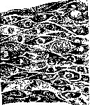
Figure 2. Scleroma bacilli in epithelial cells. In the third stage, the number of Mikulicz cells increases, while plasma cells remain few. In the fourth stage (cicatricial shrinking), vacuolar cells disappear and a large amount of collagenous connective tissue mixed with elastic fibers is formed. Blood vessels are obliterated, but Russell bodies can still be found in places. The epithelium undergoes desquamation, metaplasia from cylindrical to stratified squamous; strands of epithelium penetrate far into the depth of the submucosal tissue. The formation of true epithelial masses was sometimes observed. In contrast to some animals, e.g., mice, in which subcutaneous injection of Frisch's bacilli causes septicemia, in humans Scleroma remains a local disease in the sense that it does not give metastases, but may be accompanied by secondary changes in internal organs, including lymphadenitis (secondary infection), pulmonary emphysema, dilation of the right heart, pneumonia, lung abscess and gangrene, pericarditis, pleurisy. In 10% of cases Scleroma was combined with tuberculosis. A combination with syphilitic changes and tumors was observed. Experimental pathology. In animals, including monkeys, infection with Scleroma causes a disease quite similar to the human one, but it proceeds acutely, resembling the fresh initial stages of human Scleroma, and in contrast to the latter shows no tendency to pass into a chronic form, probably due to the easy formation in animals of protective bodies acting specifically against the ectoplasm (capsule) of bacteria; in humans, these bodies are insufficient. Bacteriology and serology. The bacillus causing Scleroma belongs to the Friedländer group (see Microorganisms). Characteristic differences of Frisch's bacilli on obliquely solidified agar: they form condensation water in a medium containing glucose and sucrose, produce acid, ferment lactose without gas. Salicylic acid in a 1% alcohol-water solution kills Scleroma bacilli in 45 seconds; 3% carbolic acid in 7 seconds, sublimate at a dilution of 1:1,200 in 5 seconds. The Bordet-Gengou complement fixation reaction was successfully developed and introduced into diagnostics in 1909 as applied to scleroma by Goldzieher and Neuber, and in our country by G. E. Zhukov. Subsequently (1917) Neuber showed that only in very weak dilutions can the serum of scleroma patients bind with ozena-like antigens as well, but this plays no role in practice. In a serological study of relatives of scleroma patients, it turned out that most of them gave a positive reaction, therefore they had to be considered potentially ill as well (Feldmann). According to Elbert, to obtain specific amboceptors it does not matter whether to immunize via capsular or capsuledeprived bacteria (mutants). The agglutination method for the differentiation of scleroma bacteria was applied by Kraus in 1897 and Julianelle in 1926. Precipitation experiments did not yield convincing results. Allergic reactions were tested in various variants, with the most definite results given by the intracutaneous test, in which 0.1 cm3 of scleroma antigen causes an edematous hyperemic rim with a diameter of several mm, replaced after 6-8 days

Figure 3. Change in the shape of the soft palate from scleroma infiltrates.
with a dense infiltrate protruding 1-2 mm above the skin surface and lasting for many weeks. Vaccines from other capsular bacteria (Friedländer, ozena) did not cause a reaction and did not affect the fate of scleroma changes. Clinical symptomatology and complications. Typical cases are characterized by the so-called scleroma triad, consisting of narrowing of the nostrils (by infiltrate or already from its scarring), choanal "curtains", and subglottic stricture of the larynx (Fig. 3-5). In other parts, the mucous membrane of the upper respiratory tract is in a state of chronic catarrh, very often of a dry form, with crusts and stench (the scleroma odor somewhat resembles the ozena odor). Patients are most often in the prime of life, are natives of localities unfavorable for Scleroma, complain of difficulty in breathing (general or nasal only) and hoarseness of the voice. If the process also captures the visible parts of the face, external objective signs are added, and then often the main complaints of patients are concentrated on external disfigurement. To this more or less typical clinical picture, symptoms of its further spread to neighboring organs (mouth, lip, nasolacrimal duct, Eustachian tube and tympanic cavity, trachea, bronchi) may be added. Atypical cases are considered those where the scleroma infiltrate is primarily localized not in the places of the "triad", but somewhere else, e.g., on the skin of the external nose (Fig. 6), in the paranasal sinuses, on the tongue or in the larynx (but not in its subglottic space), or where the infiltrates have an appearance uncharacteristic of Scleroma, e.g., covered with deep ulcers, rather resembling a cancerous tumor or similar in shape to a fibroma. When localized in the ear (otoscleroma), the process can manifest itself, as Voyachek's case shows, by polypoid growths completely filling the external auditory meatus, but originating from the tympanic cavity; at the same time there is pus discharge from the ear and reduced function with signs corresponding to the middle [ear]

Fig. 4. Stricture of the nasal entrance from scleroma infiltrates.


Figure 5. Subglottic infiltrates of the larynx.
Figure 6. Scleromatous changes of the external nose. [{ with otitis. Histologically, these polyps presented a typical picture of scleromatous infiltrates. Purely atrophic processes that clinically give no suspicion of scleroma, or destructive processes on the palate, tonsils, or nasal septum, when at first glance any other possibilities seem more probable, are difficult to diagnose. The same applies to the cicatricial stage of the process, especially if it is localized in the deep sections (e.g., in the bronchi). Secondary infection (perichondritis) or applied therapeutic measures (surgery, X-ray) can complicate the clinical picture. The general condition in scleroma is often quite satisfactory in terms of general nutrition, sleep, etc., but patients are frequently exhausted and lose stability, both nervous and physical, mainly in cases where there are respiratory disorders. Course and outcomes. Scleroma is characterized, as stated, by extreme slowness of the course; cases have been described where patients lived with a diagnosed disease for more than 50 years. An acute course of scleroma is less frequently observed, when, for example, several months after the first symptoms, respiratory tract stenosis occurs. Death ensues from asphyxia, more or less gradual, from complications on the part of other organs, or from incidental diseases. The diagnosis in typical cases is made by rhinoscopy, anterior and posterior, and laryngoscopy; often by the characteristic odor one can suspect scleroma even from a distance, especially if the patient is a young man, a native of a scleromatous locality, the onset of the disease was gradual, and its course chronic. The diagnosis is confirmed by bacteriological and serological examination and biopsy. In unclear cases, these methods come to the fore and help to differentiate scleroma from syphilis, tuberculosis and lupus, leprosy and other granulomatous forms, neoplasms, and from vulgar ozena. Prophylaxis. At the 1st (Copenhagen) Otolaryngological Congress in 1928, an international commission was formed to study all epidemiological issues of scleroma. A draft international agreement on the fight against scleroma was presented by Prof. Barraud. Its main theses are the compulsory registration of all scleroma cases according to a special questionnaire, mandatory courses of treatment in a special scleroma hospital, and subsequent periodic check-ups of the condition. The project is subject to development and approval at the International Bureau of Hygiene in Geneva. In the USSR, a committee for the study of scleroma was formed (Chairman Prof. L. I. Sverжевский [Sverжевский / Sverжевский]); the problem of scleroma is being developed in otolaryngological institutes, with the Leningrad institute having a stronghold in Smolensk, and the Kharkov institute having a scleroma department in Kiev. In the BSSR, the basis for studying scleroma is Minsk. Observations of sporadic cases are conducted by individual otolaryngological clinics. Special questionnaires have been developed for registration. The question of to what extent scleroma patients should be isolated from the rest of the population remains unresolved. In any case, due to the probable contagiousness of scleroma in connection with its negative impact on working capacity and ozena-like symptoms, it has long been included in the list of diseases for which conscripts are considered unfit for military service. Treatment. In principle, one can distinguish several lines along which the fight against the individual manifestations of scleroma is conducted. 1) Surgical destruction of infiltrates and mechanical expansion of strictures can yield effective results at first, especially since the respective procedures are relatively well tolerated by scleroma patients. This category of therapeutic methods includes, for example, the dilation of strictures of the respiratory tube with bronchoscopic instruments. However, after a more or less prolonged period, infiltrates recur in the old or adjacent places, and stenoses also resume, often even in deeper sections of the respiratory tube, which requires repeating the indicated procedures with the risk of less and less effect. 2) Roentgenization, which is considered by many to be the most acceptable method. According to Kordatova (from the clinic of Prof. L. I. Sverжевский), fresh infiltrates resolve under the action of rays, denser ones partially disappear, partially scar, while scars remain unchanged. The long-term results of radio- and roentgenotherapy are still insufficiently known, and furthermore, the negative aspects of actinotherapy—inflammation, edema, perichondritis, sequestra, fistulas, and ulceration, occasionally with a fatal outcome—are little taken into account. 3) Opinions on diathermy are mixed: some French authors (according to A. A. Zhukovsky) achieved good results, others saw no noticeable advantages. 4) Vaccine therapy is also assessed differently. According to A. M. Puchkovsky, it is necessary to distinguish the stages of the disease, and vaccine therapy is suitable mainly in fresh cases. 5) Of the medical methods, hopes were pinned on Tartarus stibiatus (A. M. Puchkovsky), but its effect was noticeable far from in all cases. 6) Trials were made with injections of fibrolysin, malaria inoculation, autohemotherapy and protein therapy, parenteral administration of ichthyol, but without a favorable result. 7) Purely symptomatic treatment consists of measures to combat stenoses of the respiratory tract, especially the larynx and underlying sections. Among these measures, intubation, bronchoscopic dilation, laryngostomy, and tracheotomy play the main role.
Related articles
Mentioned in
Cite this page
“Scleroma.” Soviet Medical Encyclopedia. English translation of Bolshaya Meditsinskaya Entsiklopediya, 1st ed. (Moscow, 1928–1936), ed. N. A. Semashko. https://sovietmedicalencyclopedia.pages.dev/article/scleroma/