Blennorrhea
Historical document, translated for reference. It reflects medical knowledge of the 1920s–30s and is not medical advice.
Summary
Blennorrhea refers to mucopurulent discharge, most commonly affecting the conjunctiva of the eyes. This article details the causes, clinical manifestations, complications, and treatment approaches for both neonatal and adult forms of blennorrhea, with particular emphasis on gonococcal infections.
Encyclopedia article (1928–1936)
BLENNORRHEA, blennorrhea (from Greek blennos-mucus and rrhein-to flow), denotes literally a discharge of mucus, but in practice most often refers to a purulent discharge, pyorrhea, associated with mucopurulent catarrh of mucous membranes; thus one speaks of blennorrhea of the nose, of the conjunctiva of the eye. The incorrect application of the term to inflammation of the skin coverings is incorrect: for example, one speaks of blennorrhea of the navel. The greatest significance is attached to blennorrhea of the conjunctiva of the eyes. This process in its typical form manifests either as blennorrhea of newborns or as blennorrhea of adults. Blennorrhea of newborns (blennorrhea neonatorum). The cause of blennorrhea of newborns in most cases is infection of the newborn with gonococcus from the mother's gonorrheal genital passages during passage of the child's head through the birth canal. The gonococcus can at this time penetrate into the child's eye during the act of birth itself, or, having gotten onto the surface of the eyelids, eyelashes, etc., it can be transferred when the eyelids are opened, or during careless cleansing of them in the very first moments after birth (it can also be transferred at a later time through infected objects, through water, etc.). Sometimes infection can occur even in utero: the child is born with manifestations of pronounced blennorrhea, and in some cases even with complications of blennorrhea from the cornea. This happens most often in prolonged labor, in premature rupture of the membranes (such cases have long been described in the literature by Koch, Kruckenberg, Parishov, Liroff, Mogah and others). In the usual infection of the child during birth, blennorrhea manifests after a certain incubation period, most often on the 2-3rd day after birth; cases of illness on the 4-5th day usually do not result from infection of the child's eyes during birth, but from the introduction of infection from outside, from careless handling of the child's eyes. With this method of introducing gonococcal infection, epidemics of gonoblennorrhea sometimes occur in obstetric institutions. The disease gonoblennorrhea more often affects both eyes simultaneously, since the conditions for infection are created during birth that are the same for both eyes. In late infections, illness of one eye may first occur (it is usually easily transmitted to the other as well). Besides the gonococcus, other microbes (pneumococcus, streptococcus, bacillus coli, Koch-Weeks bacillus, etc.) can give the picture of blennorrhea of newborns, but non-gonorrheal blennorrhea usually arises at later periods after birth and has a milder course than gonoblennorrhea. Non-gonorrheal blennorrhea is encountered in general less frequently than gonoblennorrhea. Thus, according to the statistics of Kroner, out of 92 cases of blennorrhea, the gonococcus was found in 62 cases; Widmark, among 103 cases, found the gonococcus 64 times (cited according to Achenfeld); Gabrielides presents a summary table covering 843 cases of blennorrhea of newborns: of these, the gonococcus was found in 491 cases. Besides microbial forms, in some cases with the clinical picture of blennorrhea of newborns, special intracellular inclusions are found in the epithelial covering of the conjunctiva of the eye, morphologically similar to Prowazek-Halberstadter inclusions in trachoma. This form of blennorrhea German authors call Einschliissblennorrhoe (blennorrhea with inclusions). Intracellular inclusions in blennorrhea are encountered in rarer cases together with the gonococcus; in the vast majority, however, they are found in the absence of any microbes. This form of blennorrhea, as numerous studies have shown, is encountered quite frequently; its pathogenesis has still not been clarified: some authors (led by Linder) consider it as a special form of trachomatous disease of children, due to infection from the genitals by transfer of inclusions; others (Heymann, Gebb, Lohlein and others) consider it as a special form of disease of the conjunctiva, not identical with trachoma (Axenfeld). Blennorrhea with inclusions differs from ordinary gonoblennorrhea by a long incubation period (5-9 days); it also differs in its course, because, starting acutely with a picture resembling gonoblennorrhea, it subsequently proceeds chronically and as a rule ends without any dangerous complications from the eyes. Clinical picture and course of gonoblennorrhea of newborns. The process begins after incubation in 2-3 days after birth with hyperemia of the conjunctiva, rapidly increasing: the eyelids swell, become tense, their skin reddens, the child cannot open the eyes, and when opened, a liquid, similar to meat slop, secretions flows from the palpebral fissure; the conjunctiva of the eyelids, besides hyperemia, shows infiltration, greatly thickens, chemosis appears. The surface of the conjunctiva is smooth, shiny, but sometimes films form on it, as in pseudomembranous conjunctivitis. This period, characterized by intense filling of the vessels, cellular infiltration and serous impregnation of the conjunctiva, is called the period of infiltration; after a short time, it passes into the second period - the period of pyorrhea, expressed by the appearance of abundant purulent secretion and decrease in tension of the eyelids - they become soft, pliable and can be opened. The secretion changes sharply: it acquires a purely purulent character, accumulates in abundant quantity in the conjunctival sac and is abundantly discharged from the palpebral fissure, while the secretion collects at the edges of the eyelids in the corners of the eyes (sometimes it also spills onto the surface of the facial skin). The mucous membrane takes on a dark red color and loses its smooth surface: it becomes velvety due to the beginning hypertrophy of the papillary bodies; the edema of the conjunctiva of the eyeball decreases and gradually passes. The process then passes into the last stage - the stage of so-called papillary hypertrophy, or chronic blennorrhea. In this period, with decrease of the purulent discharge, the conjunctiva becomes more and more uneven: in the area of the conjunctiva of the cartilages of the eyelids and transitional folds - abundant growths of papillary bodies are observed, folding of the conjunctiva appears. The period of papillary hypertrophy can continue for weeks, depending on the severity of the case, and sometimes even for months. Having become chronic, the process gradually goes toward resolution. Papillary growths gradually decrease, flatten, the secretion turns from mucopurulent into catarrhal, and then disappears altogether. The conjunctiva finally takes on a normal appearance; only in exceptionally rare cases, after a severe form of gonoblennorrhea, insignificant scar changes remain in the conjunctiva. Complications of gonoblennorrhea. Dangerous in the course of gonoblennorrhea and in its outcomes is not the disease of the conjunctiva itself, but complications from the cornea. This complication occurs most often between the 5th and 14th days, i.e., usually already in the stage of pyorrhea, rarely - in the first days of the disease and even more rarely - in the final stage of the process. In exceptional cases, the child is born already with damage to the cornea. The disease of the cornea usually manifests in two forms. In some cases, the lesion of the cornea begins with the formation on the periphery of the cornea of a superficial purulent infiltration, rapidly breaking down into a marginal ulcer (this ulcer is located parallel to the edge of the cornea); at the beginning the process can be delimited, often covered by the overhanging chemotic conjunctiva of the eyeball, but more often the ulcer begins to spread rapidly both over the surface of the cornea and into its depth, and the purulent process captures the entire cornea, rapidly causing its destruction. In other cases, initially the cornea is affected in the center, in the area of the palpebral fissure, where a superficial clouding with loss of the epithelial covering appears; this defect in a short time becomes more pronounced, takes on a yellowish-gray color; further, the ulcerative process goes both in depth and in width, especially in the center, and in a short time the matter can lead to perforation of the cornea, after which the purulent keratitis usually stops in its progression (in reverse development, scar changes of various extent and form remain). In more favorable and at the same time exceptionally rare cases, a limited central scar clouding of the cornea remains, sometimes in combination with anterior capsular cataract (due to contact at the height of the ulcerative process of the lens capsule with the posterior surface of the perforating limited corneal ulcer), more often in the form of leucoma adhaerens, staphyloma, and when the purulent process has captured the entire cornea, which happens more often, then a complete corneal opacity forms, either flattened or elongated. All such consequences of keratitis in gonoblennorrhea are inevitably accompanied by impairment of vision (very often and complete loss of it). In view of the fact that gonococcal blennorrhea of newborns is usually a bilateral process, complications from the cornea are also usually observed in both eyes. Therefore, the consequences of this process - blindness in both eyes - are so often encountered. How frequently the cornea is affected in gonoblennorrhea of newborns is evident from statistical data: Haupt among 45 cases of blennorrhea encountered complications from the cornea in 13 cases, Gabrielides out of 64 cases had them 18 times.
Among other complications observed in the course of gonoblennorrhea, it is necessary to note inflammation of the joints of endogenous origin. Such joint damage usually manifests itself in the 3rd-4th week after the onset of B. (but was observed even on the 5th day of the process). The joints
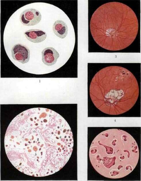
Fig. 1. Purulent infiltration of the conjunctiva: 1. Purulent exudate in the conjunctival sac; 2. Brownish purulent discharge: the conjunctiva is swollen, thickened, and covered with a fibrinous coating; the discharge is abundant and contains numerous gonococci; 3. Purulent discharge with a coal-like coating on the 3rd and 4th days. Fig. 2. Purulent discharge on the 2nd day. Fig. 3. Purulent discharge on the 3rd day. Fig. 4. Purulent discharge on the 4th day. Fig. 5. Purulent discharge on the 5th day. are affected either multiple or the damage is limited to any one joint. Most often the knee joint, joints of the hands, etc. are affected. Sometimes periarticular abscesses also develop. The connection of the joint disease with gonoblennorrhea was proven by a number of studies, in which the gonococcus was found, in addition to the secretion of the conjunctiva, also in intra- and periarticular exudates. Gonorrheal inflammation of the joints in newborns proceeds favorably. Sometimes in children with gonoblennorrhea, gonorrheal stomatitis develops, which in most cases also has a benign character. Its origin needs to be explained by the entry of the gonococcus onto the mucous membrane of the oral cavity from the conjunctival sac through the lacrimal passages or through direct transfer by the child's fingers. As for the course of B. of non-gonorrheal origin in newborns, it depends on the nature of the pathogen, but in general, unlike gonoblennorrhea, the clinical picture appears significantly less severe, the process resolves faster and does not give complications from the cornea. Diagnosis of B. Recognition of B. does not present difficulties if there is already an expressed catarrh of the conjunctiva with purulent discharge; however, the etiological diagnosis of the disease cannot always be made based on the clinical picture, and only particularly severe forms of gonoblennorrhea—with pronounced phenomena of infiltration of the conjunctival tissue, with the formation of expressed chemosis of the conjunctiva, with a huge amount of purulent secretion and, especially, in the presence of complications from the corneal membrane—make the diagnosis of gonoblennorrhea possible even without bacteriological research. For accuracy of diagnosis, microscopic examination of the purulent secretion is nevertheless necessary. For the detection of gonococci in the pus of the conjunctiva in the vast majority of cases, only bacterioscopic examination is sufficient. The Neisser gonococcus is well recognized in a smear stained by Gram (see table, fig. 5), since usually it is found in the pus in large quantities, located intracellularly and extracellularly, and in gonoblennorrhea of newborns it is found there, as it were, in a pure culture. For the differential diagnosis of the gonococcus, practically only the catarrhal micrococcus of Pfeiffer (Pfeiffer) or the meningococcus of Weichselbaum (Weichselbaum) can be considered; but these microbial forms, morphologically similar to the gonococcus, are encountered in the conjunctiva in general extremely rarely, especially in newborns and for the picture of gonoblennorrhea they are in any case not characteristic. But still there are cases in which for differential diagnosis the obtaining of cultures is required, and even with respect to the meningococcus serological reactions. In recent years, by the research of Lindner (Lindner), the fact of the regular finding of the gonococcus in the early period of the process of B. in the epithelial covering of the conjunctiva, especially of the eyeball, has been established, and its presence here is sometimes confirmed earlier than it can be detected in the purulent secretion; in some cases it persists in the epithelial covering for a long time. Therefore, sometimes for diagnosis or for clarifying the effect of therapy, it is useful to examine scrapings of the epithelium for the presence of gonococci (with staining, e.g., by Giemsa). Microscopic examination of the purulent secretion allows to detect in cases of B. of non-gonorrheal character other kinds of microbes; in infections of the conjunctiva, the diagnosis of the pathogen is possible only by means of bacterioscopic examination—this or that microbe is encountered one after another (especially the Koch-Weeks bacillus, pneumococcus, staphylococcus, etc.) as if in a pure culture; but there are cases where it is impossible to do without bacteriological research; this especially applies to the establishment of diphtheria of the conjunctiva, which can sometimes manifest in the form of B., and the recognition of which, in the frequent presence in the conjunctiva of the diphtheria group bacillus—B. xerosis, requires, first of all, differentiation from this bacillus. In cases of B., where no microbes are found (in the so-called amicrobic B.), it is necessary to conduct research for the presence of intracellular inclusions (see Trachoma). These inclusions are found in B. of newborns with great constancy and in abundant quantity (see table, fig. 1).-Prognosis. In prognostic respect, the most formidable process is B. of gonorrheal origin; the prognosis is incomparably more favorable if it is established that B. is caused by other microbes, especially, by Koch-Weeks bacilli, pneumococci, staphylococci; a favorable prognosis in terms of danger to the organ of vision is also given by B. with inclusions. Especially bad in terms of prognosis is considered gonoblennorrhea in those cases when an extremely expressed stage of infiltration is observed and, especially, excessive chemosis of the conjunctiva of the eyeball, causing disturbance of nutrition of the cornea and promoting its damage. When complications from the cornea appear, the prognosis becomes extremely serious, and in the picture of a purulent keratitis prognosis pessima is set. Prevention. B. of newborns represents a suffering in which the importance of prevention is manifested with particular clarity. This suffering, to which blindness is owed by a very large number of blind people both in the USSR and in the West (according to Pokrovsky, B. of newborns causes blindness of childhood age in 18.4% in the USSR), can be in the vast majority of cases reliably prevented by appropriate preventive measures. The basic preventive measure, naturally, would be the prevention of gonorrheal diseases of the sexual pathways; in this respect, energetic struggle with venereal diseases has great importance for the struggle with the spread of gonococcal diseases of the conjunctiva of the eye. But, besides this, there is another powerful preventive means, the so-called method of Credé (Crede, 1881), consisting in that to the child immediately after birth, after cleansing the eyelids and face from the existing mucus, 'smear', a drop of 2% solution of argol is instilled into each eye. This method proved, despite the skepticism manifested at first, extremely effective. According to the observations of Credé himself in the Leipzig obstetric institution, before the introduction of Credé prevention, gonoblennorrhea occurred in up to 10.8% of cases; after the introduction of the method the percentage fell to 0.2-0.5; such favorable results are noted everywhere. Objections to the application of the Credé method boil down to the fact that after cauterizing the delicate connective membrane of the newborn with argol, sometimes conjunctivitis (Argentumkatarrh of the Germans) are observed with mucous or mucopurulent secretion (in rare cases with prolonged course). On the basis of this, attempts were made to replace argol with less irritating compounds of silver, as protargol, sofol, argyrol, etc.; of these means particularly good results are obtained from the application of 5-10% solution of protargol and sofol. The most reliable preventive means remains and at present time the Credé method; the non-dangerous catarrhs of the conjunctiva observed after it cannot be taken into account when it comes to preventing the most dangerous infection of the eye. Thus, the preventive significance of the Credé method is indisputable; the question consists only in that its application should be compulsory in every case of childbirth; therefore it is necessary the widest information about the obligatory application of this method not only of the medical personnel, but also of the masses of the population, especially of women-mothers. The Credé method is effective only as preventing the development of gonoblennorrhea in case of infection during the act of childbirth; subsequent infections by the introduction of infection from outside can be prevented only by careful cleanliness of everything coming in contact with the child's eyes. It is also necessary to note that the Credé method is not preventing the development of B. with inclusions; this is testified to by a number of observations where after the correct application of this method amicrobic B. with inclusions developed. Treatment of B. The basic principles of therapy of gonoblennorrhea are, as in other infections of the connective membrane, the destruction of the pathogen by the action of bactericidal means, the freeing of the conjunctival sac from the purulent secretion accumulating abundantly in it at the height of the process, the creation of the most favorable conditions for the struggle of the organ, resp. organism, with the penetrated infection.
The first indication is fulfilled by cauterizing the conjunctiva with a 1-2% solution of lunar caustic, but the use of this remedy is inadvisable in the first stage of the process when there are pronounced phenomena of infiltration, vascular stasis, and when the secretion is scant; when the period of pyorrhea sets in, cauterization of the conjunctiva with lunar caustic is an extremely valuable therapeutic agent. Cauterization is usually performed once a day; only with very abundant purulent discharge is it necessary to perform a second cauterization during the day, or even better, to instill a 5-10% solution of sofol or protargol. In the period of infiltration, the instillation of protargol or argyrol is also appropriate. The cleansing of the conjunctival sac from purulent secretion is accomplished by systematic washings with various disinfectants, of which solutions of mercuric cyanide (1 : 3,000-5,000) and potassium permanganate (1 : 2,000) are particularly commonly used. These solutions are used to wash the eye every x/s-1 hour depending on the amount of secretion, with care taken not to damage the cornea. When complications from the cornea occur, treatment of the primary disease with lunar caustic cauterization should not be stopped; only in this case it is necessary to take all precautions to protect the cornea during all manipulations, carefully covering it with everted eyelids, and neutralizing after smearing or instilling lunar caustic by subsequently instilling 1% sodium chloride. The treatment of the corneal inflammation itself is conducted according to the usual rules for treating purulent keratitis. When the process passes into the stage of papillary hypertrophy, the use of astringent agents is indicated. In addition to such local treatment of gonoblennorrhea, protein therapy, mainly in the form of milk therapy, has become widely used in the last decade. Milk is administered from the very first days of the disease intramuscularly, in small doses, from 0.2 to 0.5 cubic cm at a time, with intervals of 1-2 days; usually a favorable effect is already observed after 2-3 injections. Many have also seen good success with the use of paraspecific therapy in the form of antidiphtheritic serum. Nonspecific therapy for gonoblennorrhea in newborns is currently recognized as a very valuable auxiliary agent, which should be used along with the usual local therapy. As for specific therapy for gonoblennorrhea in the form of the use of antigonococcal serum or vaccine, to date such therapy does not give reliable results. B. of adults of gonorrheal origin (blennorrhoea adultorum). Gonoblennorrhea in adults, also being an acutely occurring infection of the conjunctiva due to infection with the gonococcus, passes through the same clinical stages as gonoblennorrhea in newborns, but differs from the latter primarily in the mode of infection. In it, the infection is usually carried by the patient himself, suffering from a gonorrheal disease of the genitourinary tract, to his own eye by means of fingers or contaminated objects; in this case, the infection manifests itself through a very short incubation period, sometimes after a few hours, and rarely is the incubation longer than 1-2 days. Due to the conditions of infection, gonoblennorrhea in adults occurs in one eye, more often the right one, and with certain preventive measures, the process is limited to one eye. The disease, starting acutely, increases in intensity extremely rapidly and often passes within 1-2 days from the stage of infiltration to the stage of pyorrhea. Despite the general similarity of clinical symptoms and course, gonoblennorrhea in adults differs, in general, in greater severity of the process compared to B. in newborns. In this form, extreme degrees of eyelid tension, infiltration of the conjunctiva with a huge increase in the volume of the conjunctiva, and especially pronounced edema of the conjunctiva of the eyeball, forming a fold hanging over the edge of the cornea, are more often encountered. Here, the formation of conjunctival membranes resembling the picture of pseudomembranous conjunctivitis is more frequently observed. In the stage of pyorrhea, an enormous amount of purulent discharge is often noted here. Furthermore, in gonoblennorrhea in adults, patients complain of severe pain in the eye area; opening the tense eyelids is also very painful; the parotid glands on the corresponding side often become swollen; sometimes there is a febrile condition, but the most dangerous is the complication from the cornea, which occurs more often than in gonoblennorrhea in newborns. The appearance of complications from the cornea is sometimes noted in the very first days of the illness, still in the stage of infiltration, but more often it is observed in the stage of pyorrhea; and the earlier this complication appears, the more severe its course. As with gonoblennorrhea in newborns, the corneal lesion here begins either from the periphery or in the center; but in either case, it can in a short time lead to destruction of the cornea and subsequent blindness. Gonoblennorrhea in adults is sometimes complicated by extrabulbar processes; thus, in a number of cases, lesions of the joints of the same metastatic nature as in gonoblennorrhea in newborns are noted. The diagnosis of gonoblennorrhea in adults is made by microscopic examination, by confirming the presence of gonococci in the conjunctival secretion (in its scrapings, usually in enormous quantities). The clinical picture itself in its typical form, especially when there is a complication from the cornea, also allows for diagnosis with great probability here, but undoubtedly, there are blennorrheal forms of conjunctivitis due to other causative agents (streptococcus, diphtheria bacillus, etc.), which can cause confusion. It should also not be forgotten that gonococcal infection of the conjunctiva in adults can also give a picture of moderate conjunctivitis; in such cases, the etiological diagnosis can only be made by bacteriological examination. The prognosis of the process is clear from the description of the course of the infection—in general, it is always very serious. The main points for prediction are: the degree of expression of the stage of infiltration and, especially, the condition of the conjunctiva of the eyeball—the stronger the chemosis, the more pronounced the infiltration of the conjunctival tissue, the more data there is to speak of the high virulence of the existing gonococcus, resp. the weak resistance of the body, and the greater the danger of fatal corneal lesions leading to blindness.—Prevention. The extremely dangerous infection of the conjunctiva with gonococcus in adults first of all requires informing every patient with gonorrhea about the danger of introducing purulent secretion into the eye. From this, it is imperative to indicate to such patients the need for careful maintenance of the cleanliness of their hands and surrounding objects to prevent the possibility of transferring the infectious agent from the genital tract to their own eyes or the eyes of others. Sanitary-educational measures, as well as the entire fight against venereal diseases, play a major role here. Under ordinary conditions of infection, gonoblennorrhea in adults, as already indicated, occurs in one eye; the physician's task is to take all measures to prevent the disease of the second eye. Therefore, when an infection occurs in one eye, it should be considered mandatory to seal the healthy eye with a hermetic dressing. The most convenient form of such a dressing is a dressing with a watch glass, the edges of which are glued together with cotton and collodion (see figure). Such a dressing allows observation through the glass of the condition of the eye, and does not deprive the patient of the ability to use the healthy eye.—Treatment of gonoblennorrhea in adults is carried out according to the same rules as for B. in newborns; due to the greater danger of corneal damage, it is necessary to pay special attention here not to traumatize the cornea in all therapeutic measures, the chemosis of the conjunctiva has a beneficial effect.

Preventive dressing in Blennorrhoea adultorum.
one should strive to weaken it (often in this regard, scarification of the chemotically reactive conjunctiva is effective), and carefully cleanse the conjunctiva of the accumulating purulent secretion. Nevertheless, the primary means, and even to the present day, are cauterizations of the conjunctiva with a 1-2% solution of argyrol, 1-2 times a day, prescribed at the beginning of the pyorrhea stage; during the infiltration period, only cautious washing of the conjunctival sac with warm solutions of disinfecting fluids (mercuric cyanide, potassium permanganate, etc.) and instillation of drops of 5-10% protargol, sofrol, 15-25% solution of argyrol are permissible. With the appearance of corneal disease, the treatment of the conjunctival process should not be discontinued here either, as was already indicated when considering the treatment of B. in newborns. Attempts at treatment with specific serum or vaccine have not given satisfactory results, although recently some have noted the successful use of serum, in addition to subcutaneous injections, also in the form of instillations into the conjunctival sac. But in gonoblenorrhea in adults, non-specific therapy in the form of protein therapy has incomparably greater importance than specific therapy. It is believed that the success of treatment with proteins in gonoblenorrhea in adults is higher than in B. in newborns; but here too this therapy should be used as an auxiliary means to local treatment, since only the combined method gives the best success. Milk is administered in the treatment of gonoblenorrhea in adults, in doses of 1 to 3-5 cubic cm, intramuscularly, with intervals of 1-2 days, and 4-5 injections are usually sufficient. Such therapy noticeably shortens the course of the process and sometimes remarkably rapidly changes the clinical picture, transforming the infiltration stage into the pyorrhea stage and during the latter changing the character of the purulent secretion, as well as causing a relatively more rapid disappearance of the gonococcus from the epithelial covering of the conjunctiva and secretion, which results in less frequent corneal disease. In complications from the cornea, protein therapy often beneficially affects the course of this process. In addition, protein therapy more consistently alleviates, and sometimes completely eliminates, the pains that bother patients. Thus, in the therapy of gonoblenorrhea in adults, it is necessary to combine local therapy with protein therapy.
Related articles
Mentioned in
Cite this page
“Blennorrhea.” Soviet Medical Encyclopedia. English translation of Bolshaya Meditsinskaya Entsiklopediya, 1st ed. (Moscow, 1928–1936), ed. N. A. Semashko. https://sovietmedicalencyclopedia.pages.dev/article/blennorrhea/