Esches Operation
Historical document, translated for reference. It reflects medical knowledge of the 1920s–30s and is not medical advice.
Summary
This article from the 1928-1936 Soviet Great Medical Encyclopedia describes Esches operation, a surgical procedure proposed in 1844 for treating upper eyelid entropion caused by trachoma. It details the technique of dividing and repositioning the ciliary margin of the eyelid and discusses its historical context and variations.
Encyclopedia article (1928–1936)
ESCHES OPERATION, against entropion of the upper eyelid in trachoma, was proposed by Esche (an ophthalmologist from Nizhny Novgorod) back in 1844, but in textbooks and even in large manuals this operation is incorrectly described under the name "Esche-Arlt" (Arlt), although in fact it differs sharply from the operation of the latter author. Esche himself describes its performance as follows: having pulled down the upper eyelid with the thumb of his left hand, the author pierced it through its entire thickness from the conjunctival side with a triangular knife curved along the plane, which was punctured through the cartilage 3-4 mm above the incision, passing parallel to the ciliary margin.
so that the plane of the knife was parallel to the edge of the eyelid. Having obtained in this way a small incision through the entire thickness of the eyelid, Esche uses a sharp hook
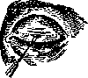
Figure 1.
the line of incision is designated, introduced into this incision (Fig. 1), pulls back the ciliary margin, which makes it possible to introduce one blade of Cooper's scissors into it, with which the incision is enlarged in both directions parallel to the ciliary margin, as a result of which the entire separated ciliary margin hangs down, maintaining connection with the rest of the eyelid at its corners (Fig. 2). Next, a second incision is made on the eyelid parallel to the first, going however only to the cartilage, i.e., through the skin, tissue, and muscle layer, and connected at the ends with the first. Then, using scissors, the outlined skin-muscle
flap is removed.
The width of the flap varies depending on the size of the entropion. After stopping the bleeding, 5-6 sutures are applied so that the needle is passed through the skin edge of the wound on the eyelid, and then guided between the skin-muscle layer and the cartilage of the severed ciliary margin and brought out right above the eyelashes (Fig. 2). The sutures are tied so that not only is the ciliary segment moved away from the cornea, but the eyelashes look forward and up. The sutures are not cut, and their ends are glued with plaster to the skin of the forehead. A bandage is applied, changed daily, for 5-6 days, then the sutures are removed, along with the bandage. Subsequently, this operation was performed with the modification that a Jaeger plate was placed under the eyelid or Snellen or Knapp forceps were used, correspondingly, the excision of the ciliary margin is performed with a scalpel from the anterior surface of the eyelid, while the skin, muscle, and cartilage with conjunctiva are immediately dissected. In this way, it is easier to excise a uniformly wide ciliary segment than with scissors. Otherwise, the operation ends in the same way as in the original method. This method was widely used in the Volga-Kama region and yielded good results.
K. Orlov.
Related articles
Cite this page
“Esches Operation.” Soviet Medical Encyclopedia. English translation of Bolshaya Meditsinskaya Entsiklopediya, 1st ed. (Moscow, 1928–1936), ed. N. A. Semashko. https://sovietmedicalencyclopedia.pages.dev/article/esches-operation/