Blendes
Historical document, translated for reference. It reflects medical knowledge of the 1920s–30s and is not medical advice.
Summary
An overview of diaphragms and blending devices used in X-ray diagnostics during the 1920s and 1930s to improve image sharpness by eliminating scatter and stray radiation.
Encyclopedia article (1928–1936)
BLENDES [from German Blende - diaphragm (in optics)], diaphragms used in X-ray diagnostics (fluoroscopy and radiography) to obtain sharp images. Unclear X-ray images are produced for two reasons: 1) due to radiation originating not only from the tube focus (glass radiation, stem radiation), and 2) due to the appearance of scattered X-rays in the human body having various directions. Blendes serve to eliminate these phenomena. Their simplest variety is the iris blende, consisting of a series of lead plates which, by means of a special lever, are moved together and apart, similar to the diaphragm in photographic lenses. With the help of such a blende, it is possible to pass a beam of rays that is truly necessary to obtain an X-ray image of a specific part of the human body. From Fig. 1, which schematically depicts such a blende, it is seen that part of the glass radiation nevertheless passes through it and obscures the X-ray picture. To eliminate this phenomenon in radiography, Albers-Schönberg (see) designed his compression blende (Kompressionsblende). It is a metal cylinder lined on the inside with lead, which is pressed by means of a special device against the part of the body to be radiographed. Thanks to this arrangement (see Fig. 2), rays not originating from the tube focus are absorbed by the blende, and the radiograph is not obscured by them. This blende has the further advantage that the body part being imaged can be firmly pressed with it, which, on the one hand, leads to a steadier position of the subject, and on the other hand, to a reduction in the volume of the imaged body part, the consequence of which is a decrease in the amount of scattered rays. Varieties of this blende are the Rosenthal and Robinson blendes. All these blendes (except the iris one) also reduce the amount of scattered rays originating from the organism, but only
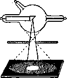
Figure 1.
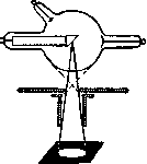
Figure 2.
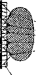
at a limited (compressed) site. To eliminate them in more extensive areas, Bucky adapted his Wabenblende, which is placed between the screen and the object under study. It is a metal frame, measuring 30x40 cm, inside of which in the longitudinal and transverse directions thin copper strips 4.5 cm wide are arranged. These plates are located at a certain inclination, corresponding to the path of the primary X-rays, thanks to which the entire blende appears as if consisting of individual small cells, each of which forms a truncated pyramid. The continuations of the side planes of these truncated pyramids converge at one imaginary point located at a distance of 70 cm from the blende, where the tube focus is also located. Fig. 3 shows its external appearance, and Fig. 4 shows a schematic representation, from which it is seen that only primary X-rays (dashed line) pass through the blendes, while scattered rays (solid lines) originating from the body are absorbed by it. These blendes are used in fluoroscopy, while in radiography, an image of the grid from the plates is obtained on the plate,
[ILLUSTRATION]

Figure 4.
which obscures the picture. For radiography, the Bucky-Potter rollblende is used. It has only a single row of strips, between which thin wooden plates are placed, fastening the strips together. The metal strips are arranged so that they form a segment of a cylinder, and each strip is located along the radius of the cylinder. In view of this, the planes passing through these plates intersect along a single straight line, which is located at a distance of 60 cm from the blende, where the tube focus is also located. This entire system of plates during exposure is automatically set in motion around the imaginary axis of the cylinder by means of a special device. The speed of rotation depends on

Figure 5.
the duration of the exposure and can be regulated within the range of 2 to 150 seconds. Thanks to such an arrangement, the image of the grid does not appear on the plate. Fig. 5 shows the external appearance of such a blende, and Fig. 6 shows such a blende together with a special device for placing the tube, allowing a certain focal distance (60 cm) to always be maintained, and a special canvas belt for compressing the imaged area. A blende suitable for fluoroscopy and radiography is Bucky's Drehblende. In it, the metal plates are arranged along the radius of a circle;
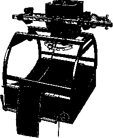
FIG. 6.
the tube is located on the axis passing through the center of the circle, perpendicular to it, at a distance of 60 cm from the blende. By means of a special motor, the plates are driven into rotational movement around this axis. The speed of rotation can be regulated arbitrarily, so that the image of the blende on the screen or plate does not appear. Fig. 7 shows the grid of the blende, and Fig. 8 shows its external appearance. A modification of such a blende is the Akerlund blende, consisting of lead plates serving to absorb scattered rays and arranged

Figure 8.
of the ray so as not to obtain an image of the circles on the photographic plate. The lead plates are driven into rotational movement, the speed of which depends on the exposure and is regulated automatically. When using such blendes, the exposure time increases due to the absence of scattered rays.
M. Kanikov.
Related articles
Mentioned in
Cite this page
“Blendes.” Soviet Medical Encyclopedia. English translation of Bolshaya Meditsinskaya Entsiklopediya, 1st ed. (Moscow, 1928–1936), ed. N. A. Semashko. https://sovietmedicalencyclopedia.pages.dev/article/blinds/