Bacteria
Historical document, translated for reference. It reflects medical knowledge of the 1920s–30s and is not medical advice.
Summary
This article provides a comprehensive overview of bacteria, covering their morphology, biology, distribution, and historical study. It details their basic forms, cellular structure, and prevalence in various environments.
Encyclopedia article (1928–1936)
BACTERIA. Contents:* General morphology of bacteria........6 70 Degeneration of bacteria............675 Biology of bacteria..............676 Acidophilic bacilli ........... 677 Pigment-producing bacteria.......681 Luminescent bacteria..... .......682 Sulfur bacteria................684 Pyogenic bacteria.............685 BCG bacillus (BCG)......685 Bacteria (from Greek bacteria-rod), single-celled living organisms, by their morphological and biological characteristics belonging to the plant world. The main milestones in the history of the study of B. are as follows: first saw them in the microscope Kircher (Kircher) in the 17th century (1683); Antonie van Leeuwenhoek (Anto-nius Leeuwenhoeck) first gave their image; in 1786 Friedrich Müller (Fried-rich Muller) isolated bacilli, spirilla, and vibrios; in 1876 Ferdinand Kohn introduced the term "bacteria".-B. were first studied mainly in putrefying liquids, which created the idea of their spontaneous generation from putrefying organic matter. This idea was destroyed by Pasteur in 1862, proving that fermentation processes are caused by B. In 1897 Buchner (Buchner) showed that fermentation can be caused not only by a living microorganism but also by the enzyme contained in it. In 1876 Koch introduced dense nutrient media for growing B., which made it possible to obtain them in a pure form.- Distribution of B. The main environment in which B. are found are soil and water; from there they are passively carried into * Groups of bacteria not included in the present article should be looked up under the corresponding word; for example: nitrogen-fixing bacteria-Nitrogen-fixing bacteria, putrefactive bacteria-Putrefactive bacteria; individual species: Vibrios, Bacterium coli commune, Gonococcus, Spirochetes, etc.
I air by air currents. If a Petri dish with nutrient agar or gelatin is left open in a room, then in 1 minute 1-10 B. will settle on the surface of these media. In 1 cubic meter of air in the center of Paris there are 330-1,540 B., In good drinking water there are at most 100 B. in 1 cubic cm:, in wastewater in 1 cubic cm there are up to 1 million. Good, poor in B., milk contains in 1 cubic cm 1,000-7,000 B., in contaminated milk there are 1-200 million. In "normal" soil, in 1 g of dried soil-up to 100 million B.; sandy, poor in organic substances, soil contains much less of them (about 100,000 in 1 g); conversely, humus-rich soil in 1 s contains hundreds of millions of B. One g of fresh human or animal excreta contains 20-40 billion B., and this bacterial mass constitutes approximately half of the dry matter of the excreta. General morphology of bacteria. Size of B. The diameter of a bacterial cell is generally equal to 1 µ; for comparison, it can be noted that the diameter of a plant cell of higher plants is 10-90 µ. The longest forms of B. are found among spirilla; thus, Spir. rubrum reaches a length of 100 µ with a diameter of 1 µ.; relatively long are B. of soil; the smallest are cocci. Among the shortest B. can be named: Spir. parvum (0.1-0.3 µ x 1-3 µ), Bac. mu-risepticum (0.2 - 0.3µ x 1 µ), the influenza bacillus and others. Short forms are thus also found among spirilla and bacilli. The question of the size of B. is closely related to the question of their filterable forms. Theoretically, it is difficult to imagine that a living cell could be infinitely small; one must think that only in rare cases can B. be found smaller than the minimal visible size in the microscope, and in all probability, it is usually a matter of certain, short-lived stages of existence of B.-Basic forms of B.-cocci, rod and spirally curved form - spirillum. The primary form is the cocci as corresponding to the form that a liquid takes when left to itself and not experiencing any effects from the surrounding environment. From the perfectly spherical form of the cocci, deviations are not uncommon: sometimes one pole sharpens and takes a lanceolate shape. Sometimes the cocci is flattened on one side or takes an elliptical shape. Bacilli have a cylindrical, sausage-shaped form with rounded ends, with deviations not being uncommon: rods with pointed or sausage-shaped swollen ends, slightly curved forms, etc. Spirilla are spirally curved forms; transitional from rods to spirilla are vibrios (see Figure 1). The bacterial cell is surrounded by a capsule, the existence of which was long disputed, because when staining B. the capsule cannot be seen. It is noticeable, however, when examined in the dark field of view, as a light line sharply delimiting the B.; it can often be seen on stained preparations as a light belt surrounding the B. Undoubted proof of the existence of the capsule was given by A. Fischer (Fischer) in his studies on plasmolysis; placing B. in a liquid with a higher osmotic pressure than in their protoplasm, he obtained extremely clear pictures: the protoplasm shrinks, Figure 1. Schematic representation of various types of bacteria. takes the form of round or elliptical formations and moves away from the capsule; the latter thereby sharply stands out as a line (see Figure 2). Chemically, the capsule of B. consists not of cellulose (as is the case in higher plants), but of hemicellulose or hemicellulose-like (pectin-like) substances; products of its decomposition are dextrose, galactose, arabinose. In addition to the capsule, B. is also surrounded by a mucous capsule; these capsules exist in any case in very many B. The layer of mucus around the bacterial cell arises from the secretion of mucus by the capsule; more rarely its cause is the swelling of the capsule. It is easiest to verify the existence of a capsule by adding B. to not too diluted ink; the layer of mucus in the form of a light belt borders the dark from ink bacterial cell. Chemically, the capsule, like the capsule, consists of hemicellulose. The significance of the capsule is that, due to its presence, B. has a definite shape: naked protoplasm, by its consistency, would inevitably have to take a spherical shape. The capsule of B. protects it from drying out and protects it from direct contact with various particles. Speaking of the mucous layer surrounding B., it is necessary to remember the following: what in medical microbiology

Fig. 2. Schematic representation of plasmolysis of bacteria. is described under the name of capsule, is most often the capsule + capsule.- Protoplasm (cytoplasm) of B. is little studied. It is known, however, that in them, as in plants in general, the outer layer of cytoplasm forms a membrane of great importance as a regulator of metabolism in the cell; this membrane (Plasmamem- bran) represents a hydro-colloidal system. Previous researchers denied the existence of a nucleus in B. or assumed that chromatin is diffusely distributed in the protoplasm. According to the research of Arthur Meyer (Arthur Meyer), however, it is necessary to admit the existence of a typical nucleus. In Bac. tumescens (found in soil) the nucleus has a size of 0.2-0.3 µ (with a cell size of 1.7x7 µ); these ratios correspond to those observed in plants in general. According to Kirchenstein (Kirchenstein), the nucleus of the tubercle bacillus has the appearance of the smallest grain; this researcher succeeded, using a special staining method, in observing the division of the nucleus. Vacuoles can be counted among the normal components of the bacterial cell. In a young cell there are none; later they appear, gradually increasing and often merging with each other; they contain water with mineral substances or various cellular inclusions.- The organ of movement of B. are flagella. The number and arrangement of flagella in motile bacteria differ in great constancy; in this respect
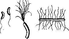
a
b
c
Figure 3. Flagella: a-monotrich; b-lophotrich; c-peritrich. Bacteria are divided into monotrichs, which have one flagellum located at one of the poles of the bacterium; lophotrichs, which have a bundle (of 5-40) flagella at one or both poles; peritrichs, which have numerous flagella located over the entire surface of the body, surrounding it on all sides. The flagella pass through the membrane and mucous layer of the bacterium and are connected to its cytoplasm, consisting, apparently, of the same substance as the plasma membrane of the bacterium. The length of flagella is not always the same: in B. subtilis the flagella reach a length of 6-12 μ (with the size of the bacterium being 0.7x2-8 μ). Generally, the relative length of flagella is greater the smaller the bacterium; the thickness of flagella is only about 0.05 μ. Not all bacteria possess independent movement, and therefore not all have flagella as organs of this movement. It can be assumed, however, that many bacteria have lost the ability to move due to abnormal artificial conditions of existence. Proof of this is that certain, undoubtedly immobile, bacteria under certain conditions possess flagella and can move. Thus, in young forms of tubercle bacilli, the ability to move can be demonstrated; the same ability is possessed by young forms of some varieties of Azotobacterium (see fig. 3). - Spores. Many bacteria have the ability to form so-called spores. This ability is possessed by bacilli and (only as a rare exception) cocci (sarcinae). The process of spore formation consists in the fact that first, at a certain place in the bacterium, a thickening of its body occurs; in this thickening, a section of protoplasm gradually becomes more and more differentiated, and, surrounded by a dense membrane, forms a spore. A mature spore is a round or oval, strongly light-refracting formation, surrounded by a dense, difficult-to-penetrate-by-fluids membrane; because of this, spores with great difficulty take on dye. The spore is usually located in the middle of the bacterium; sometimes it occupies one of the poles of the bacterial cell. Soon after the final formation of the spore, the bacterium that produced it is destroyed, and the spore becomes free. The meaning of spore formation is that spores are characterized by sharply expressed resistance; the bacterium forms a spore when its living conditions are unsatisfactory, in particular, when the bacterium lacks food. Upon again finding itself in favorable living conditions, the spore gives rise to a new bacterium; it loses its strongly expressed ability to refract light, the membrane bursts in one place or another, and through the resulting opening a new individual grows (see figure 4). - Bacterial inclusions. The bacterial cell can contain various inclusions, which in most cases represent reserve nutrients. Of carbohydrates, glycogen in the form of droplets is encountered; sometimes starch (staining with iodine in blue color) and others. Fatty substances are also often found inside the bacterial cell, and usually where there is fat, there are no carbohydrates, and vice versa. The following data illustrate this position. 21 bacteria were studied, and it turned out that only carbohydrates were contained in 8 bacteria, only fats in 10, and fats and carbohydrates together in 3. The amount of fat in the bacterial cell is very different; thus, B. mallei contains 39.3% of it, while the diphtheria bacillus only 1.6%. The fat of tubercle bacilli is of great importance; here, however, the matter is not about fats in the strict sense of the word, but simultaneously about so-called waxy substances. The third substance, often encountered in bacteria as a reserve, is protein. Unlike carbohydrates and fats, which cannot be morphologically differentiated within the cell, protein forming the so-called volutin is encountered in bacteria in the form of grains ('metachromatic bodies of Babes-Ernst'; see figure 5). - Coloring substances of bacteria are of two kinds:

Figure 5. Volutin (grains) in the protoplasm of Spirillum volutans.
some are secreted outward, coloring the surrounding environment, others are secreted into the bacterial cell, coloring the cytoplasm in the corresponding color. The most well-known examples of bacteria of the first category can be - B. pyocyaneus (rod of blue pus), which secretes a blue coloring substance, soluble in water and chloroform; B. prodigiosus, which secretes prodigiosin in the form of small grains; in an acidic environment it is ruby-red, in an alkaline environment it canary-yellow, etc. A pigment is associated with the bacterial cell in the group of purple bacteria; usually here one has to speak of a red or green pigment.
Evolutionary forms of Bacteria are observed in those bacteria that are in unfavorable living conditions, in which most often forms with club-shaped swellings, thickenings, as well as branching forms develop. However, in the latter case, it usually goes about false branching; true branching forms, observed in spirilla, tubercle, diphtheria, and glanders bacilli, can hardly be attributed to degenerative evolutionary forms (see figure 6).
Systematics of Bacteria. There is no scientific classification of bacteria; their division into groups is based mainly on morphology.

Figure 6. Evolutionary forms of a bacillus.
signs. If we limit ourselves to B. in the strict sense, excluding all thread-like forms of microbes, then the simplest classification will be as follows: A). Eubacteria: 1. Coccaceae (streptococci, micrococci, staphylococci-division in one, two and three planes). 2. Bacteriaceae (B.-without flagella, bacilli-with flagella); bacilli are divided into: a) those without spores; b) spore-bearing, in which the spore-bearing bacillus is not thickened; c) spore-bearing, having the form of clostridia, in which the spore-bearing bacillus is spindle-shaped thickened; d) spore-bearing, having the form of plectridia, in which the spore-bearing cell is thickened at the pole and resembles a drumstick, and e) pseudomonas, with flagella located at the pole-monotrichous. 3. Spirillaceae (vibrios, spirilla). B). Mycobacteria, characterized by irregular body shape and often observed branching. They include the rods of sap, tuberculous and paratuberculous bacilli, diphtheria bacilli. Close to bacteria are Chlamydobacteria, which are thread-like forms, unbranched or forming false branches (for example, Cladothrix, Leptothrix, etc.). Degeneration of B. In contrast to involutionary forms, which sometimes possess both viability and virulence, degenerative forms of bacteria are distinguished by reduced growth energy and decreased resistance. The morphological features of degenerative forms are very different. Thus, pneumococci on artificial nutrient media can form long, irregular, rod-shaped forms; in old cultures of anthrax bacillus, long, curved threads are formed, with apparent branching; in these two cases, growth energy is preserved, while the energy of reproduction (resp. division) is reduced. In other cases, growth energy is reduced while the ability to reproduce is preserved; as a result, small, irregular, coccal formations are obtained. The final stage of degeneration is the disintegration of the microbe into small grains. An example can be provided by the bacilli of tuberculosis: in old cultures, as well as under certain conditions in the body, they turn from solid rods into forms that represent a series of grains; along with such granular rods, freely lying grains are also encountered. These grains are "living" grains, because under favorable conditions, typical rods arise from them. The final product of degeneration of the tuberculosis bacillus is a yellow-brown and black pigment. Degenerative forms arising in a living organism are not always identical to those appearing in artificial cultures. Thus, the plague bacillus in a bubo gives a form in the form of irregular lumps, rings, etc.; in pneumococci in the body, the body sometimes disappears before the capsule, so that at a certain moment empty capsules or with remnants of the body are encountered. Degenerative forms are not always dead forms; fully preserved microbes can be dead, and on the other hand, from the grains into which the microbe has disintegrated, new, fully developed bacteria can arise. Biology of bacteria. Reproduction of B. occurs by transverse division. In young cultures, under favorable conditions, two successive divisions occur in 17 min.; in old cultures-significantly longer. B. equipped with flagella possess independent movement. Its speed is small: according to Lehmann and Fried, the average speed in rapidly moving species (cholera vibrio) does not exceed 30 µ in 1 sec. Being particles suspended in liquid, B. also exhibit the ability to molecular (Brownian) movement. With respect to temperature, B. are divided into: 1) psychrophilic (minimum 0°, optimum 15-20°, maximum 30°; example-many species of aquatic B.); 2) mesophilic (minimum 9-30°, optimum 28-38°; maximum 43-50°; almost all pathogenic B. and most saprophytes belong here); 3) thermophilic (minimum 40-49°; optimum 50-60°; maximum 60-75°; B. of hot springs and others belong here). B. are little sensitive to cold; the low temperatures observed in nature cannot kill them; they are sensitive to high temperature: most B. (of course, except thermophilic ones) perish from the action of 56° within 1 hour; at 80°, 1 minute is sufficient. Wet heat in combination with increased pressure has a particularly destructive effect. Light B. (except "purple" ones) is not needed. Sunlight acts on them destructively, artificial light is also harmful. In a dried state, B. remain alive for some time, and in this respect there are large differences between different species of B.: meningococci and cholera vibrios, when dried, die within a few minutes or hours, while staphylococci remain alive for several years. The chemical composition of B. varies greatly depending on the species of bacteria, the composition of food and the temperature of cultivation. On average, water in B. is 85%. In the dry residue, 40-70% is protein, 10-30% is carbohydrates and 3-30% is mineral components; there is little fat (see above). Food must absolutely contain nitrogen, oxygen, carbon, sulfur and phosphorus, and among metals-potassium and magnesium. According to their ability to assimilate necessary nutrients from simple substances or from complex compounds, B. are divided into: 1) prototrophic, capable of assimilating them in elementary form (e.g., C or N as such); 2) metatrophic, assimilating elements only from their compounds (autotrophic-from inorganic compounds: C from CO2 etc., N from nitric acid etc.; heterotrophic-from organic compounds); 3) paratrophic, capable of living only in a living animal organism. All these properties have either an obligatory or a facultative character. With respect to oxygen, B. are divided into aerobes and anaerobes. Strictly obligatory anaerobes do not exist at all; all anaerobes can grow with slight access to oxygen. It should be noted that in certain cases anaerobes can live under aerobic conditions; excess food (mainly peptone), the presence of adsorbing substances, etc. can influence in this direction. B. can exhibit diverse chemical action. Putrefaction is a process caused by B. and consisting in the decomposition of organic substances (mainly proteins), with the formation of foul-smelling, gaseous products. In the process of putrefaction, anaerobes (Bac. putrificus) mainly participate, and among aerobes-proteus, hay bacillus, colon bacillus, etc. Products of putrefaction: peptone, albumoses, amino acids, indole, skatole, phenol, fatty acids, etc. In the processes of fermentation, i.e. in the transformation of carbohydrates, B. also participate. The most important are-alcoholic fermentation (yeast, but also various B.), i.e. the breakdown of sugar into alcohol and carbonic acid; lactic acid fermentation (Kruze streptococcus, long lactic acid bacilli, etc.), i.e. the formation of lactic acid from milk sugar, etc.; depending on the composition of the nutrient medium (on the presence of sugar in it), B. are capable of forming acid (acidophilic bacilli). They are also highly characterized by oxidative ability, e.g. the transformation of alcohol into acetic acid, ammonia-into nitrous, nitrous acid-into nitric, sulfur-into sulfuric acid, ferrous oxide-into ferric oxide, etc. This ability is expressed differently in various species of B. An example of the reducing ability characteristic of many B. is denitrification, i.e. the transformation of nitric acid into nitrous, ammonia and free N. B. have the property of forming enzymes (ferments). Such are diastase, sucrase, lactase, proteolytic ferment, lipase, urease, oxidases, reductases, catalase. Pathogenic microbes have the ability to secrete toxins (exo- and endotoxins). The moment determining the significance of B. is their role in the processes of putrefaction (nitrogen cycle), fermentation, as well as their role in the pathology of animals and humans. With respect to the animal and human organism, B. are divided into parasites, growing and multiplying only in living tissues, and saprophytes, living exclusively in dead organic substrate. Facultative or conditional parasites are B., which are characterized by a dual way of life-parasitic and saprophytic (for the methodology of studying B.-see Staining of bacteria and Bacteriological analysis).
V. Lyubarsky. Pigment-forming bacteria, microorganisms colored in various shades of all possible colors by means of colored metabolic products they form. These products can be insoluble or soluble in water. In the first case, the bodies of B. are colored, while the medium remains colorless; in the second case, on the contrary, the bodies of B. are more or less weakly colored or completely colorless, while the medium is colored by the dissolved pigment. According to their biochemical features, pigment B. differ greatly. Among them there are representatives of all possible groups: agents of putrefaction, fermentative organisms, pathogenic microbes, etc. Most colored B. produce pigment only with access to air; under anaerobic conditions, if they grow at all, they give colorless races. Scattered sunlight has no effect on pigment formation; direct sunlight,
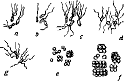
Figure 7. Pigment bacteria: a-Bact. fluorescens liquefaciens; b-Bact. pyocyaneum; c-Bact. syncyaneum; d-Bact. prodigiosum; e-Staphylococcus; f-Sarcina lutea; g-Bact. violaceum (magn. 1,500).
Paralyzing B., also stops the production of pigments by them. Among colored B. of various shades, the following can be named: Bact. fluorescens liquefaciens, Bact. pyocyaneum, Bact. syncyaneum, Bact. prodigiosum, Staphylococcus, Sarcina lutea, Bact. violaceum and others (see figure 7). Pigmenting B. differ in the nature and properties of the pigments they form. Bacteria luminous, or photobacteria, form in their bodies photogenic substances that emit light upon oxidation; luminescence is never observed under anaerobic conditions (in the absence of air). Most luminous B. belong to marine species; freshwater B. are less common. To cause meat or fish to luminesce, it is sufficient to immerse them in a 3% solution of table salt and leave them in a cool place at 9-12°. Luminescence usually occurs after several days. The luminescence of photobacteria represents a unique form of energy release during the oxidative processes taking place in the body of B. The stronger the flow of air to B., the brighter the luminescence. That is why the sea glows more brightly where the wheels of a steamer leave a foamy trail on the water. In a tall cylinder with a culture of luminous B., usually only the upper part of the liquid glows; in the deeper layers

Figure 8. Culture of Photobacterium italicum on fish agar, photographed in a dark room without external illumination. Exposure-2 days.
the light quickly goes out due to lack of oxygen; but if the liquid is shaken vigorously, it lights up evenly throughout its entire mass. In Fig. 8 is depicted a culture of Photobacterium italicum, photographed in a dark room in its own light without external illumination. The luminescence of photobacteria usually begins on the second day after infection at 15° (this is generally the best temperature for most photobacteria). The appearance of a dense nutrient medium with individual colonies of photobacteria resembles a picture of the starry sky. Having reached its greatest intensity, the luminescence of the culture in the following
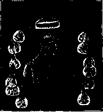
Figure 9. Bust of Claude Bernard, photographed by Dubois when illuminated by 13 "living lamps" containing luminous bacteria.
days gradually weakens, however, sometimes lasting for weeks.-In their nature, bacterial light resembles the luminescence of phosphorus in the dark or moonlight; differing in characteristic shades in different species, it is sometimes bluish, sometimes slightly green, and almost white. The intensity of light of luminous B. is relatively small; however, already with the luminescence of one test tube with a culture of photobacteria, one can count the reading of the second hand of a pocket watch. Despite the poverty of light of photobacteria in chemical rays, it is possible to take photographs; thus, in Fig. 9 is depicted the bust of Claude Bernard, photographed when illuminated by 13 "living lamps" containing cultures of photobacteria. Luminous bacteria are very sensitive to adverse effects (bright sunlight, high temperature, chemical poisons), but cold almost does not affect them: by freezing a luminous emulsion of photobacteria, one can obtain luminous ice. The luminescence of various fish and meat products is much more common than is generally thought. From a sanitary point of view, luminous products should be considered completely safe, since photobacteria are not pathogenic for humans, especially the commonly occurring on meat and fish species Bact. phosphoreum, which does not grow at temperatures above 30° and therefore cannot develop in the human body. The luminescence of meat rather serves as a guarantee that putrefactive processes have not yet begun in it, since B. of putrefaction would immediately suppress the development of photobacteria. Among fishermen there is even a belief that herring is good as long as the "phosphorus" remains on it, i.e., as long as it luminesces. Sulfur bacteria, in the broad sense of the word, are bacteria that participate in the cycle of sulfur in nature; in the narrower sense of the word, sulfur bacteria, or thiobacteria, are the group of bacteria studied by S. N. Vinogradsky that oxidize hydrogen sulfide with the formation of water and sulfur (2H2S + O2 = 2H2O + S). Sulfur is deposited in the body of thiobacteria in the form of semi-liquid droplets (see figure 10)-a sign by which thiobacteria can be easily distinguished under a microscope. The sulfur deposited as a reserve substance is then further oxidized with the formation of sulfuric acid: S + 2H2O + 3O2 = 2H2SO4. This oxidative process plays the role of a respiratory act in thiobacteria. The biological significance of thiobacteria lies in the fact that they convert hydrogen sulfide, which is poisonous to plants, into sulfuric acid, which is well absorbed by plants in the form of salts. Sulfur bacteria can almost always be found in /31 Figure 10. View of the filament Beggiatoa alba: a-in a liquid rich in H2S (the filament is filled with droplets of sulfur); b-after being for a day in a liquid not containing H2S (the filament contains only individual droplets of sulfur); c-after another two days (there are no droplets of sulfur, the protoplasmic content lagging behind the filaments is visible). - Magnification 900. ponds and swamps, on the bottom of which animal and plant residues undergo decay with the release of hydrogen sulfide. Sulfur B. are also found in sulfur springs and sources, covering their bottom with a fluffy carpet of white, reddish, and violet color. In waters containing sulfate salts, for example, in sea and lagoon water, especially in shallow coastal areas of the sea where hydrogen sulfide is almost always present in solution, sulfur B. are found in abundance. They are divided into two large groups- colorless and colored purple. The colorless species include filamentous ones, which are divided into three main genera: Beggiatoa, Thioploca and Thiothrix. The group of purple bacteria is much more diverse. They were first observed and described by Ehrenberg; they were systematically classified by S. N. Vinogradsky. Purple sulfur B. are more often found in waters with a high content of hydrogen sulfide. They differ sharply in their attitude toward light: while sunlight acts destructively on colorless sulfur bacteria, purple B. develop on the side of the glass vessel facing the light. V. Omeliansky. Bacteria pyogenic, capable of causing purulent processes in tissues and organs when they penetrate the body. In various types of suppuration in pus, pyogenic bacteria are found in two types of microbes: grape-like coccus (Staphylococcus pyogenes) in its three varieties: 1) orange, or golden (Staphyl. pyog. aureus), 2) lemon-yellow (Staphyl. pyogenes citreus) and 3) white (Staphyl. pyog. albus) and chain coccus, Streptococcus pyogenes. These two types of microbes are called, properly, pyogenic bacteria: but besides them, other microbes can also cause purulent inflammation, as, for example: Bact. pyocyaneus, causing the blue-green color of pus, Bacterium coli commune-the intestinal bacillus, diplococcus Frankel's, Gonococcus Neisser's and other microbes. It should be borne in mind that the indicated pyogenic bacteria can (especially streptococcus) also give non-purulent forms of inflammation, just as non-purulent bacteria (e.g., Bact. typhi) can cause purulent inflammation. These phenomena depend on the various immunobiological states of the organism itself (see Inflammation).
A. Calmette. The Calmette-Guérin bacillus (BCG), the tubercle bacillus of bovine type, was passaged by Calmette for 13 years on glycerinated potato with ox bile (a total of 230 passages made); as a result the bacillus lost its virulence and ability to produce tuberculin. When subsequently passaged onto glycerinated potato without bile, the bacillus regained its lost ability to produce tuberculin, but remained avirulent. Thus, according to Calmette, BCG is a tuberculin-producing but avirulent bacillus of bovine tuberculosis. Into this original definition of BCG given by Calmette, research of subsequent years has introduced much that is new. Works by Russian authors (chronologically - Togunova, Tsekhnovitzer, Korshun with his colleagues), as well as later German works by Kraus, Gerlach, Selter, Uhlenhuth, Bruno, Lange showed that upon administration of BCG into the organism of experimental animals (guinea pigs, rabbits), specific tuberculous changes arise in it, which, however, have certain peculiarities: they are atypical, not prone to generalization, remain local and are capable of complete reverse development, so that several months after administration of the bacilli, the changes they caused disappear, undergoing complete resorption. Passages are usually negative: only Korshun and Gerlach report positive results. Thus, according to these studies, BCG should be considered apathogenic, i.e. incapable of causing tuberculous disease; it is, however, not avirulent but of low virulence. The immunizing properties of the BCG vaccine have been proven by Calmette and his colleagues (Guerin, Negre, Boquet, Vilber and others) by experiments on cattle and monkeys; the former were vaccinated with subcutaneous injections of BCG; part of the latter were also vaccinated with subcutaneous administration of BCG vaccine, while another part were vaccinated per os. The vaccine protects these animals both from subsequent infection with a lethal dose of virulent bacilli and from infection under natural conditions, i.e. when living with other individuals of the same species suffering from tuberculosis; the experiments of Vilber on monkeys are especially convincing. Morphologically, BCG bacilli differ little from true tubercle bacilli; they are acid-fast but are longer, thinner and granular. On glycerinated potato without bile, the growth of BCG does not differ from the growth of ordinary tubercle bacilli. A good medium for growing BCG is Sauton's synthetic medium; its composition is as follows: asparagine - 4 g, pure glycerin - 60 g, citric acid - 2 g, dibasic potassium phosphate - 0.5 g, magnesium sulfate - 0.5 g, ammonium citrate of iron - 0.05 g and water - 940 cubic cm. On this medium BCG grow like tubercle bacilli, but the resulting film is thicker, whiter and rises higher up the flask wall. The BCG vaccine is an emulsion of bacilli; the best liquid for preparing the suspension is a liquid of the following composition: pure glycerin 40 g, pure glucose 10 g, distilled water 1,000 cubic cm; for the same purpose Sauton's liquid can be used, diluted four times. The vaccine is prepared so that in one ampoule, which represents one dose, there are contained in 2 cubic cm of liquid 0.01 BCG; this corresponds to 400 million living bacilli. Having convinced himself of the harmlessness of BCG and its immunizing properties, Calmette proceeded to vaccinate children. Only newborn children are vaccinated in the first 10 days of their life; the vaccine is given per os three times every other day (e.g., on the 4th, 6th and 8th, or 3rd, 5th and 7th, or 5th, 7th and 9th day of life) in the dose mentioned above (0.01). The reasons for vaccinating newborns are as follows: first, in such children the intestine has a pronounced ability to absorb microbes, and second, at a later age natural infection of the child with a minimal amount of tubercle bacilli and the associated natural immunization may already occur. Among adults, only representatives of peoples living in primitive conditions ('primitive' peoples) who have not come into contact with tubercle bacilli can be objects of vaccination; such adults should be vaccinated not through the mouth but under the skin. As of December 1, 1927, 52,000 children had been vaccinated in France; not only children from tuberculous families but all children whose parents expressed such a desire were vaccinated. Of the 21,000 children vaccinated as of February 1, 1927, 969 are from tuberculous families and remained in them. Of these 882 children who were under observation from 1 to 2 years after vaccination, the overall mortality during this period was 8.9% and mortality from tuberculosis was 0.8%. The latter figure speaks of the undeniable influence of BCG vaccination on mortality from tuberculosis, since unvaccinated children in the same conditions give a mortality from tuberculosis (during the first year) of 24-25%. Besides France, BCG vaccination is carried out in French colonies, Belgium, Holland, Poland, Norway, Greece; it has also been started in the USSR, and in our country, unlike France, only newborn children from tuberculous families are vaccinated for the time being.
Related articles
Cite this page
“Bacteria.” Soviet Medical Encyclopedia. English translation of Bolshaya Meditsinskaya Entsiklopediya, 1st ed. (Moscow, 1928–1936), ed. N. A. Semashko. https://sovietmedicalencyclopedia.pages.dev/article/bacteria/