Urethra
Historical document, translated for reference. It reflects medical knowledge of the 1920s–30s and is not medical advice.
Summary
This article from the 1928–1936 Soviet Great Medical Encyclopedia provides a detailed anatomical description of the male and female urethra. It covers the structural divisions of the male urethra, its function as a conduit for both urine and semen, and the clinical significance of its various segments and glands.
Encyclopedia article (1928–1936)
URETHRA. Contents: Anatomy.....................174 Research methods..............178 Pathology....................183 Anatomy. The urethra, or urinary conduit, is a continuation of the urinary bladder; its initial part is located in the pelvis, and the rest is outside of it. The urethra is a tube lined with mucous membrane, with a length (which varies significantly) averaging 18–22 cm. Strictly anatomically, this structure is not a canal, because it assumes the shape of one only during urination or ejaculation; at rest, the mucous folds of the urethra are directly adjacent to each other. In functional terms, the male urethra is not exclusively a urinary conduit, as it is only such for a short section immediately after exiting the bladder; starting from the seminal colliculus to the external orifice (orificium ext.), the urethra serves as a reservoir and conducting channel for seminal fluid and for the secretion of numerous glands embedded in its mucosa, i.e., it is a true excretory duct of the urogenital apparatus (sinus urogenitalis). The female urethra differs sharply from the male in this respect, as it serves exclusively for the excretion of urine, having no connection with the genital apparatus. The male urethra (urethra virilis) begins at the fundus vesicae with a wide ostium vesicale and reaches the head of the penis (glans penis), ending there in the form of a slit surrounded by two perpendicular lips—labia urethrae. Along this extent, the urethra, shortly after exiting the bladder, pierces
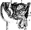
the prostate gland, then the diaphragma urogenitale, and continues further to the ventral side within the corpus cavernosum; consequently, the urethra is divided into three sections: the prostatic part (pars prostatica), the membranous part (pars membranacea, s. isthmus), and the cavernous part (pars bulbosa-cavernosa). Civiale and other authors divide the urethra into the fixed part (pars fixata), to which the prostatic and membranous parts belong, and the mobile part (pars mobilis, s. pendula) with the cavernous part: the boundary between them is formed by the ligamentum suspensorium. The urethra forms two curvatures along its length: the first, curved downward, bypassing the symphysis of the pubic bones—curvatura praepubica, and the second, curved upward toward the root of the penis—curvatura infrapubica (Braus). Consequently, the direction of the urethra resembles a horizontally positioned Latin letter S (Fig. 1). By lifting the penis up toward the abdomen, one can straighten one curvature, and then the direction of the urethra will correspond to the Latin letter U with one short limb. The prostatic part of the urethra. Its length corresponds to the height of the prostate gland—3–4 cm (Braus); the upper and lower ends of this section are narrower than the middle, which represents the widest place of the urethra—1.25 cm. This entire section is surrounded by the prostate gland, the muscle tone of which at rest compresses the walls of the canal so tightly that instead of a lumen, a narrow slit is formed. On the posterior wall is located a longitudinal ridge—crista urethralis. The largest elevation of this section is the seminal colliculus (colliculus seminalis), on the sides of which are the orifices of the ejaculatory ducts (ductus ejaculatorii) (Fig. 2). Between them, at the apex of the colliculus, is a narrow opening—a passage into the utriculus prostaticus, or uterus masculinus (a derivative of the Müllerian duct in men; Broman). The excretory ducts of the prostate gland open in the niche near the crista urethralis and on the surface of the seminal colliculus; they can serve as pathways for infection from the urethra into the prostate gland and vice versa, just as the vasa deferentia serve for the epididymis and seminal vesicles. The urethra, piercing the prostate gland obliquely from behind and above forward and downward, approaches the anterior surface of the gland quite closely, and due to this intimate connection of the gland tissue with the wall of the canal, abscesses of the former easily find their way into the urethra. The mucous membrane of the prostatic part of the urethra forms longitudinal folds, which stretch during the passage of urine. Its epithelium is identical to the epithelium of the bladder. The tunica propria is very rich in elastic fibers, which are an active factor in the propulsion of semen; the bladder sphincter prevents the entry of semen into the bladder; insufficiency of this muscle is one of the causes of male infertility. The membranous part of the urethra is so named because its walls are formed only by its own layers—mucous, submucous, and muscular membranes. This interval between the prostatic and cavernous parts is the shortest and narrowest section (hence the name isthmus) and is not surrounded by anything (Fig. 3). It adheres along its entire length to the muscles of the soft pelvic floor, bends around the lower edge of the pubic bones, and pierces the dense fascial ligament (lig. triangulare urethrae), which is in connection with the deep fascia of the perineum.
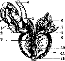
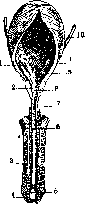
Figure 2. Figure 3. Figure 2. Prostatic part of the urethra: 1 and 7—vesicula seminalis; 2 and 8—ductus excretorius; 3 and 9—ductus ejaculatorius; 4—ductus deferens; 5 and 6—ampulla duct. def.; 10—utriculus prostaticus; 11—colliculus seminalis; 12—urethra. Figure 3. Frontal section through the urethra: 1—orificium ureteris; 2—prostata; 3—corpus cavernosum penis; 4—glans penis; 5—fossa navicularis; 6—pars cavernosa; 7—pars membranacea; 8—colliculus seminalis; 9—trigonum Lieutaudi; 10—ureter. This triangular fascia, as well as the contraction of the muscle fibers of the membranous section, can be the cause of so-called "spasmodic stricture." The mucosa, the epithelium of which changes from squamous, as in the bladder, to columnar, is also arranged here in folds and is surrounded by a thick layer of circularly arranged striated muscles—sphincter urethrae membranaceae. It is precisely here that muscular resistance is observed during catheterization. The cavernous part represents the longest section of the urethra; it is located in the lower unpaired cavernous body of the penis, reaching up to the external orifice. The corpus cavernosum urethrae, surrounding the urethra, does not have a significant difference from the cavernous bodies of the penis. Under the pubic bones, at the very beginning of the cavernous part, there is a thickening—the bulb (bulbus corporis cavernosi urethrae) (Fig. 4). The cavernous part is dilated at the proximal end, as well as at the distal end (fossa navicularis). The bulbous part is distinguished by the presence of a large number of excretory orifices of the acinous glands of the urethral mucosa; in this same section are the excretory orifices of Cowper's glands (see). The most distal part of the urethra, the fossa navicularis, is wider, and here there are a large number of racemose mucous glands (glandulae Littre) (see Littre glands). On the upper wall, corresponding to the proximal end of the fossa navicularis, there is a relatively deep bay (lacuna Morgagni), sometimes covered by a membrane; often on the dorsal wall of the fossa navicularis, a semilunar transverse fold is encountered (valvula fossae navicularis); the presence of the latter requires the introduction of instruments into the urethra so that their end is directed ventrally to avoid traumatization of the mentioned fold; subsequently, the end of the instrument must be directed dorsally so as not to touch the fossa bulbi and the seminal colliculus. All the mentioned protrusions, as well as the bays and crypts of the urethral mucosa, which play a large role in the pathology of this section, are accessible for examination, and recently, with the introduction of contrast masses—also for radiography (see below). Blood supply of the urethra. Arterial supply is obtained from branches of the artery.
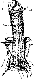

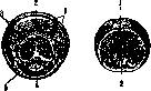
Figure b. Cross-section of the penis. a-through the body: 1-v. dorsalis penis cutanea; 2-v. dors. penis; 3-a. dors. penis; 4-n. dors. penis; 5-tunica albuginea; 6-a. profunda penis; 7-septum penis; 8-corp. cavernosa penis; 9-corp. cavernosum urethrae; 10-urethra; b-through the posterior part of the glans: 1-praeputium; 2-glans penis; 3-corp. cavernosa penis; 4-corp. cavernosum urethrae; 5-urethra; c-through the anterior part of the glans: 1-fossa navicularis urethrae (Morgagni); 2-septum glandis. Pudenda communis: the bulbo-urethral artery for the bulbous and cavernous parts reaches the glans of the penis, the perforating branches of the deep artery of the penis - along the lateral surfaces of the penis, and the terminal branches of the dorsal artery of the penis for the fossa navicularis (Fig. 5). All these branches anastomose with each other in the cavernous part and form a wide arterial network around the urethra. The veins of the cavernous bodies of the urethra collect partly in the fossa urethralis and partly in the fossa navicularis and, forming superficial plexuses, connect with the dorsal vein of the penis, surrounding the body of the penis before this (venae circumflexae). The veins of the prostatic and membranous parts drain into the plexus of Santorini. The lymphatic vessels of the mucous membrane of the urethra exist in the form of a network, which is connected by means of perforating branches with the lateral plexus of lymphatic vessels of the integuments of the glans and is thus connected by the lymphatic vessels of the mucous membrane of the anterior section of the urethra and with the inguinal glands. The efferent lymphatic vessels of the remaining sections of the mucous membrane of the urethra and the cavernous tissue have been poorly studied. The nerves of the urethra originate on both sides from the cavernous plexus of the sympathetic nerve (plex. cavernos. sympath.) and from the urethral branches of this same plexus, which also receive spinal elements from the sacral nerves. All of them, before converging into the cavernous bodies, collect in the fossa urethralis together with the veins and arteries (Rauber-Kopsch). The female urethra (urethra muliebris) can be compared with the membranous part of the male urethra: morphologically it corresponds to the pars pelvina urethrae virilis (Fig. 6). Figure 6. Sagittal section of the female pelvis: 1-cavum Retzii; 2-urinary bladder; 3-urethra; 4-vagina; 5-rectum; 6-uterus. The length of the canal varies from 2.5 to 4 cm. The female urethra passes a short distance from the internal bladder orifice to its external orifice under the clitoris between the labia pudenda in an arc (corresponding to the posterior surface of the symphysis). Upon exiting into the vagina, the external orifice of the urethra is surrounded by slightly elevated, ridge-like edges. The canal descends along the anterior wall of the vagina, heading from behind and above forward under the pubic bones. It is strengthened by a ligament similar to the triangular ligament of the male urethra (lig. triangulare urethrae), but is located at a greater distance from the symphysis pubis. The mucosa of the female urethra consists of squamous epithelium and forms numerous longitudinal folds, although fewer than in the male urethra. On the dorsal side, there is a crista urethralis. The connective tissue is rich in elastic fibers and numerous veins - spongy bodies (corpora spongiosa). Near the exit orifice, there are narrow passages on both sides (Skene's ducts) 1-2 cm long - paraurethral ducts (ductus paraurethralis). These are crypts lined with columnar epithelium. The muscular layer (tunica muscularis) consists of smooth muscle tissue. R. Herzenberg. Methods of examination. The male urethra 1) is a canal for the excretion of urine from the bladder; 2) serves as an apparatus that retains urine in the urinary bladder; and 3) performs an important task - the expulsion of semen and the mixed secretion of all the sex glands located both in the canal itself and in the cavity of the small pelvis (see above). The female urethra performs only the first two functions. During an erection, the male urethra undergoes physiological changes: it stretches by virtue of its elasticity, following the dimensions of the erect penis. Normally, the lumen of the urethra is closed, and the production of its glands is so insignificant that their discharge only moistens the mucosa of the canal and does not appear externally. The pathological physiology of the urethra manifests itself as a disorder of the act of urination, a disorder of the act of conducting urine through the canal, the discharge of pathological secretion from the glands of the canal, and a disorder of the act of erection and ejaculation. Clinical examination of the urethra possesses a number of methods. By inspecting the external orifice of the canal, one can determine the color of the mucosa of the nearest section of the urethra, which normally has a pale pink color. In pathological cases, the mucosa changes its color from whitish-gray to cherry-red depending on the stage and intensity of the inflammatory process. In addition, phenomena of edema and infiltration of the sub-mucosal tissue can be detected here. In the first case, the lips of the canal (labia urethrae) take on a glassy sheen; in the second, palpation reveals a hardening of the tissues. Inspection of the external orifice makes it possible to find here polypous or condylomatous growths protruding from the lumen of the canal. Of very great importance in some diseases of the canal (gonorrhea) are the accessory, more or less long passages (paraurethral - ductus paraurethralis), pockets, depressions - cryptae, etc., located at the external orifice. The shape of the external orifice - orificium externum of the urethra - and the size of its lumen play an important role: a slit-like, large, easily opening orifice is more likely to receive an infection than a narrow, poorly elastic one. The latter, in turn, complicates the introduction of instruments into the canal. Palpation is performed with the patient lying on their back. With the left hand, the penis is held by the glans, and with the right, the entire canal is palpated up to its bulbous expansion. The posterior part of the canal is examined with a finger inserted into the rectum, with the patient lying on their back, in the knee-elbow position, or on their side. The female canal is palpated through the vagina. Upon palpation of a normal canal, the latter is determined as a soft, uniform cord. Hardening of the urethra speaks of its infiltration, which is often observed in inflammatory processes. Sometimes these thickenings are localized in limited areas of the canal and possess cartilaginous density (strictures), and sometimes individual nodules are felt, ranging in size from a millet seed to a pea (blocked or infiltrated glands of Littre and sinuses of Morgagni - littreitis et morgagnitis). In the presence of foreign bodies in the canal (catheters, stones, accidentally introduced bodies), they are also determined by palpation. The combination of palpation together with inspection makes it possible to establish the presence of pathological discharges from the canal. By squeezing it from the bulb to the external orifice, one can obtain either a few drops of retained urine (in post-stricture sacs and diverticula) or squeeze out a discharge of thicker consistency, the macroscopic examination of which makes it possible to judge its character (mucous, mucopurulent), as well as its consistency and color, the presence of blood, etc. Microscopic examination of the discharge is performed on a preparation evenly spread on a glass slide and has the purpose of studying the character of the formed elements of the discharge (epithelium, pus globules) and the flora. The presence of discharge indicates an inflammatory process in the canal, and the flora indicates the etiology of this process. In some subjects, under the influence of sexual arousal, hypersecretion of the glands of the canal appears, which manifests itself by the appearance of transparent, viscous discharge - urethrorrhea ex libidine. Treatment is carried out by prescribing mild astringent washes and atropine. When examining the female urethra, attention is also paid to the presence of paraurethral passages, the state of the Skene's glands surrounding the canal, discharges from the canal, etc. In the case of a minimal amount of discharge in the canal, it is possible to determine its origin with the glass test. Several glass tests have been proposed, bearing the names of their authors. The Thompson test with two glasses. By examining the urine voided by the patient sequentially in 2 portions, one can establish three facts: 1) both portions are cloudy or with impurities, 2) the first portion is cloudy, the second is clear, 3) both portions are transparent, but in one or the other there are impurities of threads, crumbs, or flakes. In the first case, there may be a lesion of both the entire canal and the overlying organs - the urinary bladder and renal pelves. With a small amount of urine in the bladder (not over 100 cm3) and abundant suppuration in the anterior canal, the urine does not have time to wash the anterior canal, and part of the turbidity will be in the 2nd portion of the urine. In the second case, there is undoubtedly a lesion of the anterior canal. In the third, the presence of local lesions of the canal in one or another of its segments is clear. Usually, a perplexity arises: why does a lesion of the posterior part of the canal affect the urine in the 2nd portion, whereas it would seem that the 1st portion should have washed the threads from the entire canal. But it must be kept in mind that upon the contraction of the powerful muscular apparatus surrounding the posterior urethra, upon the excretion of the last portion of urine, the contents of the excretory ducts of the glandular apparatus of the posterior canal enter it. This simple Thompson test was subsequently subjected to changes and additions.
Jadassohn and Kollmann first thoroughly wash out the anterior urethra and then ask the patient to urinate into 2 or 3 glasses. Some authors (Baradulin) combine the glass tests with massage of the Cowper's glands, prostate, and seminal vesicles, the introduction of a catheter into the bladder, etc. Luys recommends a four-glass test; some, before urination, introduce a dye into the anterior urethra, whereby all impurities from it are stained one color or another. By taking into account the localization of the pathological process and the quantity and quality of the voided urine, one can find sufficient objective diagnostic data in the glass test. No less important is the glass test in the case of bleeding from the urinary tract. If all the urine is stained with blood, this indicates bleeding located above the external sphincter. Involuntary discharge of blood from the urethra, as well as macroscopic or microscopic admixture of blood in the first portion of urine, indicates the source of bleeding in the anterior urethra. An admixture of blood in the last portion indicates so-called terminal hematuria, i.e., the localization of the bleeding focus in the posterior urethra or in the area of the bladder neck. The nature of the impurities in the urine in the form of threads, crumbs, and flakes can rarely indicate the localization with precision, but rather the gradation of the inflammatory process in the urethra. Heavy threads that quickly fall to the bottom indicate a purulent process. Light, transparent threads suspended in the urine, dispersing in it and gathering in the form of a cloud, indicate a mucous character. In desquamative processes of the urethra, small flakes are usually visible in the urine, quickly settling to the bottom of the glass. An admixture of prostatic fluid gives the urine an opalescence. The secretion of the seminal vesicles is noticeable in the form of small grains (like boiled sago), sometimes with a slight pearly luster. Turbidity in freshly voided urine often depends on the precipitation of phosphate salts. It clears up very quickly upon the addition of a few drops of acetic acid. All other changes in the urine do not depend on the state of the urethra and require a more thorough examination of the entire urinary tract and clinical examination of the urine for their interpretation. The stream of urine during the act of urination also makes it possible to have some judgment about one or another pathological process in the urethra. Urination in a sluggish stream usually speaks of an acute inflammatory process in the urethra; delayed urination with straining in a thin stream indicates an existing sharp narrowing of the urethra. Splashing of the stream occurs with the loss of elasticity of the urethra or infiltrates of one degree or another and localization. This is also indicated by the bifurcation of the urinary stream, although the latter also happens with the sticking or gluing together of the lips of the external opening of the urethra by dried discharge from it. Examination of the urethra with instruments. Probing the urethra with an instrument must be of the nature of palpation and is never performed forcibly. Rough examination causes spasms of the muscular apparatus surrounding the urethra and can give a false impression of obstacles in its lumen. By probing the urethra, one determines the presence of narrowed areas, their localization, extent along the length, and the elasticity of the tissues forming them. In the presence of foreign bodies, they immediately make themselves known by the hardness of their consistency. The instrument for examining the caliber of the urethra is the bulbous bougie of Guyon. It has bulbs at its ends, one of which is olive-shaped or bulb-shaped, and the other conical with steep shoulders. Sometimes these bougies are marked in centimeters for more precise localization of the obstacle in relation to the external opening. For examination, a No. 23 bougie is taken, which should pass freely through a normal external opening and the entire lumen of the urethra. Even in a normal urethra, the bulbous bougie encounters physiological obstacles that must be taken into account

by any examiner. The first obstacle is the narrow external opening of the urethra. Following it, on the upper wall, one may encounter a strongly developed Guérin's fold—valvula Guérini (a defect of embryonic development), therefore here the bougie must be directed along the lower wall of the urethra. Further, the head of the bougie may be delayed in the navicular fossa, which in some subjects may be particularly deep. To overcome this obstacle, it is sufficient to pull the penis, and the fossa of the bulb is smoothed out. Immediately, the head of the bougie encounters the resistance of the external sphincter of the urinary apparatus, which is easily overcome by a light pressure of the instrument. After passing 2-3 cm beyond the sphincter, the last obstacle is encountered—the high seminal colliculus, but the head of the bougie passes through it easily and, without encountering resistance from the internal sphincter, penetrates into the urinary bladder. If the bulbous bougies taken for the first examination encounter an insurmountable obstacle, it is important to establish whether this is the result of a sharp spasm. The latter can be eliminated by introducing anesthetic agents into the urethra (5-10 cm3 of 2% Sol. Novocaini, 5% Sol. Alipini, with the addition of 5 drops of 0.1% Sol. Adrenalini, etc.). After this, a new attempt at examination is made. If a bougie of a certain caliber does not pass, then

Figure 8.
Take instruments of a smaller caliber until a suitable one is found. It is necessary to keep in mind that the best sensations of infiltration and cicatricial strictures of the urethra are obtained during the withdrawal of the instrument. Sometimes, even by simple observation of the examiner's hand, one can see how the head of the bougie jumps over obstacles in the canal. A more detailed determination of the caliber of the urethra along its entire length can be performed with the urethrometer of Otis (Fig. 7) or Kollmann (Fig. 8), which have expanding branches and a dial on the handle indicating the caliber of the canal. These instruments have not acquired practical significance. Determination of the caliber of the urethra serves as an indicator for further manipulations in the canal: selection of appropriate instruments for endoscopy, dilation of strictures, and surgical intervention. The introduction of straight or curved (according to Benique, Guyon, Dittel, or Nitze) metal bougies into the urethra makes it possible to more easily palpate indurations and infiltrates of the wall on a hard basis, just as one palpates the soft tissues of the limbs on a hard bone basis. X-ray examination is of no small importance for the diagnosis of diseases of the urethra. Ordinary fluoroscopy or radiography makes it possible to judge the presence, position, and size of foreign bodies in the urethra (stones, catheters, nails, etc.). The introduction of contrast agents into the cavity of the canal followed by a snapshot (urethrography) makes it possible to ascertain changes in the lumen of the canal: stricture, accessory passages, diverticula, fistulas, etc. Urethrography is performed with the patient in a lateral position with the lower leg tightly pulled to the abdomen, and the upper leg extended and slightly thrown back. In this position, the course of the urethra is not covered by the pelvic bones. The canal is filled through a syringe with a 5-10% solution of collargol, or a 10-20% solution of sodium iodide or bromide, 10% iodipin, or lipiodol. To determine the state of the posterior urethra, preliminary filling of the bladder and taking a snapshot at the moment of the act of urination must be done. Methods of examining the urethra using lighting devices—see Urethroscopy. Pathology of the urethra. Malformations of the urethra can be subdivided into two groups: a) having only pathological-anatomical and b) clinical significance. All malformations of the urethra must be linked to the embryonic formation of the urinary organs, which is quite complex in its course. Most often, malformations of the urethra are localized where the most complex moments of formation occur in the embryo: the canal of the glans penis, the place of transition of the anterior urethra into the membranous part of the canal, and the region of the bladder sphincter. These are the sections of the urethra in which the process of formation of the urethra in the embryo must overcome septa. Here, partial underdevelopment of the canal—atresia—sometimes forms. In newborn children, it is necessary to keep in mind the possibility of adhesion (or rather fusion) of the inner layer of the prepuce with the glans penis and the formation of a film covering the external opening of the urethra. A symptom of these anomalies is the impossibility of urination in newborns. Diagnosis is made by inspection or probing. Treatment—incision with a scalpel or piercing the membranes of the external opening with a probe. If they are located deep, it is sometimes necessary to resort to high section of the bladder with subsequent interventions in the canal. Congenital strictures must also be attributed to these malformations. They are localized in the same places as atresias and are caused by the same reasons. Their shape is annular, cylindrical, valve-like, and in the form of cords. The symptoms of congenital strictures and treatment do not differ from acquired strictures (see below). Congenital narrowness of the external opening of the urethra has some clinical significance: by hindering the free and full flow of the urine stream, it leads to dilation of the canal from increased urine pressure. The bladder compensates for this with hypertrophy of its muscle with the formation of trabeculae, and then it weakens, and atony of the bladder develops. A narrow external opening prevents the introduction of instruments of the appropriate caliber. To eliminate it, the operation of dissection—meatotomy (meatotomia)—is resorted to. It is performed with a special instrument.

Figure 9.
with a meatotome (Fig. 9), a scalpel, or scissors. The lower wall of the canal is incised for a length of 1/2-1 cm, and then the mucosa is sutured to the outer covering of the glans with 2-3 silk sutures. The patient urinates on his own. The dressing is changed after urination. The sutures are either removed on the 5th-6th day or come out on their own. - Congenital dilatations of the canal manifest in the form of sinus-like protrusions of the lower wall of the urethra (urethrocele) or saccular dilatations with a narrowed entrance—diverticula. The pathogenesis of these defects of the canal has not yet been definitively established. In view of the fact that they have been found in embryos even before the onset of the physiological functions of the urethra, it must be assumed that they are defects of embryonic development. Accessory urethra (urethra accessoria) belongs to the same defects of embryonic origin. This accessory urethra runs parallel to the course of the normal urethra for some distance; its peripheral end may be blind, open on the glans below or above the normal urethral opening, or on some part of the lower surface of the penis. The central end may also be closed or communicate with the urethra along its length. Given a closed peripheral end and an open central one, conditions are created for the development of diverticula. Diverticula rarely make themselves known in childhood. They are more often noticed in adolescence and in adults. Characteristic is the filling of the diverticulum sac with urine during the act of urination and its emptying upon pressure. A filled sac hinders the act of urination, and after it ends, the symptom of urine dripping remains. Diagnosis is confirmed by urethro- and roentgenoscopy. It is necessary to consider the possibility of stone formation in the diverticula. Treatment: removal by operative means—excision. The same applies to accessory tracts if they are sources of chronic or recurrent infection in the urethra (in gonorrhea). - The formation of accessory (paraurethral) tracts is explained by a developmental defect similar in its origin to an accessory urethra. Usually these tracts are blind; their openings are located near the external urethral opening, and sometimes on the skin of the lower surface of the penis or in one or another place on the glans of the penis. Under normal conditions, they are not noticed and acquire clinical significance only during gonorrheal inflammations, being a source of reinfection for the patient himself or his partner. - Cysts of the canal formed in one or another of its sections should be classified as developmental anomalies. Cysts cannot be attributed to the blockage of the glands of Littre and Cowper, since the final development and function of the latter occur significantly later, almost by the period of sexual maturity. Developmental defects in the form of a lower cleft of the urethra (hypospadia) and an upper cleft (epispadia)—see Hypospadia and Epispadia. Surgical diseases of the urethra are usually classified as 1) injuries of the canal, 2) strictures, 3) dilatations, 4) tumors, 5) stones and foreign bodies, 6) prolapse of the canal, and 7) fistulas. - Traumatic injuries of the canal can arise from the action of violence from the outside and from the side of the canal lumen. The former include falling and striking the perineum against an oncoming object, then blows to the perineum with a foot, a horse's hoof, and gunshot wounds. The site of injury is the bulbous and membranous parts of the urethra, as they are less mobile compared to the pendulous part and cannot escape the traumatizing object. Ruptures of the membranous and prostatic parts occur more often from coarser impacts: being run over by a carriage, a blow by an automobile or tram, crushing by buffers, falling from a height, being buried by a collapse of earth, a building, etc. They are usually combined with a fracture of the pelvic bones and a rupture of the strong pelvic fasciae. According to the extent of destruction, urethral ruptures should be divided into complete and partial. The former are especially unpleasant in that the torn-off sections of the urethra separate and expose the surrounding cellular tissue, where urine can penetrate during urination, and a urinary infiltrate forms with all its consequences—phlegmon of a gangrenous-putrid character. In the subsequent course, the rupture site scars over, and a traumatic stricture of the canal forms. Symptoms of rupture: bleeding from the urethra, swelling in the perineum and hematoma, disorders of urine excretion. Upon the addition of infection—fever with shaking chills, sometimes phenomena of sepsis, etc. Upon the opening of phlegmonous foci to the outside, fistulas form. - Treatment: in cases of urethral trauma without visible indications of rupture, expectant treatment—rest and cold on the urethra. In case of reflex urinary retention, to avoid introducing infection, puncture of the bladder is preferable to catheterization. In cases of obvious urethral ruptures with incipient urinary infiltration, and even better—before it, it is necessary to immediately divert urine through a high bladder incision, and then expose the urethra, find its edges, and suture with catgut sutures (circular suture) over a catheter, which is immediately removed. The operation is performed according to the type of external urethrotomy (urethrotomia externa) or resection of the urethra (see below). Punctured, incised, and bitten wounds of the canal are for the most part only of casuistic interest. Usually they are associated with one or another sexual psychopathy. Gunshot wounds of the canal are not uncommon in wartime. In these cases, there are more often wounds of the posterior part of the canal with a wound of the pelvis and sometimes damage to the pelvic bones. Wounds of the anterior freely hanging part of the urethra are encountered comparatively less often due to the ability of the canal to slip away from the wounding projectile. Their course is similar to the course of traumatic urethral ruptures and requires urgent surgical intervention. Separately, one must consider so-called constrictions of the urethra: tying with a thread (often as a prank in children), putting rings, nuts, rubber rings on the penis in adult men for the purpose of masturbation or delaying semen during intercourse, increasing orgasm, etc., constriction of the canal with a narrow opening of the foreskin (paraphimosis). These injuries of the canal cause stasis, hyperemia, nutritional disturbance with subsequent gangrene of the urethral wall and the formation of a fistula. - Treatment consists of the immediate removal of the constricting object and subsequent surgical treatment of the complications that have arisen. Injuries of the canal from the side of its lumen arise most often during the introduction of foreign bodies or medical instruments into it. The former are of casuistic interest, are encountered more often in women and less often in men, as a result of masturbation. In the casuistics of these cases, hairpins, pins, needles, pencils, pen nibs, etc., have been described. The wounds produced by them rarely reach large sizes, because pain stops the continuation of the violence. Wounds by medical instruments—bougies, metal catheters, lighting devices, dilators—are in most cases the result of rough or unskilled introduction of these instruments. Often the cause of injury is unskilled selection of the instrument—a dilator for coarse, callous strictures, a bougie not corresponding in caliber to the existing stricture, unskilled selection of the catheter curvature in prostate hypertrophy, etc. But the main cause of violations of the integrity of the canal is the rough overcoming of obstacles encountered in the path of the instrument. More often, pathological conditions of the urethra—cicatricial strictures and hypertrophied prostate—contribute to injuries of the urethra. Here the instrument can create a false passage (via falsa). The mechanism of the formation of a false passage appears in the following form: the instrument encounters obstacles to entering the posterior part of the canal, abuts against the bottom of the bulbous sac, bores through it, and goes into the thickness of the perineum; in cases of prostate hypertrophy, especially in its soft form, the instrument embeds into the thickness of the prostatic urethra, ruptures it, and penetrates through the gland via a through-passage into the bladder. During the forceful passage of bougies or dilators through a stricture not corresponding to the caliber of the instrument, the instrument can cause a rupture of the strongly stretched section of the stricture. Injuries of the canal can also be encountered at any point along its length in the presence of pockets and valve-like strictures; Signs of a false passage or injury of the urethra are bleeding to one degree or another and pain, disturbance of the act of urination, swelling of the penis. More precisely, the diagnosis is made with the help of urethroscopy. Mild injuries of the canal, as well as false passages, if they are not infected, usually heal very easily with rest and the administration of urine-disinfecting agents internally. Urinary infiltration of tissues in this case is a rare complication, because the opening in the mucosa is not stretched by the pressure of urine in these cases, but on the contrary, is compressed and covered. In case of urinary retention, one can resort, under the strictest asepsis, to catheterization with soft instruments or, in case of failure, to puncture of the bladder. In case of severe complications, the creation of a fistula on the bladder and corresponding surgical intervention on the urethra are inevitable. Injuries of the urethra from the introduction of cauterizing agents into it—strong solutions of sublimate, carbolic acid, iodine, silver nitrate, etc.—are produced accidentally, by mistake, or for the purpose of preventing venereal diseases. After them, gangrene of the mucous membrane usually develops, followed by scarring and narrowing of the canal, often along its entire length.
In acute cases, it is necessary to resort to one or another agent neutralizing the action of the cauterizing agent (e.g., for corrosive sublimate and silver nitrate, irrigation with a physiological solution). Developed cicatricial strictures are treated by the appropriate surgical method (see below). Injuries to the urethra during erection can arise spontaneously, i.e., upon the loss of elasticity of the urethral tube: the latter cannot follow the erect penis and the mucosa tears. External violence to the erect penis—a blow, contusion, compression, twisting, etc.—usually, along with damage to the corpora cavernosa (fracture of the penis), leads to deep injuries of the urethra. The first symptom of such a lesion of the canal is bleeding from the canal (urethrorrhagia), and then, with deep injuries, urinary infiltration and even gangrene of the peripheral part of the penis. Injury to the urethra in women, besides the external and internal violence mentioned above, can also occur on the basis of the act of childbirth. During the passage of the fetal head, the urethra is pressed against the pubic symphysis and undergoes traumatization. Sometimes this traumatization is limited to hemorrhage into the tissues, tearing of the mucosa, and sometimes complete crushing develops not only of the canal itself but also of the muscular layer between it and the vagina. Under the condition of urinary extravasation into the tissues, an infiltrate and an abscess form, which breaks through into the vagina and leads to the formation of a urethro-vaginal fistula. Narrowings of the urethra (stricture—strictura) are more often not congenital, but of acquired origin. The main causes of narrowing are gonorrhea and trauma to the urethra. The former is observed in 80–90% of all strictures, the latter accounts for 10–20%. In both cases, it is a matter of the development of connective tissue in the submucosa of the canal and the formation of a scar. Besides these persistent strictures, narrowings of the canal can be of a temporary nature, e.g., acute swelling of the submucosal tissue of the canal during inflammatory processes in it or convulsive spasms of the canal muscles—sphincters. It is also necessary to keep in mind the reduction of the canal lumen from compression by processes developing both in the canal itself and outside it (prostate hypertrophy, tumors, abscesses, foreign bodies, scars around the canal, etc.). Gonorrheal narrowings of the canal, according to Thompson's statistics, most often develop in the bulbous part of the canal (67%), near the navicular fossa (17%), and in the remaining pendulous part of the canal (16%), i.e., where the infection remains longest and most persistently. Strictures of the posterior canal and in the female urethra are rare. In the majority of cases, gonorrheal strictures are multiple. Their form is ring-shaped, cylindrical, valve-like; the lumen ranges from thread-like to so-called wide strictures (strictura lata). The lumen of strictures in their longitudinal section is rarely straight, more often tortuous or zigzag. Depending on the composition of the tissues forming the stricture (infiltrate, young connective or scar tissue), when palpated with a bougie, the stricture gives a sensation of soft resistance, low compliance, and finally, dense-elastic constriction of the bougie head (callous, calous strictures). Histological examination of strictures shows that the development of scar tissue prevails in them. At the same time, in strictures of gonorrheal origin, along with the developed scar, there is the presence of inflammatory infiltration, which over time also turns into scar tissue. In connection with this, the formation of a gonorrheal stricture proceeds at a slow pace, but at the same time, there is no guarantee of the cessation of scarring either in time or in the spread of the process. The question of why strictures develop in some subjects who have had gonorrhea and not in others cannot be considered resolved at the present time. There exists an opinion that the treatment of gonorrheal urethritis with strong cauterizing chemical solutions (silver nitrate) is to blame for this. Observations seem to confirm this opinion, since with the widespread adoption of the method of treating gonorrhea with large irrigations, the number of strictures has significantly decreased. An opinion was also expressed about a congenital individual predisposition of the organism to the development of scar tissue. The development of connective tissue is often dependent on the degree and nature of the infection, which gives grounds to believe that the intensity of scar development is proportional to the virulence of the infection. It is also necessary to reckon with the localization of the infection in the glands of the canal, with the development of periglandular infiltrates and their subsequent scarring. Traumatic strictures, unlike gonorrheal ones, develop faster. Their form is more often in the shape of a cord or a gutter; they sometimes extend from the bulbous part through the sphincter into the posterior part of the canal. Traumatic strictures very quickly take on their final appearance, and microscopic examination of them shows the absence of small-cell infiltration in them. In view of the fact that injuries to the canal are usually single, traumatic strictures are also single. Symptomatology of strictures. In the initial stages, strictures are usually little noticeable even to an attentive patient. Subsequently, patients note a lengthening of the period of the act of urination, narrowness of the stream of urine, its spraying, and the loss of that arc-like path of the urine stream which it assumes with a free canal and preserved expulsive force of the urinary bladder. In connection with the development of an obstruction, patients have to strain, i.e., increase the automatic work of the bladder by pressure on it from the abdominal wall. A change in the character of the urine with strictures is not mandatory. If inflammatory changes of the mucosa have subsided by the moment of the development of the stricture and the inflammatory process is nested only in the deeper submucosal tissues, the urine may not contain any impurities (threads, crumbs, turbidity, etc.). In connection with the presence of a narrowing in the urethra, and consequently an obstacle to the free outflow of urine, the urinary apparatus (urinary bladder) and renal pelves strive to compensate for this state with hypertrophy of their muscles. Subsequently, the muscles weaken, atony of the urinary reservoirs sets in, stagnation of urine in the pelves and the urinary bladder, and even before the development of catarrhal phenomena, patients experience disorders of urination in the form of frequent urges to urinate with clear and aseptic urine. This phenomenon is explained by the constant overfilling of the urinary bladder. With the development of a stricture in the region of the sphincter of the urethra, a symptom of urinary incontinence develops. Under the influence of conditions favoring hyperemia of the pelvic organs (constipation, intercourse, alcohol, etc.), swelling of the tissues and complete retention of urine occur. Some changes in the act of ejaculation also occur: semen may be retained in the canal, ejaculation gives painful sensations, bloody semen appears, etc. Under the influence of increased pressure on the walls of the canal in the sections lying above the stricture, dilation of the lumen of the urethra occurs with the formation of cavities in which urine stagnates. The pressure of the urine can be so high that the thinned mucosa cannot withstand the pressure and develops cracks through which urine penetrates into the surrounding tissues and causes urinary infiltrates. With the addition of pyogenic infection, catarrhal processes of the mucosa of the entire urinary tract very easily develop, and the penetration of infection into the urinary infiltrate leads to the development of periurethral abscesses with their opening into the canal or through the skin with the formation of urethral fistulas. The presence of strictures with stagnation of urine in the upper tracts contributes to the stone-forming process. The penetration of infection into the seminal ducts or the prostate creates conditions for the emergence of purulent epididymitis and prostatitis with a frequent outcome in an abscess. The diagnosis of a urethral stricture is made, besides the presence of the above-described symptoms, on the basis of instrumental examination of the canal. Examination begins with thicker numbers of bulbous bougies (No. 23–20), and in case of obstruction, one moves to thinner numbers until one reaches the number that passes through the stricture. It is necessary to keep in mind that with multiple gonorrheal strictures, their caliber is not uniform, and one has to select a series of bougies until one manages to penetrate to the urinary bladder. Taking into account that the course of the narrowed part of the canal is often tortuous, has depressions and valves, it is necessary to keep in mind the impossibility of passing even a small number of a bulbous bougie. In such cases, one has to resort to the introduction of filiform bougies (see). The end of such a bougie has only a small bulbous thickening, and it is easier for it to pass through a narrow opening. The curved shape of the end of the bougie makes it possible, by turning it around the longitudinal axis, to find the course of the narrowed part of the canal and form an idea of it. In the presence of pocket-like depressions, valves, etc., in the narrowed part of the canal, an examination with a bundle of filiform bougies is performed. The bundle, sufficiently moistened with a lubricant, is introduced into the canal to the place of narrowing, and then, one by one, an attempt is made to penetrate further with one or another bougie. In this case, the lateral bougies will occupy all the depressions of the canal, and one of the central bougies will stand against the opening, and in this way, the entrance to the deep parts of the urethra can be found. With the help of a bulbous bougie, one can almost accurately establish the location of the strictures, their number, arrangement along the length, caliber, and the state of the tissues. A more detailed idea of the stricture is given by X-ray examination with filling of the urethra with a contrast liquid.
It makes it possible to more accurately identify the state of the canal's lumen with all its variations. Examination with the help of a urethroscope adds little to the data indicated above: one only receives an impression of the state of the mucosa (a matte coloring speaks for its cicatricial degeneration). Treatment of canal strictures aims at the elimination of clinical symptoms and their radical treatment. Proceeding from the view that a gonorrheal stricture develops at the site of a submucosal infiltrate, the majority of urologists conclude the treatment of gonorrhea with urethroscopy of the canal and, in the presence of infiltration, conduct treatment by bougienage. The same system of methodical bougienage of the canal is conducted when a stricture has already developed, but with the presence of infiltration of the submucosal tissue of the canal. The action of bougienage in these cases boils down to the resorption of the infiltrate, since the bougie is a massaging instrument that causes an alternation of hyperemia and anemia. To avoid complications during bougienage (hemorrhage, creation of a false passage), it is necessary with narrow strictures to begin with elastic bougies and, upon reaching a caliber of 30-32 on the Béniqué scale, to switch to metal bougies or to begin with them, but screwing on plastic guides. The caliber of the first bougie must correspond to the caliber of the stricture, and subsequent sessions must be conducted every 2-3 days after the reaction caused by the bougie has passed and in strict order of numbers. Forced bougienage with several bougies in one session is undesirable, as it can cause complications—tears of the strictures. Bougienage must be especially cautious in the presence of urinary tract infection. Here, the most formidable complication is possible—urinary fever. Clinically, it proceeds in the form of a sharp, shaking chill with a rise in temperature to 40°, which lasts for several hours, and sometimes days. Urinary fever is explained by the entry of infection into the blood and is dangerous due to the possibility of the development of septicopyemia. Its treatment boils down to the use of disinfectants internally: Urotropin 0.5 three times a day, Salol in the same dose, Benzonaphthol 0.3 twice a day, or intravenously: 40% Urotropin 5 cm3, Electrargol 5 cm3, 1% Rivanol 5-10 cm3, 1% Trypaflavin 5 cm3. With fully developed strictures, treatment is also conducted by bougienage, but it achieves only one goal—stretching the already formed scar—and cannot guarantee that in a few years a narrowing of the canal will not occur again. To such
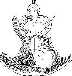
Figure 11. Median incision—for external urethrotomy. Arcuate incision with a solid line—for external urethrotomy. Arcuate incision with a dashed line—for recto-urethral fistulas.
methods of treatment, which give only a clinical effect, belongs the method of electrolysis of strictures. Its essence boils down to the dissection of cicatricial tissue with the help of an electric current of 10-20 mA, conducted to the scar by special tips. Such a current causes aseptic necrosis of the cicatricial tissue. After electrolysis, methodical bougienage is conducted. To such palliative surgical interventions also belongs the operation of internal urethrotomy. It aims to dissect the cicatricial segment of the canal so that subsequently the scar is deprived of the ability to constrict the lumen. The operation is performed with one of the instruments designed for this purpose. Most often, the Maisonneuve urethrotome is used (Fig. 10). The operation is performed under local anesthe-
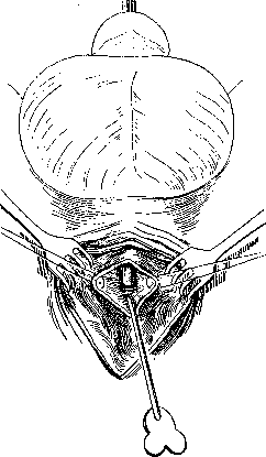
Figure 12. Exposure of the urethra before narrowing. Head of the catheter before narrowing. Probe inserted into the central end.
sia of the urethral mucosa. The instrument is inserted with an elastic guide, and the guide must coil in the filled urinary bladder. Then, a knife is inserted along the groove of the instrument, with which the narrowed part is dissected. Dissection is carried out along the upper or lower wall of the canal; some dissect one lateral wall when moving in and the second when moving back. Then the knife and instrument are removed, a straight rod is screwed onto the guide, and a special elastic catheter No. 18-20 with a truncated end is inserted along it. The catheter is held in the canal for 2-4 days. 7-8 days after the operation, when the wound has managed to be covered with epithelium, bougienage of the canal begins. The operation of internal urethrotomy does not always lead to a permanent cure and results in recurrences of canal scarring, but the clinical effect is very pronounced. Indicated
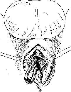
Figure 13. Insertion of a catheter into the central end of the urethra.
for internal urethrotomy are gonorrheal strictures, in which methodical bougienage is impossible due to complications or when there are indications for rapid dilation of the canal (bladder distension). More radical operations include external urethrotomy and resection of the canal. They are used for certain forms of gonorrheal and always for traumatic strictures. Of the gonorrheal strictures, those that give deep scarring of tissues—callous strictures—are proven for these operations. The operation of external urethrotomy is performed under general or spinal anesthesia. The patient is in a supine position with raised legs, as in any operation on the perineum. An elastic bougie or metal guide (itinerary) is preliminarily inserted into the urethra up to the obstruction. This is especially important during operations on the perineum, where the urethra lies deep among the scarred tissues and is found with difficulty. The skin incision is made along the midline of the perineum—from the base of the scrotum to the anus (Fig. 11, 12, and 13). Gradually dissecting the tissues, one reaches the urethra and performs a section of its lower wall along the guide. If the guide could not be inserted into the lumen of the stricture, then the canal is dissected at the end of the bougie, the urethral wound is stretched, the entrance to the stricture is found, and the section is continued further either under visual control or on an inserted grooved probe. When the incision reaches the posterior, post-strictural segment of the canal, its patency is checked with a catheter. In the presence of strictures located in the membranous part of the canal, their incision is performed either with one of the urethrotomes or continued gradually along the course of the wound. With free patency of the posterior segment of the urethra, a catheter is passed through it into the bladder and secured in the wound with one of the sutures. If the posterior segment cannot be found, then a high section of the bladder is performed, a retrograde bougie (Guyon-Farrabeuf; Fig. 14) is inserted through it into the posterior part of the urethra, and the section of the posterior segment of the canal is made on it. During the operation, the surrounding cicatricial tissue is removed as much as possible,

abscesses are opened along the way, and fistulas are excised, and the urethra is freed from the scars constricting it. After cleansing the wound and the appearance of granulations, the catheter is passed through the entire urethra into the bladder and left for the entire time until the wound is completely closed. The operation is calculated on the formation of a cicatricial tube around the inserted catheter. There is also a calculation that the formed scar will be softer than the previously existing stricture. In the presence of remaining
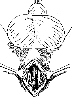
Figure 15. Liberation of the urethra. Stretching of the stricture during catheter insertion.
infection in the tissues and the organism's tendency to develop cicatricial tissue, these calculations are often problematic. Therefore, the operation of external urethrotomy has currently given way to resection of the canal, the author of which is Marion. Resection of the urethra. The patient is preliminarily drained of urine through a high section of the bladder and the application of a siphon. This operation is performed simultaneously with clean urine, and preliminarily with infected urine. In the latter case, resection of the canal is started after the infection has been overcome. A median longitudinal incision is made, and if the stricture is lo-
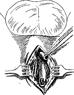
Figure 16. Stricture and scars resected. Peripheral end prepared. A retrogradely inserted probe protrudes from the central end of the urethra.
resides in the membranous part of the canal, a transverse arcuate incision is added from one ischial tuberosity to the other (Figures 15, 16, 17, 18, and 19). The urethra is freed, its peripheral end is severed with a circular incision and secured with ligatures. Then, the entire narrowed part is gradually excised up to the healthy central segment, which is freed by a circular incision and also grasped with Kocher clamps. The anterior segment of the canal is mobilized from the cavernous bodies of the penis and surrounding tissues to such an extent that it can be freely pulled toward the posterior segment. The latter is also slightly freed from the surrounding tissues. Upon freeing the segments of the canal, all scar tissue is carefully removed, a soft or elastic catheter, as thick as possible, is inserted through the external opening of the canal to the bladder, over which the edges of the urethral segments are sutured with fine catgut; usually, 5-6 interrupted sutures are applied. The skin wound is closed tightly. On the 12th-14th day, a permanent catheter is inserted, and the bladder fistula gradually closes. The section of the canal that can be excised sometimes reaches 8 cm. The success of the operation depends on sufficient mobilization of the anterior segment of the urethra, which facilitates bringing the ends together without excessive tension. Strictures of the urethra in women are encountered significantly less frequently than in men. Gonorrheal strictures are an extreme rarity here. The majority of strictures in women are of traumatic origin due to birth trauma, trauma from contusions, ulcers of the external opening, cauterization of polyps and caruncles, etc. The anatomical substrate of the strictures is the same as in men. Among the complications, one usually observes dilation of the post-strictural part of the canal in the form of a sinus bulging into the vagina (urethrocele), and insufficiency of the bladder muscle, manifested by frequent urges, pain in the lower abdomen, etc. Treatment: dilation with short bougies and urethrotomy. With strictures of the canal, acute urinary retention often occurs due to swelling of the scarred area. Measures taken in such cases are reduced to reducing hyperemia: cleansing the intestines, heat to the perineum, suppositories with morphine, pantopon, and antipyrine, a warm bath in which the patient attempts to urinate. Sometimes it helps to leave a filiform bougie passed through the stricture in the urethra. It would seem paradoxical that a patient is able to urinate with a bougie blocking the lumen of the stricture, but in practice, this measure is successful, apparently by softening the walls of the narrowing. If all these measures do not yield success, one has to resort to
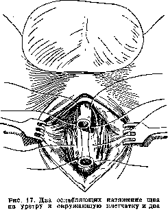
suture on the ends of the urethra.
to capillary puncture of the bladder, or the creation of a perineal or suprapubic fistula to the bladder. Stones of the urethra. Primary stones of the urethra are among its rare diseases. They form only under conditions of urinary stasis, i.e., in the presence of dilations, sinuses, and diverticula, or upon the settling of salts around foreign bodies retained in the canal (remnants of a catheter, blood clots, pus, mucus, etc.). Secondary stones, or rather those passing from the upper urinary tract, are stopped in the urethra only by virtue of their size. If this happens, they can grow here due to the deposition of salts on them. In the posterior urethra, stones can originate from the bladder or prostate gland and, protruding into the lumen of the urethra, are in essence only their continuation. The composition of the stones is the usual one for all stones of the urinary tract. Characteristic symptoms for a stone of the urethra cannot be noted, and only upon finding
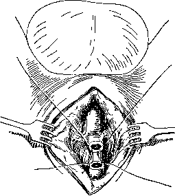
Figure 18. The tension-relieving sutures are tied, thanks to which the connection of the ends of the urethra occurs without tension.
them in the posterior urethra are there all the usual symptoms of a disease of this part of the canal; in the anterior urethra, they impede the outflow of urine. Diagnosis is made on the basis of palpation through the external coverings and probing of the canal; urethroscopy and X-ray confirm the diagnosis precisely. Removal is performed through a urethroscope for small stones; usually, however, one has to resort to the operation of cutting the canal. Foreign bodies can enter the urethra from above: ligatures after operations on the overlying urinary tract or uterus, fragments of catheters from the bladder, bullets, feces, and worms in cases of fistulas, etc., but more often foreign bodies are introduced through the external opening of the canal. In casuistic descriptions of such cases, all sorts of objects are encountered (pencils, wax candles, buttons, peas, etc.). These objects are in most cases introduced for the purpose of masturbation, less often for the purpose of self-mutilation (in the Tsarist army), and even less often as hooliganism and mischief in children. With the spread of urethroscopy, it is very common for cotton swabs used for wiping the mucosa to be left in the canal. Often, pieces of soft catheters remain if a dried-out and brittle rubber catheter is used for catheterization purposes. In all such cases, patients initially usually hide their condition and turn to a doctor only when the foreign body causes phenomena of severe inflammation or urinary retention. Depending on the nature of the introduced object, various complications may occur: injury, rupture, pressure sore with all the resulting complications. Diagnostics is not difficult and is made on the basis of the anamnesis. Treatment is the same as for stones of the urethra. Tuberculosis of the urethra in the form of a primary lesion of the anterior part of the canal is considered an extremely rare disease. It can arise sexually through intercourse with an infected woman. Clinical observations of this form are very rare, and it is difficult to establish typical pictures. Tuberculosis of the posterior part of the urethra is encountered significantly more often. Here the process is of a secondary nature, i.e., it spreads from the urinary bladder, and also from the prostate gland and seminal vesicles. Tuberculous infection initially causes infiltration, specific nodules, and then ulcerations with a grayish-yellow base and red edges. The symptomatology is the same as in general for diseases of the posterior part of the canal and the neck of the urinary bladder. Early diagnosis is difficult and is usually made when the process has already developed, in comparison with the presence of tuberculosis of the urogenital system. Treatment is extremely difficult and is reduced to general climatic or physiotherapeutic (quartz lamp) treatment. Local endourethral treatment—scraping, cauterization, application of medication—is problematic and
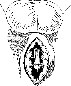
Figure 19. Circular suture of the urethra.
yields little success. Sometimes an effect is obtained by removing the primary focus (prostate, seminal vesicles, epididymis, and kidney). Syphilis of the urethra is observed in all stages. A primary syphilitic chancre can be located in the very initial part of the canal, behind the lips of the external opening, and in the navicular fossa. It is characterized by a cartilaginous infiltrate that does not produce abundant discharge. Ulcerations are frequent due to irritation by urine. Subjective sensations are insignificant. The absence of gonococci and the presence of spirochetes allow for differentiation from gonorrhea. The onset of secondary signs is exhaustive for diagnosis. Secondary forms of syphilis of the canal are extremely rare and are described in the form of papular and herpetiform syphilides. They cause discharge from the canal and simulate urethritis. Tertiary gummatous syphilides of the canal are characterized by the formation of individual nodules or by cylindrical hardening of the canal walls, often along its entire length (syphilome cylindrique Fournier). The recognition of forms of syphilitic involvement of the urethra is based on the same principles as in the disease of other organs: a thorough anamnesis, the Wassermann reaction, etc. Treatment is specific. A soft chancre of the urethra is more often a process spreading from the affected glans penis. Ulcerative forms are characteristic of this disease, and consequently, an abundant, thick, and sticky discharge. Finding Ducrey's bacilli in it clarifies the diagnosis. Treatment follows the usual principles for soft chancre. Medicinal substances are very conveniently introduced in the form of urethral suppositories prepared with cocoa butter.
Tumors of the canal, as in other organs, can be benign and malignant. The former include benign growths of the urethral mucosa. These growths occur in the form of acute condylomas and appear as cauliflower-like villous growths of a red or grayish color, sitting on a stalk. Mostly they grow near the external opening, although they can spread throughout the entire canal. Somewhat similar to them are papillomas of the canal with the same villous surface, but sitting on a broad base. The histological structure of these two forms is identical: they consist of a connective tissue base and are covered with squamous epithelium. Etiological factors for their occurrence are considered to be preceding inflammatory processes in the canal or prolonged irritants—instrumental treatment, cauterization, etc. Polyps of the urethra are rounded formations on a broad or narrow stalk, usually hanging in the direction of urine flow. Most often they develop in the posterior part of the canal. In their histological structure, polyps resemble an adenoma. Cysts of the urethra most often develop on the basis of obliteration of the excretory ducts of the glandular apparatus of the urethra. They appear as spherical, shiny, grayish, or yellowish formations. Depending on the location of the above-described formations, they can produce one clinical picture or another. In the anterior canal, when small in size, they do not produce any symptoms and are revealed only during urethroscopy. Large tumors can cause symptoms of canal stricture. When located in the posterior part of the urethra, tumors can cause disorders of urination or ejaculation.
Treatment is possible only with the help of urethroscopy: cauterization with an electrocoagulator, a galvanocautery loop, a Paquelin cautery, or chemical cauterizing agents (silver nitrate, chromic acid, trichloroacetic acid, etc.). Removal with forceps or curettage with a small spoon is used. For cysts, puncture with special instruments is used (see Urethroscopy). In the female urethra, special formations are often observed—caruncles. They appear as bluish-red round tumors protruding from the external opening, ranging in size from a cedar nut to a hazelnut. In their structure, the tumors resemble angiomas. They cause a sensation of pain and burning in the canal, are usually prone to bleeding, and are painful during sexual intercourse. Their treatment consists of burning with a galvanocautery or Paquelin cautery. In women, prolapse of the mucous membrane (prolapsus urethrae) of the urinary canal is also often found, which by its appearance can simulate a tumor; it is often seen in girls. The cause of this disease is the looseness of the connective tissue layer underlying the urethral mucosa. Any physical strain, increased work of the abdominal muscles, or inflammatory processes in this area provide an impetus for the appearance of the disease. Usually, the anterior section of the canal mucosa prolapses, more often the lower wall; sometimes circular prolapse also occurs. It does not cause particular suffering until the prolapsed wall becomes ulcerated. Then bleeding, pain during and after urination, difficulty in walking, and urinary disorders begin.
Treatment: in acute cases, attempts at reduction are made; in chronic cases—burning with a Paquelin cautery and galvanocautery, circular suturing, or excision of the prolapsed section with the application of sutures. Very rarely, among benign neoplasms in the urethra of men and women, myomas, fibromas, and angiomas are encountered. In view of the possibility of easily palpating the tumors, their diagnosis is not difficult. Treatment consists of excision of the tumor. Primary malignant tumors of the urethra are comparatively rare. The most favored site for them in women is the external opening of the urethra, and in men, the pendulous and bulbomembranous parts. Initial forms of the disease are diagnosed with difficulty; late forms ulcerate rapidly and simulate inflammatory diseases of the canal, which are most often (50%) cited as the etiological factor of the tumor. An accurate diagnosis can be made only by biopsy and pathological-histological examination. Treatment is surgical; sometimes radiotherapy yields very good results. Fistulae of the urethra (fistulae urethrae) can be caused by almost all the above-described diseases of the canal: inflammatory processes, strictures, foreign bodies, trauma, neoplasms, etc. The immediate cause of the development of fistulae is a breach in the integrity of the mucosa with subsequent urinary infiltration and the formation of abscesses, which seek an outlet both into the canal and through the skin. In view of the fact that a chronic inflammatory process usually precedes the fistula, the latter causes extensive development of connective tissue around the fistula. A clinical symptom of a fistula is the passage of urine through the fistula during the act of urination. The amount of urine passing through depends on the caliber of the fistula. A fistula can open in the free part of the urethra, in the perineum, on the buttocks, in the vagina in women, and in the rectum in men.

In the perineum, fistulae are often multiple, since the urinary infiltrate (abscess) is forced to overcome a rather thick layer of tissues; the formed fistula often closes, the pus seeks an outlet elsewhere, etc. When a fistula opens into the rectum, feces and gases penetrate into the urethra. Complications of fistulae are manifested in the form of eczematous processes on the skin of the perineum, scrotum, and thighs; in women, vulvitis and vaginitis develop. Constant wetting of underwear, the smell of urine from the patient, and recurring flare-ups of the inflammatory process around the fistulae and in the urinary tract bring patients to complete disability. Sometimes stones form in the fistulous tracts. Clinical diagnosis of a fistula is not difficult. The connection of the fistula with the urinary tract can be established by staining the urine (with methylene blue, indigo carmine) and observing the fluid discharged from the fistula. It is more important for the purposes of surgical intervention to establish the course of the fistula and the place of its origin in the urethra. For these purposes, a bougie is inserted into the urethra, and a thin metal probe is passed through the fistula; the meeting of the latter with the bougie will indicate the direction of the fistula and the place of its origin. The course of the probe can be recorded on an X-ray image. Direct examination with a urethroscope often does not make it possible to find the opening of the fistula in the canal, and therefore it is usually performed with a thin probe inserted through the fistula or by injecting dye. But the probe may not pass through the narrow and tortuous course of the fistula, and with force, one can create a false passage through granulation tissues, while the injection of dye during urethroscopy is extremely inconvenient. Therefore, they prefer to resort to urethrography with one or another contrast medium. Treatment
of urethral fistulae belongs to surgical operations requiring great patience on the part of the patient and persistence from the physician. Fistulae are closed plastically. With an insignificant size of the fistula openings, an attempt can be made to destroy the epithelialization of the openings by cauterization, to induce a granulation process, and to wait for the closure of the fistula. Previously, the canal is subjected to methodical bougienage in order to smooth out all strictures and folds of the mucosa and to provide free drainage for urine. In the case of labial fistulae, in the absence of infection in the canal and urine, it is necessary to freshen the edges of the fistula, separate the skin from the urethra, and apply sutures to the wound of the urethra and skin (Figs. 20 and 21). More often
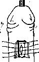
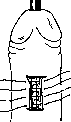
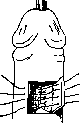
Figure 22. Skin incisions for lateral plastic surgery (solid line). Incisions according to Dieffenbach (dashed line). Figure 23. Mucous membrane sutured. Closure of the skin defect. Plastic closure of the fistula is applied, which can be performed in two ways. The fistulous opening is surrounded by a quadrangular incision and excised down to the mucous membrane of the canal. A longitudinal suture is applied to the opening of the canal, and the skin defect is closed with a flap taken from the side or closer to the perineum (Figs. 22 and 23). It is possible to close the urethral defect with a skin flap according to the Guyon-Pasteau method. A skin incision in the shape of the letter H, placed transversely on the penis. The lateral flaps are dissected. The fistula is freshened. Below it, a U-shaped skin incision is made. The flap is dissected, turned, leaving it on a pedicle, with the skin facing the lumen of the urethra, and sutured to the edges of the fistula. The lateral flaps are sutured over it. Longitudinal tightening sutures are applied to the U-shaped skin defect. In cases of fistulas opening into the perineal part of the urethra, it is most rational to perform an external urethrotomy operation with excision of all fistulous tracts and scar tissues in the vicinity of the urethra or a radical operation of urethral resection according to Marion (see above). In cases of urethrorectal fistulas, the operation according to the Albarran method is performed: an arcuate skin incision from one tuber ischii to the other with the convexity towards the scrotum. The tissues of the perineum and the tendinous cord between the urethra and the sphincter of the rectum are dissected in layers; then, by blunt dissection, the cellular tissue between the posterior urethra and the rectum is separated until the fistulous tract is reached. The latter is cleared of cellular tissue, dissected, inverted into the rectum, and closed with an invaginating Lembert suture. The urethral defect is closed with the same suture. Sutures are applied to the skin wound. In cases of fistulas of the female urethra, which occur most often on the basis of birth trauma, the crushing of tissues is so extensive that subsequent scarring shortens the urethral tube, fuses it with the vagina and the pubic symphysis. Therefore, when closing fistulas of the female urethra, it is necessary first of all to free it from scars, and then to suture the defect, thereby avoiding any tension (Fig. 24). The operation is performed through the vagina. The lower wall of the vagina is widely retracted with a speculum, then a cross-shaped incision of the vaginal wall is made, the flaps of which are widely detached by blunt dissection or with scissors. With an elastic catheter or bougie inserted into the urethra, the latter is freed from surrounding scars and adhesions. The edges of the fistula are freshened and closed with invaginating Lembert sutures in a longitudinal or transverse direction, depending on the width and shortening of the urethra. Over them is sutur-
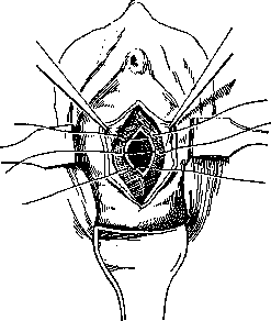
Figure 24. Suturing of the fistula after freshening its edges.
-ed the vaginal wound. The general requirement for successful surgical intervention for urethral fistulas is the asepsis of urine and tissues. Therefore, in the presence of infection in the urinary tract, preliminary preparation of the urinary tract is necessary: treatment of catarrh with irrigations, vaccination, intravenous injections of urotropin, neosalvarsan, trypaflavine, rivanol, etc. Of particular importance in all injuries of the canal, as well as a result of spontaneous rupture of the urethral mucosa in very narrow urethral strictures, are the so-called urinary extravasations or urinary infiltration. Its etiological factor is clear: rupture of the urethra and impregnation of the periurethral cellular tissue with urine, where urine penetrates under the influence of the pressure of the passing stream. If the injury occurred above the external sphincter, then the urinary extravasation goes into the posterior part of the perineum, into the pelvic cellular tissue, and from there it can rise higher into the greater pelvis and the perivesical cellular tissue. In case of injury to the bulbous and anterior parts of the urethra, the extravasation goes to the anterior half of the perineum, into the cellular tissue of the scrotum, and from there along the course of the spermatic cords to the pubis, the lower part of the abdomen, and into the cellular tissue of the skin of the penis. Urine that has penetrated into the cellular tissue decomposes very quickly, and sometimes, before one's eyes, redness of the skin forms with the development of gangrenous-putrid necrosis. Urinary infiltration proceeds like acute phlegmonous inflammation of the subcutaneous cellular tissue with sharp, shaking chills, high temperature, exhaustion, and cardiac failure. In the absence of immediate help, the patient dies within a few days with symptoms of delirium and collapse. The prognosis must be very cautious: mortality in the development of acute urinary infiltration is 35.7%. Treatment of urinary infiltration is carried out by surgical opening of all foci of urine accumulation and appropriate drainage of the wounds. To avoid further influx of urine, the latter is diverted by the application of a suprapubic fistula or boutonnière (see), depending on the location of the injury to the canal. In case of injury to the posterior urethra, it is necessary to apply a suprapubic fistula. Of course, the treatment of urinary phlegmon is an auxiliary act. The primary treatment must be considered the radical treatment of the cause that caused the urinary extravasation, such as: injury to the urethra, stricture, tumor, diverticulum, etc.
Related articles
Mentioned in
Cite this page
“Urethra.” Soviet Medical Encyclopedia. English translation of Bolshaya Meditsinskaya Entsiklopediya, 1st ed. (Moscow, 1928–1936), ed. N. A. Semashko. https://sovietmedicalencyclopedia.pages.dev/article/urethra/