Urological Instruments
Historical document, translated for reference. It reflects medical knowledge of the 1920s–30s and is not medical advice.
Summary
This article describes specialized urological instruments used for diagnosis and treatment of urinary tract diseases in the 1930s, including various catheters, cystoscopes, urethroscopes, dilators, stone extractors, and specialized surgical tools for kidney, bladder, and prostate operations.
Encyclopedia article (1928–1936)
UROLOGICAL INSTRUMENTS. In addition to the general surgical instrumentarium for the diagnosis and treatment of diseases of the urinary organs, there exist special instruments, for example bougies, catheters (see), cystoscopes (see Cystoscopy, cystoscopes), urethroscopes (see Urethroscopy, urethroscopes), dilators (see) (dilators) Ko'lman, lithotriptors (see Lithotripsy). The remaining U. i. can be subdivided into instruments used specifically in bloody urological operations, and instruments for endourethral and endovesical therapy. In kidney operations, in addition to ordinary surgical instruments, springy clamp forceps are used for grasping the fatty capsule of the kidney (Fig. 1) and kidney forceps for compressing the renal pedicle (Fig. 2), rounded or bent at a right angle (Fig. 3). If a clamp on the renal pedicle is left a long time, it is preferable to use the clamp of Fedorov with detachable handles (Fig. 6). For removing stones from the renal pelvis and calyces, special forceps without locks with pointed or cup-shaped branches (Fig. 5) or the dagger-shaped extractors of Fedorov (Fig. 4) are used. In operations on the urinary bladder requiring wide access to the bladder, for example in large bladder tumors, a special bladder mirror of Léguè (Fig. 8) is used, which allows wide separation of the edges of the bladder wound and fixing them in this state for the entire duration of the operation. For grasping the pedicle of a tumor in the bladder, forceps of Guyon (Fig. 9) with a short, strongly curved beak are used, which facilitates access to the pedicle and depth of the wound. Bladder stones at high section are extracted with forceps of Levkovich (Fig. 11) with shovel-like ends. The inner surface of the latter is made slightly spiny, which makes it possible to easily hold round and smooth stones. Multiple small bladder stones are most conveniently extracted with a special spoon with blunt edges (Fig. 7). In those cases where during bladder operations it is necessary to examine the cavity of the bladder in detail, a small specially constructed electric bulb is introduced into the bladder on forceps, which together with the wiring can be sterilized, or for the same purpose the instrument proposed by Gasparian (Fig. 10) can be used, which is a combination of a catheter with an electric bulb at the end of the beak. This instrument is introduced through the urethra and serves simultaneously for filling, washing out and emptying the bladder until it is opened. In perineal prostatectomy, a double blunt hook of Prust (Fig. 12) is used to push upward the anterior edge of the perineal wound together with the bulbar part of the urethra. In the space between both branches of the hook is placed the bulbous part of the urethra and thus it is protected from crushing when the edges of the wound are pulled strongly upward. In order to bring the prostate gland out from the depth of the wound, an instrument called desenclaveur, or tractor of Jung (Fig. 13) is introduced through the perineal urethral incision into the bladder. The instrument is introduced in a folded form (a), and then is opened (b), and by pulling it forward the prostate gland is brought into the wound. Special instruments are used for palliative operations in hypertrophy of the prostate gland in order to make a tunnel in it for the outflow of urine. Previously for this purpose the instrument of Bottini-Freudenberg (Bottini, Freu-denberg) (Fig. 14) called incisor was used: it has the appearance of a lithotriptor, in which the male branch is a branch provided at its end with a galvanic cautery knife (Fig. 14). In the handle of the instrument (a) there are two tubes (b), through which water circulates continuously in the handle for cooling the instrument during heating of the knife; the knife moves along the groove of the female branch by means of a disk (c). Behind the disk there is a contact {Щ for electric current, which is taken from the lighting network and is conducted through a transformer. Later Goldschmidt improved the incisor, adapting it to the cystoscope (Fig. 15), which made it possible to perform the operation not in the dark, but under visual control. At present the incisor is almost not used because of poor results of the operation, mainly because of postoperative bleeding. Instead of the incisor of Bottini, instruments constructed in the form of a urethroscope with a large lateral opening, called punch instruments, i.e. instruments for punching, or resectoscopes, are used. The resectoscope is introduced through the urethra so that the lateral opening is adjacent to the prostatic adenoma, and that part of the tumor which protrudes into the lateral opening is cauterized by a platinum knife introduced through the lumen of the urethroscope, which is heated by direct or alternating current. This manipulation is performed several times until a sufficient groove is burned for free outflow of urine. There are several modifications of resectoscopes, among them the most popular are the instruments of Jung


%^ш

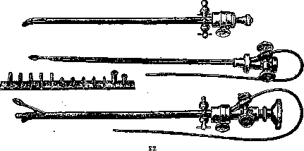
The 3rd..... -in -§ urological instruments and Kolk. The latter is shown in Fig. 16. In the USSR, resectoscopes are little used, but abroad they have recently become widely used. Some special U. i. are used in operations on the urethra; thus, for incising the narrowed external urethral orifice, a meatotome is used (Fig. 18), consisting of a handle inside which there is a knife. The meatotome is inserted into the lumen of the urethra beyond the narrowed external orifice, then by turning the screw the knife is extended from the handle to the desired width, and by withdrawing the instrument the external orifice is incised to the required caliber. For internal incision of the urethra in cases of stricture, the urethrotome of Mesonneau (Fig. 17) is used. It consists of a very thin metal bougie having a groove along the anterior or posterior wall (the urethrotome of Albarran has two additional grooves along the lateral surfaces of the bougie), along which moves a small triangular knife on a long handle. The knife is constructed in such a way that its tip is blunt and only the lateral slopes are cutting. Thanks to this, when the knife moves along the groove, it only cuts the narrowed areas, without injuring the healthy parts of the urethra, but merely pushing them aside. The urethrotome is equipped with a screw at its end, by means of which a thread-like bougie can be screwed onto it, which facilitates the passage of the urethrotome through the urethra in very narrow strictures. For external urethrotomy, an itinerarium—a grooved bougie—is used, which serves as a guide during incision of the urethra (Fig. 19). Some itineraria have an opening at their end for tying a long thread, which is passed in this way through the entire urethra when the itinerarium is withdrawn from the bladder. Among the instruments used for endovesical manipulations, we mention the stone seeker (Fig. 20)—a metal bougie with a short beak, allowing it to be freely moved in the bladder and thus to locate a stone or foreign body therein. There is a modification of the stone seeker, constructed in the form of a metal catheter with a short beak, the handle of which is equipped with a resonator. This instrument gives a louder sound when striking a stone, and at the same time through it the bladder can be filled with fluid. There is quite a large number of instruments for operations and manipulations in the bladder under visual control through an operating cystoscope. This includes an electrode for electrocoagulation of bladder tumors, consisting of a thin copper wire about 45 cm long, insulated along its entire length except for the tip. The insulation is usually a section of a ureteral catheter stretched over the mentioned wire. Through the operating cystoscope, small forceps are also introduced for grasping small stones in the bladder or at the ureteral orifice, as well as foreign bodies; small scissors—for cutting ligatures or the ureteral orifice stuck in the bladder wall; wire loops, cold and galvanocautic—for removal of bladder tumors, and sharp curettes—for intravesical biopsies. All these instruments are equipped with a thin, flexible metal sleeve, freely passing through the channel of the operating cystoscope, and a handle by means of which they are operated. Their construction is clear from Fig. 21, which shows an operating cystoscope with a set of endovesical instruments for it. Also belonging here are olives for ureteral dilation according to Joseph (Fig. 22), which are screwed onto a ureteral catheter and introduced through the cystoscope into the ureter. For removal of foreign bodies from the urethra, Coleman's forceps are used, the construction of which is clear from Fig. 23. A number of instruments have been constructed for endo-urethral therapy under visual control through a urethroscope; for example, the urethral probe (Fig. 24)—a curved metal rod, the end of which is heated over a flame, then passed through a stick of lunar caustic. The latter, melting, covers the end of the instrument with a thin layer. Having inflammatory granulations or polyps in the field of vision, one touches them with the end of the urethral probe. For the same purpose, one can use Coleman's set, which consists of several tips of different shapes and a handle for connection to an electric current (the construction is clear from Fig. 25). Urethral polyps can also be removed with the loop of Krause (Fig. 26). Inserting the thumb, index, and middle fingers into the rings of the handle, the middle and index fingers are brought close to the thumb, as a result of which the wire loop, previously placed over the polyp through the urethroscope, is drawn into the instrument and cuts off the polyp. Small granulations and polypoid growths of the urethra are curetted with the curette of Vossidlo (Fig. 27), with which one can also perform biopsies of pathological areas of the urethra. For opening abscesses and inflamed glands and cysts of the urethra, Coleman's knife (Fig. 28) is used, which is introduced through the urethroscope. In very narrow strictures of the urethra, impassable even for the thinnest metal and elastic instruments, thread-like (filiform) bougies made of whalebone are used. Manipulations on the urethra are quite painful and require prior anesthesia. The latter is performed by injecting anesthetic solutions into the urethra, which must be retained in the urethra for several minutes. For injecting anesthetic solutions into the canal, as well as medications, Lanz's syringe (Fig. 29) is used. To retain solutions in the urethra, a special clamp called a penis-clamp (Fig. 30) is used. In conclusion, we mention some instruments that were used in the not too distant past, but are no longer used today. These include bladder separators (Fig. 31), with which, before the invention of ureteral catheterization, an attempt was made to obtain urine separately from the kidneys. On the concavity of the bougie, after its introduction into the bladder, a rubber film is stretched by means of a screw, which divides the bladder along the median line into two halves. Urine from the right and left kidneys can flow out separately through tubes, which constitute the bougie-separator. The instrument does not hermetically separate the bladder, resulting in gross diagnostic errors. Also abandoned is the instrument of Lallemand (Fig. 32), equipped at the end with lunar caustic for cauterizing the urethral mucosa and seminal colliculus blindly. It has been completely replaced by the instrumentation used in combination with the urethroscope. The psychrofor of Winteritz is almost never used, which has the appearance of a hollow bougie with a partition along the median line, through the openings of which cold water is passed for the purpose of cooling the posterior urethra with a constant current of water. At present, the instruments of Arzberger (Fig. 33) and Winteritz (Fig. 34), constructed on the same principle, introduced per rectum and intended for cooling the prostate gland, are also not used.
Related articles
Mentioned in
Cite this page
“Urological Instruments.” Soviet Medical Encyclopedia. English translation of Bolshaya Meditsinskaya Entsiklopediya, 1st ed. (Moscow, 1928–1936), ed. N. A. Semashko. https://sovietmedicalencyclopedia.pages.dev/article/urological-instruments/