Lithotripsy
Historical document, translated for reference. It reflects medical knowledge of the 1920s–30s and is not medical advice.
Summary
Lithotripsy is a surgical procedure for crushing bladder stones using specialized instruments called lithotripters. The article describes the historical development, design, and various types of lithotripters and evacuation instruments used in the 1930s.
Encyclopedia article (1928–1936)
LITHOTRIPSY (lithotritia, from Greek lithos-stone and Latin terere-to rub; litho-tripsia, from Greek tribo-to rub; lithothrypsia, from Greek thrypto-to grind; litholapaxia, from Greek lapaxis-removal), an operation in which, with the help of special instruments, lithotripters, stones in the urinary bladder are crushed and brought to such a finely ground state that they can be completely washed out of the bladder. - Lithotripsy was first performed by the French surgeon Civiale in 1824. He worked with a very primitive lithotripter. Since then, mainly by American, French, and English surgeons (Bigelow, Guyon, Thompson), the lithotripter has been greatly improved. The lithotripter currently in use took its final form 40-50 years ago. This can serve as proof that its design has basically reached almost perfection. The steel from which lithotripters are made must be hardened enough so that they cannot break or bend during operation. The technique for manufacturing these instruments is therefore a highly specialized matter. The best lithotripters, satisfying all requirements, are manufactured by French firms. In the USSR, the most popular are Collin lithotripters from Paris. The lithotripter consists of two steel rods-branches (Fig. 1). One branch has the shape of a groove and is called the female branch. A well-polished second branch, shaped like a flattened rod and called the male branch, easily slides along it. The front ends of both branches are bent upward in exactly the same way at an angle
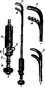
a little more than straight. In the closed state of the instrument, i.e., when both branches form one solid rod along their entire length, they form its beak, into which the stone is caught and by which it is crushed. In the beak, the tip and the heel are distinguished - the place where the branches bend. The tip is slightly smaller than the heel. The parts of the branches that form the beak are called spoons. They are made more powerful than the main rod, as they are the point of application of force during lithotripsy. They have a slightly flattened, rounded shape along the edges. In some lithotripters-spoon lithotripters-the spoons are solid plates with rough mating surfaces and a small hole near the heel of the female branch. In other lithotripters-terminal lithotripters-these spoons are thicker; in this case, the spoon of the female branch is perforated so that teeth are formed inside it, into the space between which the powerful teeth of the male spoon enter. The end opposite the beak of the lithotripter has a lock and screw, by means of which greater force can be applied during crushing, determined for each instrument. The screw belongs to the male branch. It ends with a strong ring head with threading on the periphery, by means of which the screw is operated. The female branch, however, forms 2 spring plates free at the ends with screw threads on their inner surface. Along these spring plates move the ends of a bracket fixed above and forming a ring on its free part. When the ring is lowered, the ends of the bracket press on the spring plates of the female branch, the screw threads of which engage with the screw of the male branch. In this position, the instrument is closed, and the free movement of the male branch in the female branch ceases. It is possible only by means of the screw. In addition, the upper end of the female branch has a handle, by which the instrument is held during operation. It is formed by a steel corrugated sheath covering part of the upper end of both branches. The larger the stone to be crushed, the thicker and stronger the branches of the instrument and the longer its beak must be. On the upper
Figure 1. Lithotripters: 1 - terminal; 2 - spoon; a - male branch; b - female branch; 3 - head part of lithotripsy: a - head; b - screw, which is part of the male branch; c - lock, connected with the female branch; d - lock bracket; e - handle. end of the lithotripter, a marking is always made indicating the size (in centimeters) of the stone that can be crushed by it. There are many numbers of lithotripters. The most
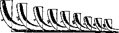
Figure 2. Collin scale, representing the beaks of terminal lithotripters manufactured by him (reduced 2nd/g times).
commonly used are the following: No. 000 corresponds to No. 12 on the Charrière scale, No. 00-No. 16, No. 0-No. 18, No. 1-No. 22, No. 3-No. 26, No. 4-No. 30. The absolute size of the beaks of lithotripters manufactured by Collin can be seen from Figure 2. Lithotripters are also manufactured in which a closed channel, similar to a catheter, is made along the entire length of the female branch, through which the bladder could be washed. Recently, lithotripters combined with a cystoscope have been manufactured (Fig. 3). However, such complication of the instrument, both
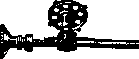
Figure 3. Joseph lithotripter.
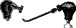
for the purpose of bladder washing and for the purpose of cystoscopy, weakens the strength of the female branch and is not caused by absolute necessity. Lithotripsy in the form in which it is currently performed (litholapaxia according to Bigelow) was introduced by the latter in 1878. The stone is crushed, the bladder is washed and freed from the smallest fragments by suction during one operation. For suctioning fragments, special instruments-evacuators-are used, consisting of metal catheters and a pump in the form of a rubber bulb. Catheters of different sizes have a beak with a very large radius of curvature and a large oval opening. Not far from the opposite end of the catheter, in assembled form (i); 2 - three catheters; a-rubber bulb; GROOVE^R™t"tеГ °S" lano conical thickening, by means of which it is firmly joined with the corresponding hole in the pump. The most common are Clover evacuators (Fig. 4), Bigelow evacuators (see Bigelow evacuator), Guyon evacuators (Fig. 5) and Duchatelet evacuators (Fig. 6). All evacuators are built on the same principle.
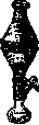

They consist of a large rubber suction bulb, in which a series of holes are made for connecting devices with a catheter, with a glass receiver collecting fragments and sand, and for pouring liquid. The original, simplest in design and convenience of use is the Clover evacuator. In it, the rubber bulb is connected to the catheter through an intermediate glass cylinder serving as a receiver for sand. All its parts in the assembled form are a continuation of the catheter along its length. Thanks to this, all movements are transmitted directly to the catheter, which greatly facilitates the washing out of fragments.
Many surgeons consider it possible to perform lithotripsy under local anesthesia, filling the bladder with anesthetic solutions, but this does not achieve either complete painlessness for the patient or the necessary rest for the surgeon. Therefore, it can only be used for small stones. General anesthesia is the rule for large stones. But here also deep sleep is required, so that the urinary bladder does not react with contraction to manipulations, especially if the case is about a disease or a very developed catarrh of the bladder, creating conditions for increased sensitivity. Ether, which is most popular for other operations, in the opinion of some surgeons, has a weaker effect on the contraction of the urinary bladder than chloroform. Therefore, for lithotripsy, it is better to use the latter.- Preparation of the patient for the operation is carried out in the usual way. Figure 5. Guyon evacuator.
Ch- Figure 6. Duchatelet evacuator.
In case of bladder catarrh and great irritability of the latter, it is necessary to first keep the patient in bed for some time and treat the catarrh, remembering however that before the removal of the stone the catarrh can only be reduced but not cured. If there is urethritis, it must be cured. Strictures of the urethra must be eliminated by dilation with bougies. Technique of the operation. A small pillow is placed under the patient's pelvis. This is especially important in adults with hypertrophy of the prostate gland. In children, one can do without it. As soon as the patient begins to fall asleep, a 5% solution of novocain is introduced into the urethra with a pipette. This addition of general anesthesia with local anesthesia of the urethra and bladder neck allows, without waiting for deep sleep, to begin careful washing of the bladder with irrigation fluid. The entire operation of L. must be performed under conditions of strict asepsis. All instruments are sterilized in the usual way; only the pumps of evacuators, the main component of which is a rubber bulb that cannot withstand frequent sterilizations, are thoroughly disinfected with antiseptic solutions. The bladder is washed with a regular metal catheter until the outflowing irrigation fluid becomes clean. It is necessary to wash the bladder and subsequently fill it with warm fluid to avoid causing undesirable contractions of the urinary bladder. After washing the bladder, 50-100-150 cm3 of fluid is introduced into it, depending on the age of the patient and the sensitivity of the bladder. The introduction of fluid must be done slowly and evenly and stopped at the slightest contraction of the bladder. Otherwise, all the fluid will be expelled from the bladder. L. must necessarily be performed with a full bladder. Only an extremely experienced lithotripsy surgeon and under exceptional conditions may allow himself to crush a stone in an empty bladder. A well-closed lithotriptor, previously greased with vaseline oil, is introduced into the bladder. The oil must penetrate between the branches so that they move quite smoothly. The oil should not reach the lock of the lithotriptor, otherwise it will be difficult to hold it and work the screw. The surgeon stands on the right side of the patient. He introduces the lithotriptor into the urinary bladder in the same way as a catheter. But whatever method is used to introduce the lithotriptor, it is necessary to pull the penis onto the instrument like a glove on a finger, and not push the instrument deep into the urethra. A lithotriptor is taken which corresponds to the presumed size of the stone, determined by the sensation from sounding, cystoscopy or X-ray. It should pass into the bladder completely freely, without violence and without direct preliminary dilation of the urethra. Sometimes in case of narrowing of the external orifice of the urethra, it is necessary to incise it with scissors. Taking the upper end of the lithotriptor with the fingers of the right hand, the surgeon initially uses it as a sound, finds the stone and determines its position in the bladder. Then, taking the instrument by the handle with the left hand, with the thumb on the lock bracket, he sets the lithotriptor in the sagittal plane at an acute angle to the plane of the table so that its beak is in the center of the filled bladder and points upward, not resting against any of its walls. Opening the lock bracket with the thumb of the right hand and thus making the branches of the instrument free, the operator, taking its head with the fingers of the right hand, begins to open the beak, pulling the male branch out of the female to a greater or lesser length depending on the presumed size of the stone, while lowering the female branch downward towards the border of the posterior and lower walls of the bladder and making small movements of the beak to the right and left. It often happens that, before reaching the wall of the bladder, the surgeon feels that the stone is already in the open beak (fig. 7). Then it is necessary again to bring the branches of the instrument together, close the lock, lowering its bracket, and begin to crush the stone with the screw. Throughout the entire operation, the closing and opening of the lock are done with the thumb of the right hand, and the work with the screw is done with the right hand (fig. 8). Under no circumstances should one begin work with the screw without being sure that the wall of the bladder has not been caught along with the stone. For this, it is sufficient to make a lateral movement with the beak of the instrument. If it is indeed caught, it either slips out during these movements or the surgeon feels that something is hindering the free movement of the beak to the right or left, and he opens the instrument and thus frees the wall of the bladder. No matter how many times during the operation it is necessary to close the instrument, one must never begin work with the screw without making these preliminary protective lateral movements with the beak. This act should become a habitual reflex for the surgeon. If the size of the lithotriptor corresponds to the size of the stone, the stone is usually caught easily and at once. Otherwise, only a small peripheral part of the stone is caught, and it easily slips out. Once the stone does not immediately enter the beak of the instrument, it must be caught. The lithotriptor is closed, lowered deeper, maintaining the same direction until a slight
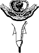
Figure 7.
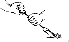
Figure 8. Stone caught, lock closed. The left hand firmly holds the handle of the instrument, its thumb rests on the ring of the lock bracket, and all the force of the right hand is directed to working the screw.
resistance against the urinary bladder at the border with its posterior and lower walls; the stone is felt, and at the same time the instrument is opened, the branches are spread apart by lifting the male branch towards the neck of the bladder without touching the latter, with the female branch immobile, and a series of lateral
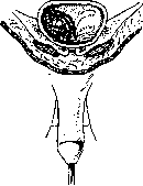
Figure 9.
movements are made. As a result of all these complex movements, the stone gets between the branches of the beak (fig. 9). It is very difficult and even impossible to describe the series of small, very varied, but in any case not rough or rapid movements with the beak of the instrument that need to be made to catch the stone. In persons with an enlarged prostate gland, in whom the bottom of the bladder is very deep, it is sometimes recommended to turn the instrument so that its beak points downward, and with the male branch fixed at the neck of the bladder, to catch the stone by moving the female branch (fig. 10). If the stone is small and brittle, it may be crushed by simple pressing of the male branch against the female with the hand. Sometimes it is sufficient to perform these manipulations several times, and the stone is crushed. In the vast majority of cases, however, it is necessary to work with the screw. Depending on the size and hardness of the stone, this work may require considerable strength and skill from the surgeon. With large and hard stones, the left hand, with its elbow bent, is firmly fixed by its muscles to the trunk and the entire hand firmly holds the lithotriptor. The right hand, in turn, uses all its direct force to bring the screw into motion to crush the stone, firmly fixing all the joints of the arm that is in suspension with its muscles and working only
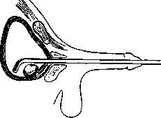
Figure 10.
with the hand. No intermediary, for example, wrapping the head of the lithotriptor in a towel for a firmer grip with the hand and therefore for the possibility of developing great force, should in no case be used. The point is that, as stated above, the lithotriptor is manufactured for a certain maximum size of stone on the condition that only the free force of the human hand is applied. When significantly greater force is applied, it may bend or even break. The right hand works the screw to the limit, i.e., until the branches of the instrument close. Then the fragments are caught in the same way and crushed. Although when working with a final lithotriptor there cannot be any jamming of stone fragments between the beaks of the forceps when the screw is tightened to the limit, it is still necessary to ensure that even the finest sand, formed during the crushing of the stone, does not accumulate there. Such a thin layer may remain there even with the screw fully tightened. If this is not paid attention to, after a series of individual crushing moments, the finest layer of sand may begin to deposit in the groove of the female branch near the very heel of the beak. Then under certain conditions this sand may become compacted and create a serious obstacle for the screw. To prevent this from happening, after a series of individual crushing moments, the lithotriptor should always be positioned so that its beak is in the sagittal plane in the center of the (necessarily) filled bladder, and a series of light, sharp blows should be made by hand with the male branch against the stationary female branch with the lock open. In the case where there is nothing between the branches, these blows produce a metallic sound. The absence of this sound indicates that there is sand between the branches. Then a series of such blows frees the instrument of sand, produces a metallic sound, and the operation is continued further. Without removing the lithotriptor from the bladder, crushing of only a small stone can be completed. After the stone has been crushed into a series of fragments that no longer require a strong lithotriptor, crushing can be continued with a thinner instrument. This is also dictated by the circumstance that after a series of insertions of the lithotriptor and evacuator catheter with subsequent washings of the bladder, the walls of the urethra may become slightly swollen, and the urinary canal, which initially passed the given lithotriptor very easily, no longer passes it so easily with subsequent insertions. Throughout the crushing process, it is necessary to ensure that the bladder remains filled. With a wide urethra and irritable bladder, sometimes, especially in women, the fluid filling the bladder constantly leaks out in small amounts. Then it is better to remove the lithotriptor, suction out the fragments, refill the bladder, and only then continue the procedure. Every time before removing the lithotriptor from the bladder, it is necessary to make sure by the method described above that there is no sand between the branches of the instrument in the beak, that the movements of the beak are completely free, that the lock is closed, and the screw is tightened to the end. Before proceeding to suction out the sand and fragments with the evacuator, the bladder must be washed through a catheter using a syringe. The evacuator catheter should be of the largest size provided it passes freely through the urethra. After removing the lithotriptor, sand and small fragments tend to enter the urethra behind it, especially with an irritable bladder. To avoid injury to the urethra by these fragments when inserting the catheter, a stream of liquid should first be passed through the urethra directly from the syringe (without the catheter), and only then the catheter should be inserted. The bladder should be washed with small portions of liquid and slowly, so that during its reverse flow, especially when the bladder contracts, the catheter does not become clogged with fragments. The latter circumstance not only disrupts the washing and suction process but can also become dangerous for the urethra when removing the catheter if a fragment gets stuck right at its opening. For this purpose, catheters for the evacuator are equipped with spiral mandrels, by means of which fragments are either pushed back into the bladder or crushed if they get stuck somewhere in the lumen of the catheter. The bladder is washed from the syringe until sand and fragments come out, and only then is suction begun when the washing liquid becomes clear. The evacuator is first filled with washing liquid, and in the Clover evacuator the intermediate glass cylinder is also filled with it. The pump and its parts are filled with liquid enough so that when the bulb is squeezed, no air enters the bladder. With the Clover evacuator, the surgeon does not need assistants. With the left hand he holds the catheter together with the receiver for fragments, and with the right hand he grips the pump bulb and squeezes and releases it. In this way he easily controls the evacuator, moving the bladder end of the catheter to any place in the bladder where fragments may accumulate. The fragments, however, accumulate mainly where the stone was located. When using other evacuators, the pump is held by an assistant who also watches the movements of the surgeon manipulating the catheter. During suction, it is constantly necessary to move the beak of the catheter to collect fragments wherever they are in the bladder. This is necessary because, as experiments have shown, the suction effect of the pump acts only in a very small space. After the squeezed pump bulb, left to itself, begins to expand, a flow of washing liquid from the bladder into the bulb appears. Fragments and sand, carried by this flow, on their way fall by gravity into the glass part of the pump intended for this purpose. The surgeon watches them with his eye and stops suction only when nothing more settles. At the end of suction, in the case where stone fragments that are too large to pass through the eyelet of the evacuator catheter remain in the bladder, a sound of very light blows against the end of the catheter is produced, which are also felt by the surgeon's hand. After a series of such alternating crushings, washings, and suctions, the bladder is freed of fragments and sand, and the operation is completed. The final act is examination of the bladder with a sound or cystoscope. For a very large stone, especially if it turns out to be very hard (oxalate), sometimes special techniques have to be resorted to. After grasping the stone with the lithotriptor and tightening the screw to the limit, one waits for several minutes. There are cases when during this time the stone cracks and breaks into pieces. In the same cases where this also does not help, a metal hammer is used. The stone is grasped, and the lithotriptor is held with the left hand so that the fingers fix both its branches in this position above the clamp with the lock open, and with the right hand a series of weak but short, sharp blows are made with the hammer against the head of the lithotriptor. In order for the bladder part of the lithotriptor to remain stationary in the center of the bladder, the holding left hand should find a firm support for its forearm on the anterior superior iliac spine. Under the influence of such hammer blows, the molecular structure of the stone changes, so that with subsequent work with the screw it is then crushed. If this also does not help, attempts at crushing should be stopped and a high incision of the bladder should be made. However, it must be firmly understood that it is better to underestimate the possibility of crushing the stone than to overestimate it. For large and very hard stones, a surgeon beginning his practice in lithotripsy had better perform a high incision of the bladder. Lithotripsy should be performed exclusively with final lithotriptors; the use of scoop lithotriptors should be abandoned due to the possibility of injuring the urethra during removal of the instrument with a fragment stuck between the scoops. Of the complications encountered during lithotripsy, the most serious is damage to the instrument during the operation. The lithotriptor may break, or it may prove impossible to close it, and consequently to remove it from the bladder. The technique of manufacturing lithotriptors has reached such perfection that Alexandrov and Krasnobaev, in 880 lithotripsies they performed on children using the thinnest instruments, did not have a single case of instrument breakage. As for the situation when it is impossible to close the instrument during lithotripsy, this may depend on two conditions. Firstly, if when working with a thin lithotriptor not corresponding to the size and hardness of the stone, maximum force is applied, the branches of the instrument begin to spring strongly and may even start to diverge over their length, where in this case even a slit, however small, is formed. A grain of sand may get into this slit. In this case, the sliding of the branches becomes impossible, the lithotriptor may bend, and then of course it will be impossible to close it. Another reason for being unable to close and remove the lithotriptor may be the clogging of the groove of the female branch with the finest sand having the property of cement, as mentioned above. Such sand is given by urate stones rich in mucus impurities. They crush very easily, almost into dust.
If with such a stone the bladder is irritable, and the urethra is wide in relation to the lithotriptor, and the irrigating fluid constantly flows out through it, then if the surgeon wishes to finish with the stone at once, without removing the lithotriptor from the bladder, the following may occur. In the bladder, the amount of fluid will gradually decrease, it will constantly become relatively rich in crushed sand, which has been almost pulverized into dust, and this sand may begin to deposit in the groove of the female branch and create an insurmountable obstacle to the closure of the branches. The later this circumstance is noticed, the less hope there will be of getting rid of this sand and closing the branches. To avoid this danger, one should not be tempted by the possibility of crushing the stone at once under such conditions without removing the lithotriptor from the bladder, but should vigilantly ensure that the bladder is always filled with fluid, and from time to time remove the lithotriptor and cleanse the bladder by irrigation and suction of the finest sand. It is better to introduce the lithotriptor into the bladder 3-4 times with subsequent cleansing of the bladder each time than to experience such a serious complication as an insurmountable clogging of the groove of the female branch. If this has already happened and the instrument cannot be closed, then, as in the case when it cannot be closed due to grains of sand getting between the springing branches from undue force applied to the lithotriptor, nothing remains but to perform a high incision of the bladder. Through the incision in the bladder, the beak of the instrument is protruded outward and the position is clarified. In the case where it is possible to clean the groove of the female branch from sand by one means or another under the stream of irrigating fluid, the instrument is closed and withdrawn from the bladder through the urethra. If the reason for the inability to close the instrument is the entry of grains of sand between the branches, then by striking the head of the lithotriptor with a hammer while the beak is held immobile, it is possible to join the beak's spoons, close the instrument, and withdraw it from the bladder. However, if for any reason it is impossible to close the lithotriptor even after withdrawing the beak outward through the incision in the bladder and applying the above-mentioned measures, one has to saw through the lithotriptor with a file. Individual cases of such complications, known in the literature, show that even with them it is quite possible to bring the operation to completion and the patient to recovery. For lithotripsy, one can limit oneself to a relatively small set of lithotriptors. For adults, it is sufficient to have terminal lithotriptors of Collin's numbers 0, 1, 2, and 3. For lithotripsy in children, the set should consist of terminal lithotriptors numbers 000, 00, and 0. For very small children, L. P. Alexandrov and T. P. Krasnobaev were greatly aided by a terminal lithotriptor made by the former Moscow firm Schwabe according to L. P. Alexandrov's suggestion, which corresponded in stem thickness to Collin's lithotriptor number 000 (number 12 on the Charrière scale), but had a beak with slightly narrower spoons and more rounded edges. At the Schwabe firm, this instrument was known as lithotriptor number 12. There were many cases where in very small children Collin's lithotriptor number 000 could not be passed, while the Schwabe instrument number 12 passed easily. Postoperative care after lithotripsy is very simple. After lithotripsy, there is always more or less pain during urination, often disappearing within a day and quickly passing after warm baths. In cases of great pain and sometimes developing retention of urine dependent on it, there are indications for the use of narcotic agents. In cases of persistent retention of urine, it is necessary to drain the urine with a catheter several times, and sometimes even leave it in the bladder for 1-2 days. The latter is mainly necessary when there is simultaneous hypertrophy of the prostate and persistent catarrh of the bladder that existed even before the operation. Often after the operation, urine is tinged with blood for several days and is rich in sediment with an admixture of the finest sand. This complication usually passes by itself. The temperature often rises to 38°C, but more often remains normal. Sometimes, when at the end of the operation a small fragment is left in the bladder due to oversight, it may spontaneously exit through the urethra during urination in the coming days, but it may also get stuck in the urethra. In the latter case, one has to perform external urethrotomy. In the vast majority of cases, the postoperative period proceeds smoothly. The next day the patient can get out of bed, which is especially important for the elderly, and in 3-4 days can be discharged from the hospital. Before discharge, however, it is always necessary to examine the bladder again. This examination may reveal a fragment in the bladder. Then a single introduction of the lithotriptor, even without general anesthesia, is sufficient to eliminate it. Indications for lithotripsy are in all those cases when the urethra freely passes a terminal lithotriptor corresponding to the size of the stone. Stones up to 5 and even 7 cm in size can be crushed. One should resort to high incision of the bladder only in cases where lithotripsy is not feasible. Contraindications, besides urethral stricture and size of the stone, are immobile, tightly seated stones in the folds of the bladder, stones located in diverticula, and hypertrophy of the prostate causing persistent retention of urine. Indications and contraindications are indirectly determined by the general condition of the patient. In weakened, especially elderly people, with a large stone whose crushing would require a lot of time, it is better to perform a high incision of the bladder and finish the operation in a few minutes. On the other hand, severe catarrh of the bladder and even pyelonephritis with a small stone are not contraindications. Childhood age is in no way a contraindication. Western European and American surgeons consider lithotripsy in adults the operation of choice, and the number of lithotripsies among them is relentlessly increasing. The reproach usually made against lithotripsy, that it should more often have recurrences of stone than after lithotomy, has no sufficient basis. The great experience available has not confirmed this. The results obtained with lithotripsy, evaluated by mortality statistics, are excellent. Guyon performed more than 4,000 lithotripsies and had a mortality of less than 2%. Desnos and Minet in 1898 gave statistics of 1,998 lithotripsies with a mortality of 2.75%. According to similar statistics provided by Alexandrov in 1912, out of 1,555 cases the mortality was 2.77% (this statistics included 507 lithotripsies performed by Alexandrov himself on children); Sinitsyn had a mortality of 2.7% on 179 lithotripsies. In Sinitsyn's cases, out of 5 who died, 4 had signs of pyelonephritis before the operation. Lithotripsy in children is not popular in Western Europe. In France and Germany, the age up to 5-8 years, and according to Wehner's opinion even up to 10-15 years, is considered unsuitable for lithotripsy. The experience of Russian surgeons, however, has shown that in children lithotripsy for bladder stones is also the operation of choice. Alexandrov out of 698 children with bladder stones performed lithotripsy on 574 and lost 18 patients; however, the mortality due to the operation itself was only in 9 patients, i.e., 1.6%. Most of these 574 operated patients were not older than 5 years. Krasnobaev's experience is as follows: 189 lithotripsies performed at the Model (formerly Morozov) Children's Hospital in Moscow from 1904 to 1927 gave a mortality due to the operation of 1.1%, with the last 120 lithotripsies not causing a single death due to the operation. The average age of the operated children was 4.7 years, children not older than 3 years constituted 38%, and among the latter there were 5-year-old children. Taking into account 28 upper incisions of the bladder made during the same time in the same hospital in the most severe cases, we get a total mortality for bladder stones in children of 1.4%. But even if we do not exclude from the number of those who died after lithotripsy those who died independently of the operation, still according to Krasnobaev's material we get the following figures: mortality for lithotripsy-1.7% and total mortality for all cases of bladder stones after lithotripsy and lithotomy-3.7%. These figures fully justify the position that in children lithotripsy should be the operation of choice.
Related articles
Mentioned in
Cite this page
“Lithotripsy.” Soviet Medical Encyclopedia. English translation of Bolshaya Meditsinskaya Entsiklopediya, 1st ed. (Moscow, 1928–1936), ed. N. A. Semashko. https://sovietmedicalencyclopedia.pages.dev/article/lithotripsy/