Lithotomy
Historical document, translated for reference. It reflects medical knowledge of the 1920s–30s and is not medical advice.
Summary
A historical overview of the lithotomy procedure, detailing its evolution from ancient techniques to the 1930s, including the development of various perineal and suprapubic approaches.
Encyclopedia article (1928–1936)
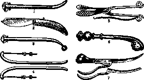
LITHOTOMY (lithotomia), an operation performed for urinary bladder stone disease, which consists of opening the urinary bladder and removing stones from it. Lithotomy is one of the oldest operations, mentioned 6 centuries before the Christian era in the medical treatise of the ancient Hindus (Susruta). In former times, lithotomy was performed not by physicians, but by specialists in this field—lithotomists, stone-cutters—who kept their art a secret from physicians and passed it on only to those close to them. Over the course of a whole series of centuries, physicians themselves avoided lithotomists, considering them ignorant. Hippocrates himself forbade physicians from performing lithotomy, but this prohibition did not have its effect, and physicians were still not strangers to lithotomy. Thus, Celsus gives the following description of the technique of lithotomy: 1. Bringing the stone down into the neck of the bladder by means of pressure with the right hand through the abdominal wall above the pubis and fixing the stone in the neck of the bladder with the index and middle fingers of the left hand inserted into the rectum. 2. An incision of all layers of the perineal tissues in the shape of a crescent in front of the anus; the ends of the incision are directed posteriorly. 3. Removal of the stone with the finger of the right hand or a special hook. The entire operation is performed with the help of two instruments—a scalpel and a hook, and therefore it was called an operation with a "small set" ("le petit appareil" of the French and "kleine Gerätschaft" of the Germans). Subsequently, there was no progress in the technique of lithotomy until the beginning of the 16th century, when an improvement appears, attributed to the physician from Cremona, Giovanni di Romanis. One of his students, Marianus Sanctus, describes the set of instruments necessary for performing lithotomy under the name of the "large set" ("le grand appareil"). The latter includes: 1) a conductor (Fig. 1), 2) a razor (Fig. 2), 3) a probe (Fig. 3), 4) wound dilators (Fig. 4), 5) stone forceps (Fig. 5), 6) a stone spoon (Fig. 6), etc. The novelty of Giovanni's technique was that he used a conductor inserted into the urethra and bladder for orientation when making an incision on the perineum to the left of the midline, whereas previously, before the introduction of the conductor, orientation was performed with the help of fingers inserted into the rectum. The technique of lithotomy was developed in detail in 1682 by François Tolet. To this same time also belongs the official recognition of this operation by the medical world and its inclusion in the field of surgery. François Tolet ended the era of lithotomists; he was also the first to perform lithotomy at the Charité hospital in Paris. Despite the official recognition of lithotomy by the medical world, this operation continues to be performed by lithotomists as before; there have been attempts by individual operators to propose their own instruments (Le Cat, Frère Côme, Frère Jacques). The lithotomes of Le Cat and Côme are based on the principle of a guillotine, the knife of which is hidden in a sheath, which serves at the same time as a conductor (Fig. 7). They already used the conductor introduced by Giovanni and Marianus, but ignorance of anatomy and the desire to avoid bleeding pushed them to invent an instrument that would be both a lithotome and a probe. Just as in France lithotomists did not disappear with the appearance of F. Tolet's treatise on lithotomy, so they have not died out to the present time in the USSR, in Transcaucasia. Since the neck of the bladder was often injured during perineal lithotomy, which entailed urinary incontinence, the thought of physicians was directed toward improving access to the urinary bladder. To Le Dran belongs the honor of developing the perineal lateral opening of the urinary bladder, in which the sphincter of the bladder is not injured; but this anatomically justified access gave significant mortality in the pre-antiseptic era, just like the original perineal method. Lithotomia perinaealis lateralis in Russia was especially widely used even before the introduction of asepsis by Bogdanovsky and Sinitsyn, but at the present time it has only historical interest (Alexandrov). High section of the bladder (suprapubic) was first (1556) accidentally performed by P. Franco when he could not remove a large stone through the made perineal incision; however, this operation, as more dangerous due to the possibility of injuring the intestine, was condemned by the authority of that time—the royal physician L. Collot. High section did not find application in France for a long time, and only a century and a half after P. Franco in Paris did Morand in 1727 again begin to perform high section, using the technique of Douglas, developed by the latter on cadavers. It is interesting to note that Morand introduced during this operation a high position of the pelvic part of the body in relation to the thoracic part, i.e., what 150 years later began to be called the Trendelenburg position. The speed of Morand's operation was amazing: the entire operation lasted 2 1/2 minutes. However, many years passed before the high section operation took its proper place. One must also mention the operation lithotomia rectalis, which did not gain distribution due to the complications caused by it (vesicorectal fistula, infection of the bladder, and ascending infection of the kidneys).
Modern lithotomy appears in the following form: the perineal method, which was used for so long in its various modifications—lithotomia perinaealis mediana et lateralis—is rarely used at the present time and only for exceptional indications, when access to the urinary bladder via the suprapubic route is extremely difficult as a result of repeated openings of the bladder and the anatomical changes that have occurred as a result of this. Some authors consider excessive obesity of the abdominal integuments to be another indication for perineal opening of the bladder. The technique of median perineal section is reduced to the following: the patient is placed in the "lithotomy position" on the edge of the table with the thighs bent toward the abdomen and spread apart, fixed with leg holders; an assistant stands to the side and fixes the metal bougie or catheter inserted into the urethra with one hand, and the scrotum with the other. Some authors recommend using a Syme catheter, which has a groove on its convex surface. The operator sits between the patient's thighs with his back to the light source, which well illuminates the operative field—the perineum. An incision 4–5 cm long is made along the perineal raphe and ends 1 cm in front of the anus. To create freer access, additional small incisions can be made from the end of the incision in both directions posteriorly in the shape of the letter Y (according to Guyon). After dissection of the fascia and perineal muscles (mm. transversi perinaei), the membranous part of the urethra (pars membranacea) is exposed, and the instrument inserted into the urethra is felt with the index finger of the left hand. The membranous part of the urethra is dissected longitudinally along the catheter for a distance of 1–1.5 cm posteriorly, immediately from the bulbous part. Through the urethral incision, guided by the groove of the catheter, a lithotome is inserted in a closed state with the concavity upward until it rests against the end of the catheter groove; at this moment, the catheter is removed first, and then, having opened the lithotome, it is withdrawn, and at this moment the prostate gland is dissected. Then, through the incision made, the bladder is examined with a finger and the stone is extracted with stone forceps. After the stone is removed from the bladder, a drain is inserted into the bladder through the wound or an indwelling catheter through the urethra. If a lithotome is not used, one can replace the Syme catheter with a simple metal bougie or catheter, dissect the membranous part of the urethra on it, and after removing the bougie, insert a sponge forceps through the urethral incision into the bladder to dilate the neck of the bladder, and then examine the bladder with a finger and extract the stone with forceps. The operation of median perineal section of the bladder, in addition to the above, may have a very limited application due to the size of the stone: without severe injury to the neck of the bladder during the extraction of the stone, only stones with a diameter of less than 2 cm can be extracted. In women, the operation of median perineal opening of the bladder is replaced by the operation of vaginal section.
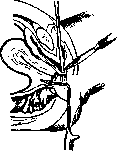
Figure 1–7. Figure 8.
of the bladder (cystotomia vaginalis, or colpo-cystotomia). The patient is placed in the "lithotomy position," and the vagina is dilated with a Sims speculum posteriorly. The wall of the vagina and bladder is incised longitudinally along the midline up to the anterior fornix, and the stone is removed from the bladder through the incision (Fig. 8). After the stone is removed, catgut sutures are applied separately to the wall of the bladder and the vagina. The disadvantages of this operation include: the impossibility of removing large stones, and the frequent occurrence of postoperative urinary incontinence and vesicovaginal fistulae (E. Wehner). Lisfranc described another approach to the female urinary bladder for stone removal, which was later developed by Pasteau, Legueu, and Cathelin—cystotomia subpubica, sive subsymphysaria. The patient is in the "lithotomy position," and the labia minora are spread by an assistant; an arcuate incision with the concavity facing downward is made halfway between the urethral opening and the lower edge of the pubic symphysis through the thickness of the mucous membrane of the vestibuli vaginae. After dissecting the lower edge of the incision, it is retracted with forceps or a stay suture. Next, the anterior wall of the urethra is dissected, the suspensory ligament of the clitoris is cut, and the neck of the bladder becomes accessible, and the bladder is opened just above it. The disadvantages of this operation include an extremely narrow access to the bladder and the ability to remove only small stones.
The most common operation for removing stones from the urinary bladder at the present time is lithotomia suprapubica, sectio suprapubica, or sectio alta. The patient is placed on the operating table with the pelvis slightly elevated, and the operator stands on the patient's left side. A catheter is inserted into the urinary bladder, the bladder is washed out and filled with sterile physiological saline, or a 3% boric acid solution, or sterilized boiled water in an amount of 300-400 cm3. To avoid irrigating the wound during the opening of the bladder with the distending irrigation fluid and thus introducing infection, the urinary bladder can be filled with air after washing. However, Rumpel insistently warns against this, as he was a witness to an air embolism that resulted in death.
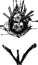
To avoid air embolism, it is recommended not to overdistend the bladder with air and to inject it with a syringe in the same amount as the liquid. To prevent the liquid introduced into the bladder from flowing out (or the air introduced into it from escaping), a clamp is placed on the catheter. In some cases (with bladder spasms and when its volume is reduced), it is recommended to perform the filling after incising the skin and the linea alba down to the preperitoneal tissue, as filling the bladder at this moment allows for a better view of its expansion and its relationship to the peritoneum (Figs. 9 and 10). To raise the urinary bladder higher toward the abdominal wall from the depth of the pelvic cavity, Peterson suggested inserting a rubber balloon (of the colpeurynter type) into the rectum and filling it with air (200-250 cm3). In women, this balloon can be inserted into the vagina, or the vagina can simply be tamponaded with gauze.
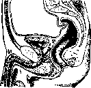
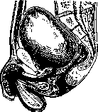
The skin incision is made from the pubic symphysis upward along the midline, up to 10 cm in length depending on the thickness of the subcutaneous fat layer. After incising the skin and subcutaneous tissue, the linea alba is located and incised, then the rectus abdominis muscles and pyramidalis muscles are retracted to both sides with retractors. With a filled bladder at this moment, the tissue of the prevesical space (cavum Retzii) is visible in the lower part of the wound, and the transitional fold of the peritoneum is visible in the upper part. The bladder wall is separated from the tissue, and at the same time, the transitional fold of the peritoneum is pushed upward with the help of a gauze sponge. At this moment, the veins of the bladder wall running from its neck along the anterior wall and the longitudinal bundles of the detrusor muscle of the bladder become visible. Through the anterior wall of the bladder, closer to the pubic symphysis, thick silk stay sutures are placed to prevent the anterior wall of the bladder from sinking into the pelvic cavity after the bladder is opened. The bladder is incised between these stay sutures, with the pointed scalpel inserted in such a way that its tip is directed toward the pubic symphysis (Fig. 11). The bladder is opened for a length of 2 cm so that a finger can be inserted into it and it can be examined. At the very moment the bladder is opened, the clamp is removed from the catheter, and the liquid is drained from the bladder. If the stone is large, the incision of the bladder wall is extended to the necessary size, and the stone is removed with stone forceps. One must try to spare the edges of the bladder wound from bruising during the extraction of the stone, as bruising of the tissues leads to their necrosis and poor healing. If there are a large number of small stones in the bladder, they are removed with a blunt spoon.
The stone may be of such a size that it will not pass through the incision in the bladder wall. In that case, one resorts to reducing the size of the stone, to crushing it, by grasping it with a strong lithotrite and striking the handle of the lithotrite with a hammer. After cleaning the bladder of all fragments, one proceeds to suture its wall. Both Russian and foreign authors have worked particularly hard on developing methods for applying the bladder suture. The requirement for the urinary bladder suture is that it must hermetically seal the bladder cavity, which is achieved as follows: the first row of sutures (catgut) is applied in such a way that the mucous membrane is not pierced and the edges of the incision are in tight contact with each other; the second row of sutures (it is possible to apply a continuous suture as well) is applied in the manner of a Lembert intestinal suture, i.e., only the muscle layers of the wall are caught in the suture, and the first row of sutures is thereby inverted (Fig. 12). To eliminate the cavity that forms in the prevesical space when the tissue is pushed away from the bladder wall, it is recommended to suture the bladder to the abdominal wall at the very pubic symphysis with one or two catgut sutures.
If the urinary bladder is not inflamed, the abdominal wall incision can be sutured tightly. In the presence of catarrhal cystitis, it is preferable to place a drain in the lower corner of the wound for 1-2 days. In purulent cystitis, the bladder wound is narrowed with sutures, a drain is inserted into the bladder in the middle of it, and it is secured there with sutures. Urine is drained into a vessel with the help of a siphon. Bladder drainage allows the bladder to be given complete rest and creates the possibility of performing abundant repeated bladder irrigations for several days in a row to treat cystitis. The drain is removed after 8 days, and the wound gradually heals. In an aseptic state of the bladder, it is possible after a tight suture to allow the patient to urinate independently, which does not hinder primary wound healing. In catarrhal cystitis, it is more expedient to leave an indwelling catheter in the bladder and perform bladder irrigations 2-3 times during the day with small amounts (about 30 cm3) of liquid to wash out mucus and blood clots from the bladder. The catheter can be removed after four days.
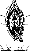
Figure 11. 1-rectus muscle; 2-peritoneal fold; 3-anterior wall of the bladder; 4-pyramidalis muscle. Figure 12.
Mortality after the operation of high lithotomy is determined differently by various authors: thus, Casper speaks of 14%, Zuckerkandl of 13.5%, Frisch of 12.7%. Such a high percentage of mortality is explained by the fact that the named authors selected severe cases for lithotomy, whereas mild cases were subjected to lithotripsy. Among those authors who perform exclusively high lithotomy (sectio alta) for bladder stone disease, the mortality percentage is significantly lower. For example, Trofimov reports 2.8% with a closed bladder suture and 4.8% with open treatment. The increase in the mortality percentage with the open method is explained by the fact that this group included exclusively more severe cases. The disadvantages of sectio alta include urinary infiltration of the prevesical tissue, dehiscence of bladder sutures, and sometimes long-unhealing urinary fistulas, which cannot always be avoided even with the most careful suturing technique. In exceptional cases, with stones of very large size, a necessity for a wide opening of the bladder may be encountered. For this, sectio alta transversalis, which was highly recommended by Ledran and Franco, provides better access; but in textbooks, it has been assigned the name of Trendelenburg. The position of the patient and the filling of the bladder are the same as in the classic high lithotomy; the transverse incision is made one transverse finger-breadth above the pubic symphysis, the aponeurosis is exposed, then the mm. pyramidales are cut transversely at the place of their attachment to the bone, which allows the index finger to be placed under the m. rectus and the latter to be sutured with threads to hold it from excessive contraction after cutting and to facilitate suturing at the end of the operation. After cutting the rectus muscles, the wound gapes widely; the peritoneum is pushed upward, and the bladder is opened widely, also transversely. Suturing of the cut tissues is performed according to general rules. In the postoperative period, it is necessary to keep the patient with the limbs drawn to the abdomen to relax the tension of the rectus muscles. Due to its greater traumatism, the operation of transverse lithotomy has not received wide distribution and has extremely limited indications in stone disease. In order to eliminate complications after classic high lithotomy, transperitoneal lithotomy was proposed. The idea of transperitoneal lithotomy belongs to Sklifosovsky, and in a clinic on a patient, this method was first applied by Rydygier. In the USSR, Solovov and Gridnev were interested in this method. The technique of the operation of transperitoneal lithotomy (sectio alta transperitonaealis) comes down to the following moments: 1) washing the bladder and completely emptying it of the washing fluid; 2) the patient is placed in the Trendelenburg position; 3) the abdominal cavity is opened by an incision along the midline from the pubis to the navel, the intestinal loops are retracted, and delimiting sponges are introduced into the small pelvis up to the plica recto-vesicalis (or vesico-uterina in women) to isolate the abdominal cavity; 4) the bladder wall is held by sutures (stay sutures) passed through its apex and is incised longitudinally (or transversely) between them; 5) the bladder cavity is dried with tampons, and 6) the stones are removed with stone forceps. Suturing of the bladder wall incision is performed according to the type of suturing of other hollow organs covered by the peritoneum, with a Lembert suture in two rows. After removing the delimiting sponges, the abdominal wound is sutured according to general rules. A catheter a demeure is introduced into the bladder. Regarding transperitoneal opening of the bladder, a number of doubts are expressed, which come down to the following: it is not always easy to protect the abdominal cavity from irrigation with urine during the operation; the prolonged contact of the peritoneum with sponges and air is not indifferent; there is no certainty in the strength of the bladder suture, and the possibility of urinary infiltration of the bladder wall is not excluded, etc. As for the indications for performing the operation of lithotomy, it is performed in all cases where lithotripsy cannot be performed. Narrowness of the urethra due to cicatricial stricture and enlargement of the prostate gland, which are difficult or impossible to eliminate, will constitute indications for sectio alta. Furthermore, stones of very large size, stones fixed in diverticula and on foreign bodies (sutures) are removed in this way. Severe cystitis with a sharp decrease in the capacity of the urinary bladder, and inflammation of the upper urinary tracts, proceeding with a febrile temperature, also indicate the removal of stones through high lithotomy.
Related articles
Mentioned in
Cite this page
“Lithotomy.” Soviet Medical Encyclopedia. English translation of Bolshaya Meditsinskaya Entsiklopediya, 1st ed. (Moscow, 1928–1936), ed. N. A. Semashko. https://sovietmedicalencyclopedia.pages.dev/article/lithotomy/