Hysterosalpingography
Historical document, translated for reference. It reflects medical knowledge of the 1920s–30s and is not medical advice.
Summary
Hysterosalpingography is a radiographic method for examining the uterine cavity and fallopian tubes after filling them with a contrast substance. It is used to determine tubal patency, diagnose tumors, and study uterine and tubal physiology.
Encyclopedia article (1928–1936)
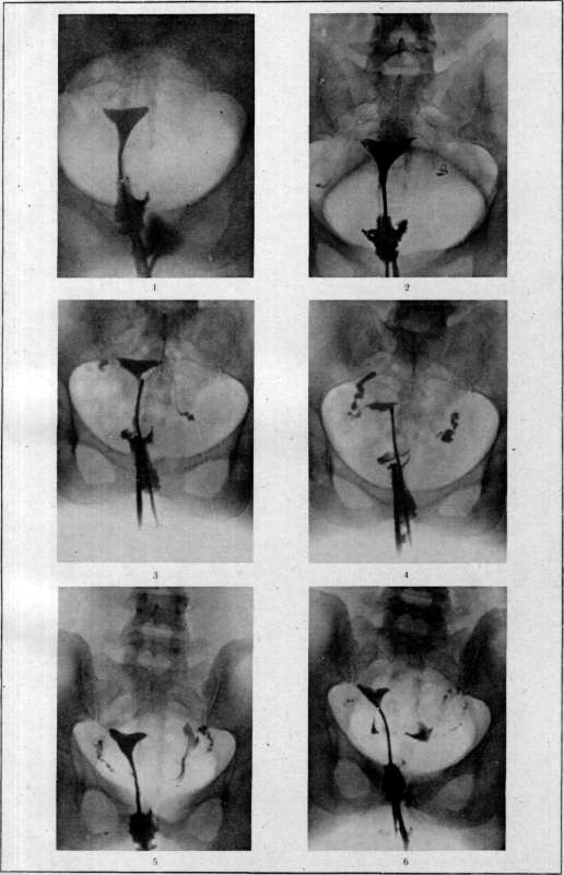
Hysterosalpingography (метросальпингография), a method of radiography of the uterine cavity and tubes filled with a contrast substance. The idea of introducing a contrast substance into the uterine cavity belongs to Rubin (Rubin; 1915), while the practical use of H. as a method is due to Kennedy (W. J. Kennedy; 1923). The method is used to determine the patency of the tubes and the site of their possible obliteration (in infertility), for the diagnosis of tumors based on changes in the normal picture of the uterine cavity (hysterography), and for the study of the physiology of the tube and uterus (peristalsis of the tubes and contractions of the uterus). Contraindications for performing H. should be: 1) the slightest suspicion of pregnancy; 2) febrile or subfebrile condition of the patient; 3) signs of local and peritoneal irritation, even if slight; 4) the presence of even slight uterine bleeding and the menstrual period. The technique of H. is not complicated, but requires the patient to be hospitalized for at least one day. After thorough examination of the patient and exclusion of the above contraindications, the bowel (enema) and bladder are emptied. The patient is placed in a chair or table and assumes the lithotomy position (with legs raised and spread apart). After thorough disinfection of the vagina and cervix, the latter is grasped with anterior and posterior lips by bullet forceps, after which a contrast substance is introduced into the uterine cavity. As a contrast substance, at present time, iodipin or lipiodol are used almost exclusively. The advantage of the latter is greater contrast, low viscosity, antiseptic properties, and at the same time almost complete absence of any local irritation. One of the most convenient devices for introducing a contrast substance is the hystermanometer of C. Beclere (C. Beclere). It is a syringe enclosed in a metal frame (fig. 6). A manometer is attached to the syringe, allowing monitoring of the pressure under which the contrast substance is introduced. At the end of the syringe there is a metal nozzle, on which a soft rubber probe (fig. d) with an oval thickening at the end is placed, which is inserted through the internal os of the uterus. An essential condition for proper technique is tight closure of the cervix, which is achieved with the help of a probe and two bullet forceps (fig. c), with which the cervix is grasped, or with the help of special forceps (fig. a) with a hollow probe-obturator inside them. In the absence of a special Beclere hystermanometer, in extreme cases, a regular 10-gram syringe can be used. The contrast substance should be introduced under pressure not exceeding 30 cm of mercury column, in an amount of 15-20 cm3. The most favorable time for H. is the middle of the intermenstrual period. By the time of introduction of the contrast substance, the cassette with the plate should already be under the patient's pelvis and the X-ray tube should be ready for the exposure. For a more accurate diagnosis, it is recommended to always take an en face, profile, and a repeat exposure after 24 hours. The best exposures are obtained when using the Buchi-Potter diaphragm, with two intensifying screens, 40-50 mA, a spark gap of 13-15 cm, and an exposure of 3-4 seconds for en face exposures and 6-8 seconds for profile exposures. In some cases, observation on the screen of the entry of the contrast substance into the uterine cavity and tubes (hysterosalpingoscopy) is extremely valuable. The results of H. can be extremely varied. The most characteristic pictures will be as follows. 1) Both tubes are obstructed, and the site of obliteration in both tubes is at the uterine end. In this case, a triangular shadow of the uterine cavity filled with contrast substance will be visible on the exposure. The tubes are not visible and (which is most important) the contrast substance is also not visible in the abdominal cavity (see separate table, fig. 1). 2) Both tubes are obstructed, but the site of obliteration of both tubes is in the ampullary part. In this case, the triangular-shaped uterine cavity and both tubes, well filled with contrast substance, are visible, which accumulates especially abundantly in the ampullary part of the tube, dilated at this point and obstructed. The most characteristic sign of true tubal obstruction is the absence of contrast substance in the free abdominal cavity, the clear filling of the tube throughout its entire length (due to the inability of the contrast fluid to flow freely into the abdominal cavity and, consequently, the emptying of the tube), and the sharp definition of the ampullary end of the tube filled with contrast substance (see separate table, fig. 2, 3 and 4). 3) Both tubes are patent throughout their entire length. In this case, the decisive sign is the presence of contrast substance in the free abdominal cavity, mainly in the bottom of the posterior Douglas space, which gives on the radiograph a picture of poorly defined, formless spots. Confirming signs of patency are the absence of accumulations of contrast substance in the ampullary parts of the tube and a weak, in places even disappearing contour of the tubes (due to the outflow of contrast substance into the abdominal cavity) (see separate table, fig. 5 and 6). In cases where the picture of the entry of contrast substance into the abdominal cavity is not sufficiently convincing, it is recommended to take a repeat exposure after 24 hours. If the tube is patent, the contrast substance flows out of the tube during this period and gives on the repeat exposure a picture of indistinct, blurred spots in the abdominal cavity. Conversely, if the tube is obstructed, the contrast substance that has entered the dilated ampullary part remains unchanged, giving a picture of sharply defined spots similar to those obtained the day before. The method of hysterosalpingography for the diagnosis of tumors is used as an auxiliary method in determining the presence of polyps, submucous fibromas, etc. in the uterine cavity. However, in this respect, the method has not received great practical value. Of great scientific interest is the method of hysterosalpingography for studying the peristalsis of the tubes and contractions of the uterus, which can be well observed by taking serial exposures.
Figure 1. Obstruction of both tubes.
Occlusion of the tubes at the angles of the uterus. The contrast substance filled only the uterine cavity.
Figure 2. Obstruction of both tubes. Occlusion of the tubes in the ampullary part. The contrast substance filled the tubes but did not penetrate into the abdominal cavity.
Figures 3 and 4. Obstruction of both tubes. Occlusion of the tubes in the ampullary part.
The contrast substance filled the tubes and accumulated in the obstructed ampullary part of the tubes. The contrast substance did not penetrate into the abdominal cavity.
Figure 5. Patent tubes. The contrast substance penetrated into the tubes and flowed out through the ampullary part into the abdominal cavity (left tube).
Figure 6. The same case, taken again after some time. Due to peristaltic movements of the tube, the contrast substance flowed out of the tube into the abdominal cavity and remained there in the form of accumulations. (Radiograms of the State X-ray Institute of the People's Commissariat of Health of the RSFSR.) Diagram for illustration of the article Hysterosalpingography.
To the article Hysterosalpingography
a-forceps with a hollow probe inside; b-hystermanometer; d-soft probe with bullet forceps (c).
In some cases, the picture of the entry of contrast substance into the abdominal cavity is not sufficiently convincing, it is recommended to take a repeat exposure after 24 hours. If the tube is patent, the contrast substance flows out of the tube during this period and gives on the repeat exposure a picture of indistinct, blurred spots in the abdominal cavity. Conversely, if the tube is obstructed, the contrast substance that has entered the dilated ampullary part remains unchanged, giving a picture of sharply defined spots similar to those obtained the day before. The method of hysterosalpingography for the diagnosis of tumors is used as an auxiliary method in determining the presence of polyps, submucous fibromas, etc. in the uterine cavity. However, in this respect, the method has not received great practical value. Of great scientific interest is the method of hysterosalpingography for studying the peristalsis of the tubes and contractions of the uterus, which can be well observed by taking serial exposures.







Related articles
Cite this page
“Hysterosalpingography.” Soviet Medical Encyclopedia. English translation of Bolshaya Meditsinskaya Entsiklopediya, 1st ed. (Moscow, 1928–1936), ed. N. A. Semashko. https://sovietmedicalencyclopedia.pages.dev/article/hysterosalpingography/