Prostate Gland
Historical document, translated for reference. It reflects medical knowledge of the 1920s–30s and is not medical advice.
Summary
This article from the 1928–1936 Great Medical Encyclopedia details the anatomy, histology, and topographical relationships of the human prostate gland. It describes its lobes, muscular and connective tissue structures, glandular parenchyma, and relationship with adjacent pelvic organs.
Encyclopedia article (1928–1936)
PROSTATE GLAND (prostata, glandula prostatica), an exocrine gland belonging to the male sex, unpaired, located beneath the urinary bladder. Anatomy and histology. Residing in the lesser pelvis and adjoining the anterior section of the perineum, the prostate gland embraces the neck of the urinary bladder on all sides and transmits through itself the prostatic part of the urethra (pars prostatica urethrae), into which it pours its secretion. In its normal state, the shape of the prostate is usually compared to a chestnut. By its obliquely cut base (basis, s. facies vesicalis), the prostate faces upward and is fused with the bladder, while its apex (apex, or "beak") is directed obliquely from top to bottom and from back to front toward the urogenital diaphragm; near the apex, the urethra emerges from the gland. The anterior, or pubic surface (facies anterior) is the shortest; the posterior surface faces the anterior wall of the rectum, with which it is connected (facies posterior, or facies rectalis). Upon palpating the gland with a finger inserted into the rectum, one can easily detect on the midline of the gland the presence of a small depression running in a vertical direction and dividing the organ into two equal lobes—right and left (lobus dexter et lobus
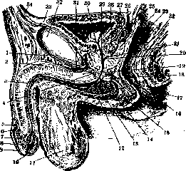
Figure 1. Median section of the organs of the lesser pelvis: 1—lig. suspensorium penis; 2—septum penis; 3—corpus cavernosum urethrae; 4 and 19—trigonum urogenitale; 5—collum glandis; 6—corona glandis; 7—glans; 8—fossa navicularis; 9—praeputium; 10—orificium ext. urethrae; 11—septum scroti; 12—m. bulbo-cavernosus; 13—ductus excretorius glandulae bulbo-urethralis; 14—pars membranacea urethrae; 15—m. sphincter ani ext.; 16—pars analis recti; 17—anus; 18—glandula Cowperi; 20 and 27—colliculus seminalis et utriculus prostaticus; 22—rectum; 23—plica transversalis recti; 24—prostata; 25—excavatio recto-vesicalis; 26—plica recto-vesicalis; 27—ampulla ductus deferentis; 28—isthmus prostatae; 29—pars prostatica urethrae; 30—orificium urethrae int.; 31—v. dorsalis penis; 32—symphysis; 33—vertex vesicae; 34—peritonaeum parietale. (After Spalteholz.) The lateral surfaces and the apex of the organ are located on the soft parts of the pelvic floor. The outer surface of the prostate is smooth only posteriorly and laterally; anteriorly it is connected by bands of connective tissue and smooth musculature with the pubic bones and the urinary bladder. The smooth surfaces of the prostate are rounded and adjoin the inner surface of the corresponding levator ani muscle, from which they are separated by the dense, shiny lateral aponeurosis of the prostate, which is connected with the endopelvic fascia. The prostate is in topographical relationships (Figs. 1 and 2) with the following organs: from above, by its base, it contacts the fundus of the urinary bladder; somewhat posteriorly, with the seminal vesicles and their ampullae; anteriorly and laterally, venous plexuses (plexus venosus pudendus et pl. vesico-prostaticus) lie against it. Loose cellular tissue, in which the venous plexuses are embedded, separates the gland from the symphysis; from below, the gland adjoins the urogenital diaphragm; posteriorly, it adjoins the ampullary part of the rectum, separated from it by a leaflet of fascia (fascia, s.
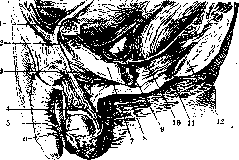
Figure 2. Organs of the lesser pelvis: 1—os pubis; 2—ductus deferens; 3—m. ischio-cavernosus; 4—epididymis; 5—penis; 6—testis; 7—ramus inf. ossis pubis; 8—perinaeum; 9—prostata; 10—rectum; 11—anus; 12—diaphragma pelvis. (After Spalteholz.) septum recto-vesicale). The prostate is connected to the urinary bladder by the following ligaments and fasciae: puboprostatic ligaments (running from the periosteum of the symphysis and from the tendinous arch of the pelvis to the anterior surface of the gland), the aforementioned rectovesical fascia, or rectovesical septum, which constitutes a sort of capsule for the gland (capsula prostatica); it is especially dense behind the latter. Here, near the rectovesical septum, the apices of the seminal vesicles and the vas deferens adjoin the base of the prostate (Figs. 3 and 4); with the latter
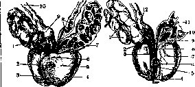
Figure 3.
Figure 4. Figure 3. Prostate gland with seminal vesicles: 1—corpus vesiculae seminalis; 2—urethra; 3 and 4—lobus dexter et sinister prostatae; 5—basis prostatae; 6—isthmus prostatae; 7—ductus ejaculatorius; 8—diverticula ampullae; 9—ampulla ductus deferentis; 10—ductus deferens. (After Spalteholz.) Figure 4. Ejaculatory ducts: 1 and 20—vesicula seminalis; 2 and 9—ductus excretorius; 3 and 8—ductus ejaculatorius; 4—urethra; 5—colliculus seminalis; 6—utriculus prostaticus; 7—basis prostatae; 11 and 12—ampulla ductus deferentis; 13—ductus deferens. (After Spalteholz.) the gland is closely connected, and they pierce the prostate obliquely from top to bottom and from back to front. Sometimes between the two usual lateral lobes a third one is also distinguished, the so-called middle lobe [lobus medius (antiprostata, s. prostata anterior)]: the section of the prostate that is enclosed between the vas deferens posteriorly and the initial part of the urethra anteriorly. The middle "lobe" is closely connected with the lateral ones. In the pathology of the gland, the middle lobe plays an important role, especially in old people during its frequent hypertrophy (lobus pathologicus). The urethra enters the middle of the base of the gland, then passes obliquely, closer to its anterior surface, and exits at the apex; thus, the greater part of the organ lies behind the urethra. The size of the prostate changes significantly with age: in children the gland is barely noticeable, then by adolescent age it enlarges and in old age reaches its greatest dimensions, sometimes reaching the size of a chicken egg or more. This variability in the size of the organ depends on the changing ratios between the amount of glandular and connective tissue in connection with age and reflects the development, state, and function of the sexual sphere. Corresponding to the indicated circumstances, the consistency of the organ is also variable and inconstant. The average transverse size of the prostate is about 4 cm, the average longitudinal size 3 cm, the greatest thickness (from front to back) 2 cm, and the weight approximately 20 g. The prostate is covered entirely by a dense connective-tissue capsule. The numerous glands constituting the parenchyma of the prostate are branched alveolar-tubular wide passages that secrete their fluid (succus prostaticus) during ejaculation. Their totality is termed the corpus glandulare. Most of these passages open into the recess between the seminal colliculus, or caput gallinaginis (colliculus seminalis, s. caput gallinaginis), and the corresponding posterior wall of the urethra, while a smaller part opens on the anterior wall of the urethra. The glands situated against the pubis are distinguished by being especially short and ampullarily dilated. The main glandular part of the prostate is formed by its lateral and posterior small glands: they are more developed than the anterior ones, highly branched, and open as punctate orifices on the seminal colliculus and in a small pocket of the latter (utriculus prostaticus, s. utriculus masculinus, s. sinus prostaticus) (Weber's organ)—a derivative of the Müllerian ducts (see Urogenital organs). This sinus is about 8–10 mm long, 1–2 mm wide at the mouth, and about 6 mm at the blind base (it may be completely absent). The number of individual glands in the prostate reaches 30–35 and more; together with their excretory ducts (Fig. 5), they surround the beginning of the urethra in the form of an incomplete wreath and open separately into the lumen of the latter, but frequently the excretory ducts of individual glands of the prostate unite (by twos or more) before emerging from the stroma of the organ into a single common excretory duct, i.e., into 15–20 united
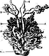
Figure 5. Prostate gland (5); 1—vesicula seminalis; 2—ductus ejaculatorius; 3—vesicula prostatica; 4—urethra; 6—ductus deferens.
ducts (ductus prostatici). All the spaces between the individual glands and their ducts are filled with connective and smooth muscle tissues, which on the surface of the prostate pass into the dense capsule surrounding the organ. Therefore, in cross-section the prostate has a cavernous appearance. The epithelium of the individual glands of the prostate is cylindrical, single-layer or double-layer, often forming protrusions toward the lumen of the duct. It contains pigment granules. The basal membrane of the epithelium is very thin. The intermediate connective tissue is abundantly permeated with elastic and individual smooth muscle fibers (collectively termed m. prostaticus); by their contraction, the latter ensure the possibility of the simultaneous emptying of the entire gland of its secretion, which is why this muscle was also called m. compressor prostatae. This motor act can also be assisted by the nearest striated musculature of the perineum (m. sphincter vesicae externus, s. urethrae membranaceae), which loops around the beak of the prostate and is connected by fibers with the m. prostaticus. Together with the bundles of the m. trigoni vesicae, the m. prostaticus constitutes the internal involuntary sphincter of the urethra (m. sphincter urethrae internus). In the lower section of the prostate, its smooth muscle fibers are replaced by striated ones and, together with the m. transversus perinei profundus, constitute the external voluntary sphincter of the urethra (m. sphincter urethrae externus). The connective tissue framework of the prostate contains a large number of elastic fibers; besides the capsule, there are trabeculae (septa), which penetrate between the individual glands and into the interior of the latter. The final development of the prostate is completed by the age of 17, when hitherto blind excretory ducts of the glands open up as a result of the transformation of their epithelium from multi-layered to single-layered. In children, the gland is very small and consists predominantly of muscle and connective tissue; they have very little glandular mass. Blood supply to the prostate is carried out by branches of the inferior vesical arteries (aa. vesicales inf.) and middle rectal arteries (aa. haemorrhoidales med.). The veins drain into the plexus venosus vesico-prostaticus—one of the most developed pelvic venous plexuses located on the sides of the bladder. Lymphatic vessels carry lymph to the nearest anterior lymph nodes of the pelvis. Nerves originate from the sympathetic and parasympathetic plexuses of the pelvis. Sympathetic ganglion cells, as well as inclusions of chromaffin cells, are frequently found in the tissue of the gland.
G. Ivanov. Physiology of the prostate gland. The secretion is a milk-colored liquid of alkaline reaction, with a specific odor depending on the presence of spermine in it in the form of a hydrochloric compound. For the first time, pure juice of the prostate gland was obtained by Eckhardt, and then by Mislavsky and Borman by electrical stimulation of the nervi erigentes. (The secretion obtained in the clinic by pressure on the prostate is of course not a pure product of its vital activity.) Upon microscopic examination of the juice of the prostate gland, the latter appears as an opalescent fluid containing a large number of small, sharply light-refracting grains, reaching up to a quarter of an erythrocyte in size, which are called lecithin or lipoid bodies. In addition, the prostate juice contains Böttcher's crystals (see Böttcher's crystals), epithelial cells from the urethra and the ducts of the prostate gland, single leukocytes, often stuffed with lipoid grains, and amyloid bodies (see). The juice of the prostate gland is produced by the glands continuously, but is normally discharged into the urethra only during ejaculation or pollution, and pathologically during prostatorrhea. All authors recognize two important physiological functions for prostatic juice: to increase the total mass of semen, dilute it, and excite vigorous movements in immobile spermatozoa. Due to its alkaline reaction, normal prostate juice is further a protective medium for spermatozoa that have entered the acidic environment of the woman's vagina. Walker proved that the activation of spermatozoa can also be achieved by a number of other fluids, such as blood serum, physiological saline, and even simply distilled water. However, Vishnevsky's studies showed that prostate juice possesses sharply specific activating properties with respect to spermatozoa. The more intense the secretory activity of the gland, the greater the activating capacity of its juice. Rohleder points out that the prostatic secretion of one individual is capable of activating the spermatozoa of another, however, the clinical observations of Fronshtein speak of the exact opposite, and it can be thought that the secretion of the prostate gland of one individual acts toxicologically on the spermatozoa of another. Under the influence of prostatic juice, spermatozoa can retain their viability in the genital organs of a woman for up to 8 days, which allowed Percy to express the opinion that the juice of the prostate gland is a nutrient medium for spermatozoa. Both by experimental data (Serrallach and Pares) and by a number of clinical observations, it has now been proved that the prostate gland possesses a specific internal secretion. Only the question of whether the prostate is a gland of internal secretion in the full sense of the word or whether the excret produced by it partially enters the bloodstream and thus the prostate is a gland with facultative internal secretion can be discussed. Pathological processes taking place in the prostate gland and altering its secretion both qualitatively and quantitatively exert an influence not only on its state, but also on the general state of the organism, on the function of other glands of internal secretion. The closest interrelationships exist between the genital glands and the prostate. The prostate gland reaches its full development by the time of puberty of the organism. Being in childhood a muscular organ containing an insignificant amount of glandular tissue, it increases in volume as it approaches the period of sexual maturity, the period of full development of the testicles due to an increase in the number of glandular elements in it. The well-known fact is that in animals castrated before the onset of puberty, an arrest in the development of the prostate gland occurs. If, however, an extract of the testicles is injected into a castrated animal, no arrest in development occurs. Castrates who lost their genital glands before the development of the prostate gland are such in the full sense of the word, whereas castrates who lost their testicles at a time when the glandular tissue of the prostate had already developed and functioned, retain erections and the possibility of sexual intercourse for a long time. This clinical observation is indisputable proof of the internal secretion of the prostate, proof that in the possibility of performing the sexual act, the internal secretion of the prostate gland plays far from the last, and perhaps even the main role. According to the experiments of Serrallach and Pares, the prostate gland affects spermatogenesis. The injection of prostate secretion into puppies showed that the vessels of the testicles of the experimental animal undergo significant dilation under such conditions, and the entire organ comes into a state of arterial congestion. On the other hand, extirpation of the prostate caused the disappearance of spermatozoa from the semen of adult animals and degeneration of the parenchyma of the testicles in the form of protein degeneration. The cessation of spermatogenesis in animals that hibernate in winter is accompanied (Hunter) by a decrease in the volume of the prostate gland. Battez and Boulet also showed by experiments on animals that the injection of prostate extracts into them causes contraction of the bladder, and the studies of Serrallach and Pares proved that the testicular extract affects the muscles of the bladder in such a way as to cause an increase in the tone of the sphincters and relaxation of the muscle emptying the bladder. Based on these experiments, it should be thought that the internal secretion of the prostate also affects the act of urination in the direction of sphincter relaxation and detrusor spasm. The internal secretion of the prostate is also not indifferent to the vital activity of the entire organism. Goetzl, Serrallach, and Pares, who fed animals "prostatim," observed their emaciation as a result of increased metabolism; at the same time, the animals became more energetic, cheerful, lively, and the intellect of the experimental puppies developed faster than that of the control ones. Excessive administration of "prostatim" led to death with phenomena of cachexia. Posner showed that the injection of prostate extract of another zoological species entails the death of the animal with phenomena of blood pressure drop. The exact same phenomena of blood pressure drop were observed by Legueu et Gallardot in cases of prostatic hypersecretion. Mendova, extirpating the prostate in dogs, observed eosinophilia, increased blood clotting, trophic disorders on the skin, etc., in them and associates these phenomena with the loss of the endocrine function of the prostate gland. His studies, however, are not entirely conclusive, since they do not delimit the disruption of the interaction between the prostate and the testicles. Clinical observations (Porosz, Burckhardt, Peyer, Ilyinsky, and others) indicate the circumstance that enhanced secretion of the prostate causes phenomena of vagus nerve irritation in the form of intestinal disorders (diarrhea), attacks of bronchial asthma, and palpitations. There is not yet a sufficient number of experimental works to be able to give an explanation of the mechanism of the occurrence of these or other clinical phenomena in connection with enhanced secretion. However, it is undoubted that under the influence of changes in the endocrine function of the prostate, these phenomena also change in one way or another. cancer). Sometimes significant pain is observed (acute and chronic prostatitis, stone); both lobes can be asymmetrical (tumor, abscess, tuberculosis, cyst). The boundaries are sometimes expressed unclearly, the prostate merges with surrounding tissues (periprostatic tumor). The second diagnostic method is the study of juice of the prostate gland: Obtaining it occurs through massage of the prostate gland. Normal juice of the prostate gland has a weakly alkaline reaction and contains a significant amount of albumin and spermine. The amount of squeezed-out juice of the prostate gland, depending on various living conditions, fluctuates within wide limits—from a few drops to several cubic centimeters. Microscopic examination of juice of the prostate gland plays a significant role in diagnosis. In inflammatory diseases, it is possible to detect an increase in the number of leukocytes and to establish the etiology of inflammation by bacteriological examination. Simultaneously, the ratio of leukocytes and lecithin bodies changes. Fluctuations of this index play a significant role in the diagnosis and prognosis of the disease (see below). In neoplasms, the juice contains Methods of examining the prostate gland. Palpation of the prostate gland is performed through the rectum in one of three positions: on the back with lower limbs spread apart and bent at the knee joints; standing, bent forward at the hip joints; and lateral lying (Fig. 6-8). Each of these positions has its merits and indications.
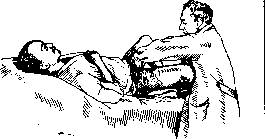
Figure 6.
examination. The latter method is most convenient for both the physician and the patient, but it does not allow for a bimanual examination. If the latter is necessary, one should resort to examination in the supine position. If one wishes to obtain secretion for examination, it is most convenient to examine with the torso bent forward. The index finger of the right hand, fitted with a rubber finger cot and well lubricated with Vaseline, is inserted into the rectum; the examiner must orient themselves regarding the size, shape, symmetry of the lateral lobes, tenderness, boundaries of the prostate gland, and consistency, and form an opinion about its relationship with surrounding organs and the state of the rectal walls, seminal vesicles, and vas deferens. In the normal state of the prostate gland, both of its lobes bulge evenly into the lumen of the rectum, separated by a groove, the entire prostate gland resembles a heart in shape, the magnitude of the sagittal diameter equals the length of the nail phalanx of the index finger, and the volume of the prostate reaches that of a small chestnut.
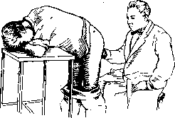
Figure 7.
Pressure on the gland causes a peculiar unpleasant sensation in the region of the head of the penis and is often accompanied by a false urge to urinate. In pathological processes in the prostate gland, the organ may decrease (atrophy of the prostate gland) or increase in volume (acute prostatitis, hypertrophy, tumor). The consistency, from normal doughy-glandular, may become soft, flabby (chronic prostatitis, atony), give fluctuation (prostatitis), or, conversely, be very dense (abscess, stone,
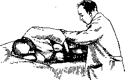
Figure 8.
usually red blood cells (Pauchet) (see Hemospermia) and a significant number of epithelial cells (Fronshtein). By means of endourethral examination, one can get an idea of the length of the prostatic urethra and consequently of the long diameter of the prostate gland, which, however, does not play a significant practical role. Endourethral examination by means of urethroscopy is indicated in long-standing chronic prostatitis as a method of diagnosis and therapy, as well as for the same purposes in stones and cysts of the prostate gland. With the eye, one manages to see the localization of pathological changes and establish their nature. Cystoscopic examination is used in all cases of changes in the volume of the prostate gland, with the exception of its acute inflammation, where it is strictly contraindicated. Often only in this way is it possible to differentiate between a malignant neoplasm and hypertrophy of the prostate gland and to exclude diseases of the urinary bladder. Cystoscopic examination, however, is not always simple, since passing the instrument through the posterior modified urethra can present great difficulties. X-ray examination in such cases can replace cystoscopy. Having filled the bladder with air and taking a picture with a soft tube, one gets a very good idea of the configuration of that part of the prostate which protrudes into the urinary bladder and of its relation to the surrounding tissues. Furthermore, X-ray examination is indicated when stone disease of the prostate gland is suspected—in the presence of inorganic concretions in it, their contours are clearly obtained, making it possible to judge the number of stones and their size. Pathology of the prostate gland. Congenital absence of the prostate gland (aplasia) is very rare and is usually observed in premature monsters, accompanied by simultaneous aplasia of the genital organs or non-closure of the anterior abdominal wall. The same can be said about partial underdevelopment of the prostate gland in the form of the absence of one of its lobes. Hypoplasia of the prostate gland is encountered, on the contrary, apparently quite often. Thus, according to English's statistics, out of 1,757 patients in the urological department managed by him, 199 showed a congenital reduction in the size of the prostate gland. Hypoplasia of the prostate gland is usually encountered simultaneously with other malformations of the urogenital apparatus, mainly with underdevelopment of the penis and hypoplasia of the testes. Patients in sharply expressed cases have the typical appearance of eunuchoids; in more mildly expressed cases, the appearance of the patients does not change. The patients' complaints boil down to disorders of sexual functions, especially regarding the ability to perform sexual intercourse. Histological examination of such cases shows the presence in place of the prostate of a small ovoid body consisting of muscular and connective tissue without an admixture of glandular elements. Upon examination, the prostate appears in the form of a pea; sometimes nothing can be palpated in place of the gland. A significant number of congenital disorders of sexual intercourse have hypoplasia of the prostate gland as their basis. Often, even before the appearance of these complaints, there are urinary disorders in the form of nocturnal enuresis, accompanied by stretching (atony) of the urinary bladder. The latter circumstance finds an explanation in a simultaneous malformation of the musculature of the region of the bladder outlet in the form of the formation of folds and "valves" here. Atrophy of the prostate gland. Depending on the etiological factor, three forms of atrophy of the prostate gland are distinguished: a) atrophy as a result of a local disease (most often of an inflammatory nature, see below), b) atrophy as a result of castration, c) senile atrophy. Atrophy as a result of castration. Experiment and clinical observations show that as a result of castration performed before puberty, an arrest in the development of the prostate gland occurs. If an animal or a human with fully formed gonads is castrated, the prostate begins to atrophy—the glandular tissue decreases in quantity, being replaced by connective tissue elements. In individuals who suffer from prostate atrophy, general complaints come to the fore, almost identical to the complaints of eunuchoids. If in castrates with a prostate that is not yet atrophied the possibility of performing sexual intercourse may exist, with prostate atrophy sexual intercourse is completely impossible due to the lack of erections. The prognosis is poor. Treatment with internal intakes of prostate extracts sometimes gives a temporary improvement in symptoms. Testicle transplantation, theoretically justified, practically gives no effect. On the contrary, according to Fronshtein's observations, such transplantations bring only a number of troubles, causing patients an increase in sexual desire in the absence of the possibility of performing sexual intercourse. Senile atrophy of the prostate gland, observed at an age later than 50 years, deserves much more attention. Regarding the frequency of this form of atrophy, there is great disagreement. While Dittel found atrophy in 31.5% of all individuals older than 52 years, Burckhardt observed it in only 1.9% of cases. Since both figures are derived from clinical material, sometimes purely accidental, and on the other hand, in view of the fact that clinical establishment of atrophy is a sharply individual matter, it is correct to consider that we do not yet have exact figures to judge the frequency of this disease. Very little has been done to clarify the pathogenesis of senile atrophy of the prostate. The most acceptable is Guyon's theory, which treats this affliction not as a purely local disease, but as a partial manifestation of arteriosclerosis characteristic of senile age. Sufficient objective evidence for this theory, however, has not been presented. Pathological and anatomical changes. The atrophied prostate is denser and more resistant than the normal one. The tissue of the gland on cross-section appears homogeneous, of a whitish-gray color. The excretory ducts of the gland are strongly stretched. In the thickness of the prostate, there are numerous fluid-filled cavities (cysts) formed as a result of secretion stagnation in the glandular passages. Stones can form in these cysts, and under the pressure of the latter, atrophy usually increases further. Upon microscopic examination, changes are observed on the part of the glandular tubes and alveoli; the thickness of their epithelial layer decreases, and the epithelium undergoes fatty degeneration. In more pronounced cases, individual alveoli completely disappear, and instead of them, narrow slits devoid of epithelium are visible in the connective tissue. The excretory ducts shrink, and the epithelium lining them atrophies. A significant development of fibrous connective tissue is observed in the thickness of the gland. In the clinical picture, disorders of urination come to the fore in the form of its frequency, first at night and then during the day, in the presence of a greater or lesser significant amount of residual water; complete bladder atony develops very quickly. Thus, the clinical phenomena in atrophy of the prostate gland resemble the symptoms existing in hypertrophy of the prostate gland, with the difference that they develop very quickly. Often, right from the beginning of the disease, a weakening of sexual potency is discovered: erections become rare, weak, short-lived, and may even be completely absent; the character of the semen changes in the sense of a decrease in its quantity and immobility of the spermatozoa. The atrophy process taking place in the prostate gland undoubtedly also involves the neck of the urinary bladder: the muscles of the latter become sclerosed, a dense, yielding fibrous ring is formed (Chetwood's disease), which of course plays an important role in the occurrence of urinary retention. 86
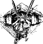
Figure 9.
However, the first clinical symptoms—dysuria and sexual dysfunction—must undoubtedly be fully explained by the impairment of the endocrine function of the prostate. Recognition of prostate atrophy is extremely simple: upon digital examination through the rectum, the gland appears smaller and denser than usual, its surface is smooth, occasionally slightly nodular. In markedly pronounced cases, only remnants of the gland are palpable in the form of a dense nodule, or no traces of the gland can be detected by palpation. Prognosis and treatment. The prognosis of such cases is directly dependent on the timing of medical intervention. In the early stages of the disease, one can attempt to achieve a halt in the development of the atrophic process through therapeutic measures by taking iodine preparations internally, applying local diathermy, and 1
specific anti-sclerotic treatment. Testicular preparations are administered internally with the aim of increasing prostate activity in the stage of prostatism without residual urine, and prostate preparations in the presence of bladder atony. In cases of developed atrophy, one often has to contend with bladder atony that has already set in secondarily and condemn the patient to permanent catheterization. Surgical treatment in the form of incision or excision of the fibrous ring (Holtzov) formed at the bladder neck in place of the atrophied prostate (Fig. 9) does not always guarantee the patient the restoration of spontaneous urination. Traumatic injuries of the prostate. Isolated traumatic injuries of the prostate gland are not observed. The location of the prostate gland in the lesser pelvis and its close connection with the bladder, urethra, and rectum are the reason that its injury is usually accompanied by the wounding of one of these organs. A distinction should be made between so-called surgical injuries, i.e., injuries as a result of a particular intervention on organs surrounding the prostate, most often the urethra, and injuries of an "accidental character" as a result of an accident. Contusions of the prostate gland are observed with simultaneous injury to the perineum as a result of a fall onto it. Clinical phenomena on the part of the urethra (rupture) usually come to the fore here, and only after their disappearance and the healing of the wound do complaints specific to the prostate gland appear in the form of pain during sexual intercourse, the explanation for which should be sought in the scars that have developed in the prostate. Resorptive treatment in the form of microclysters with iodine, diathermy, and light prostate massage leads to the softening of scars and the disappearance of subjective complaints. Gunshot wounds were observed during the war simultaneously with wounds of the perineum, rectum, and pelvic organs in a fairly significant number. Here, too, symptoms from other organs came to the fore. It can be noted, in agreement with Blum and Zuckerkandl, that injuries to the tissue of the gland itself scarred extremely quickly and significantly better than injuries to neighboring organs. Subsequently, as a rule, prostate atrophy set in with all the ensuing consequences. Surgical injuries of the prostate gland are observed during various endovesical and endourethral operations used for other reasons, most often during catheterization, bougienage, and lithotripsy. They are especially often observed in cases where there is a change in the configuration of the prostate gland as a result of pathological processes occurring in it (acute inflammation, hypertrophy). The instrument introduced into the urinary bladder encounters a natural obstacle in the region of the prostate gland and overcomes it with some force, pushing aside the prostate protruding into the lumen. The use of thin instruments with a conical end for catheterization in hypertrophics is a moment that promotes trauma and can cause the formation of a false passage—perforation of the bulging middle lobe (Fig. 10). The same can be said regarding thick metallic instruments that have to be introduced into the bladder during lithotripsy. Such wounds with uninfected urine heal quite easily on their own and are not threatening to the patient. With infected urine, on the contrary, they can cause the development of a phlegmonous process in the gland itself and the surrounding cellular tissue. Prevention is 1) the use of elastic catheters with a Mercier curve (see Catheters) in hypertrophics, 2) refraining from catheterization in acute prostatitis, and 3) if it is necessary to introduce metallic instruments into the bladder, not pushing them forcibly through the posterior urethra, but trying to bypass the obstacle by bending the pavilion of the instrument between the patient's legs as low as possible. Syphilis of the prostate gland is extremely rare, proceeding in the form of chronic prostatitis with an unclear etiology. The main symptoms boil down to frequent, slightly painful urination, a weak urine stream, and pain in the perineum. The course of the disease is sluggish, prolonged, without exacerbations. Upon palpation, the gland is enlarged, unevenly nodular, painless. In individual cases, on the contrary (Fournier, Vasilev), upon palpation examination, the gland is very large, reaching the size of an adult's fist, dense, nodular, and simulates hypertrophy or a malignant neoplasm. With conventional treatment for prostatitis, the process does not improve, which usually suggests the syphilitic nature of the disease. The presence of ulcerated processes in the bladder, the density, cartilaginous consistency of the gland, the absence of pain with significant palpable changes, the rapid development of the tumor, syphilis in
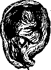
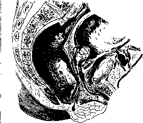
Figure 10. 8$ anamnesis and finally success from specific therapy (iodine, salvarsan) make it possible to make a diagnosis. Tuberculosis of the prostate gland. Infection penetrates into the organ either hematogenously from some primary focus in the lungs, bones, glands, etc., or with the secretion of the testicles from the latter. At the present time there is no consensus on the question of whether the testicles and their appendages or the prostate gland are affected first. One can state with certainty only that in all cases of far-advanced tubercular lesions of the testicles and appendages we usually also have a lesion of the same character on the part of the prostate gland. Predisposing factors are heredity, exhaustion of the organism, trauma, and gonorrhea. Tuberculosis of the prostate gland occurs predominantly at the age of 20-30 years, i.e., in the period of greatest physiological activity of the gland. The process usually has a focal character. Small tubercles, merging together, form caseous-disintegrating foci, which are first localized in one lobe and then can involve the entire gland. In a number of cases, the process has a tendency to self-healing through the proliferation of fibrous tissue; in other cases, the process progresses, involves surrounding tissues, and cavernous cavities are formed, giving rise to a number of fistulous tracts into the pelvic cellular tissue, perineum, urethra, and rectum. The course of tuberculosis of the prostate gland is characterized by phenomena of posterior urethritis—frequent, imperative urges to urinate, blood, and pain at the end of the act of urination. Patients develop sharp independent pains in the perineum, which worsen upon palpation. The diagnosis is established by finding tubercular bacilli in the prostatic juice or by positive results of inoculation. Upon examination, the gland is unevenly thickened, nodular, and usually at the same time there are specific changes on the part of the seminal vesicles or testicle appendages. As for treatment, some authors (Albarran, Voskresensky, Holtsov) warmly recommend active surgical intervention in the form of removal (partial or complete) of the prostate gland through a transrectal perineal incision; others (Frank, Casper, Steinberg) no less warmly advocate conservative treatment in the form of climatic treatment and the application of local radiotherapy and quartz. Very good results in the initial stages of the disease are produced by the removal of the testicle appendages affected by the tubercular process, followed by irradiation of the prostate and internal administration of large doses of potassium iodide. Hypertrophy of the prostate gland. This name is used to mean a symptom complex of phenomena observed in males over the age of 50, expressed in difficulty and frequency of urination, especially at night, and depending on the increase in the volume of the prostate gland. However, in these cases there is no truly hypertrophic process, and therefore the majority of authors prefer to speak of so-called hypertrophy of the prostate gland, emphasizing the conditional nature of the term, and view the disease as a neoplasm. Individual authors strive to introduce an oncological designation (adenoma of the prostate gland, prostatoma), however, these attempts have no solid scientific basis and only serve to aimlessly increase the terminology. The etiology of the disease has not yet been definitively clarified. Of the existing theories, the oldest is the Guyon-Lannois theory, according to which hypertrophy of the prostate gland should be considered as a partial manifestation of arteriosclerosis. According to this theory, the enlargement of the prostate gland is not the cause of the urination disorder, but the latter arises depending on a similar arteriosclerotic process in the urinary bladder. This view was soon abandoned, because in pathology the concept of arteriosclerosis is associated with the idea of sclerosis and atrophy, and by no means with the hypertrophy of an organ. According to Ciechanowski's theory, hypertrophy of the prostate gland is the result of prolonged chronic prostatitis. Inflammatory phenomena are localized in the stroma of the gland; if its central sections surrounding the excretory ducts are involved in the process, the latter are obliterated, due to which expansion of the alveoli, stasis of secretion in them, desquamation of the epithelium, etc., occur. The enlargement of the gland occurs, according to Ciechanowski, due to the expansion of the alveoli. In proof of this theory, the wide prevalence of gonorrhea is cited, which is often complicated by prostatitis, which can remain unnoticed for many years. This theory met with energetic objections mainly on the following grounds: in early cases of prostate hypertrophy, signs of inflammation are almost always absent, while the presence of these signs (infiltrates) in far-advanced cases should be attributed to secondary infection, which can easily join due to stagnation of secretion in the glands, medical manipulations, etc. The third theory, first proposed by Albarran and Halle, is the neoplasm theory. On the basis of anatomical research, these authors came to the conclusion that hypertrophy of the prostate gland is a benign neoplasm. Primarily, changes occur on the part of the glandular elements of the prostate, while changes on the part of the stroma are usually of a secondary character, although sometimes they prevail and give a fibrous form of hypertrophy of the prostate gland. This theory has the largest number of adherents, and many of them explain the cause of tumor origin differently. Some attach importance to irritation as a result of past chronic inflammation in the prostate gland, others see the cause in a violation of the internal secretion of the prostate and irritation of its glandular elements by abnormal breakdown products. The later intra-secretory theory, however, does not consider prostate hypertrophy to be a true tumor. Relying on a number of experimental works proving the dependence between the testicle and the prostate, this theory put forward an explanation according to which irritation coming from the testicle causes proliferation of the remaining glandular elements in the prostate, which has atrophied as a result of old age. Thus, a compensatory enlargement of one part of the organ is obtained here as a result of atrophic changes in another part of it. The most important result of recent research, which changed the view on prostate hypertrophy, is the establishment of the fact that the starting point of the process is not the prostate, but the rudimentary glands of the prostatic part of the canal, the so-called periurethral glands of the canal (Motz, Perearnau, Tandler, Zuckerkandl), while the prostate itself is pushed to the periphery and atrophies, forming a sort of capsule around the proliferating periurethral glands. Three groups of submucosal urethral glands are distinguished. Depending on the preferential proliferation of one or another group of glands or on their joint proliferation, various forms of hypertrophy are obtained. Adopting one or another theory, one must nevertheless admit that there is no true hypertrophy of the gland, i.e., hypertrophy and hyperplasia of its constituent elements, and if this name is to be preserved, it is only out of tradition, and therefore there is no reason to introduce the term "prostatoma". Among the etiological factors, chronic prostatitis and the change in the internal secretory function of the testicles occurring in old age undoubtedly play a large role. Gonorrhea, which is the most frequent cause of chronic prostatitis, is, however, far from being so frequently encountered in the anamnesis of hypertrophics. On the other hand, a change in the function of the testicles is observed in all old men, and hypertrophy does not develop in all of them; obviously, apart from the indicated etiological factors, an individual predisposition to the development of hypertrophy of the prostate gland is also necessary. All those conditions that promote and maintain stagnant phenomena in the pelvic organs—sexual excesses and sexual abstinence, masturbation, hemorrhoids, a sedentary lifestyle, horseback riding, chronic constipation—play the role of predisposing factors. Hypertrophy of the prostate gland is observed among the rural population half as often as among the urban population, and among the latter mainly in those whose profession is associated with a sedentary lifestyle. Assuming the weight of a normal prostate to be 20-25 g, it should be considered that any gland weighing more is hypertrophied. A case of hypertrophy of the prostate gland with a weight of 300 g has been described. Pathological anatomy. The shape of the hypertrophied gland varies depending on whether all its parts or only one of the lobes are involved in the process. The greatest increase in volume usually goes upward and forward (toward the urinary bladder). The lateral lobes bulge into the lumen of the rectum, and the groove between them is smoothed out. At the same time, a certain asymmetry of the lateral lobes and the lobe protruding into the urinary bladder is usually observed. The lateral lobes appear as separate tubercles of elastic consistency with a smooth surface. The middle lobe, protruding into the cavity of the urinary bladder, can take on extremely diverse outlines (the shape of a tongue, a spherical tumor on a wide and sometimes narrow base, etc.). Partial hypertrophy is much less common than general; usually the middle lobe hypertrophies in this case, which, protruding into the bladder cavity in the form of a valve, hangs over the internal opening of the urethra (Fig. 11). All elements comprising the gland—muscles, connective tissue, and glandular epithelium—can take part in the hypertrophic process occurring in the prostate gland.
Depending on the predominance of certain elements in the hypertrophied prostate gland, we distinguish myomatous, fibromatous, and finally adenomatous forms of prostate gland hypertrophy. Out of 100 histologically examined prostate gland hypertrophies, Albarran and Hallé found 32 adenomas, 51 myomas, 3 fibromas, and in 14 cases of clinically benign prostate gland hypertrophies, an atypical proliferation of the glandular epithelium—epitheliomas—was established. In adenomatous hypertrophy, the gland acquires very large sizes, frequently reaching the volume of an Antonovka apple, and its consistency is elastic. On cross-section, it has a lobular appearance, and in some places, cyst-like cavities filled with secretion are observed. Histological examination shows that each lobule consists of a significant amount of glandular tissue and is separated from the other by a small amount of stroma with a normal content of muscular and connective tissue elements. The epithelium lining the individual alveoli is of normal structure or is in a state of proliferation.
Fig. 11. Hypertrophy of the middle lobe.
The cavities of the alveoli are filled with cellular detritus mixed with mucous amyloid bodies. In myomatous hypertrophy, the gland does not reach such large sizes as in adenomatous hypertrophy; it is significantly denser upon palpation, slightly nodular, and on cross-section, denser areas upon histological examination turn out to consist of a significant amount of muscular tissue mixed with connective tissue elements surrounding islands of glandular epithelium that are in a state of atrophy. In fibrous hypertrophy of the prostate gland, the volume of the latter usually does not reach large sizes, the consistency of the gland is very dense, and under the microscope, the gland consists of a significant amount of connective tissue fibers and a small amount of muscular tissue. Blood vessels are numerous, and their walls are thickened. The capsule of the prostate gland as a rule is not intimately fused with the hypertrophied prostate gland. Albarran and Motz pointed out that in addition to the anatomical capsule of the prostate gland, in the case of prostate gland hypertrophy, there is also a false prostatic capsule—the «surgical» capsule. Its origin is explained by the fact that hypertrophy occurs at the expense of the glandular tissue adjacent to the urethra, while the peripheral part of the gland, which does not take part in the neoplastic process, is flattened
Figure 12. surgical capsule: 1 - glandular tissue, 2 - false capsule, 3 - excretory duct, 4 - compressed tissue of the gland.
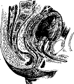
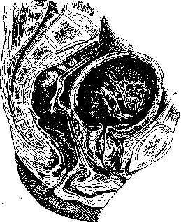
by the hypertrophied tissue, is pushed toward the true capsule, and forms a new, false surgical capsule (Fig. 12). Tandler and Zuckerkandl distinguish two main types of prostate gland hypertrophy: intra- and subvesical. The first type is characterized by deformation of the bladder orifice by a tumor protruding into the bladder (Fig. 13). In the second case, the configuration of the bladder orifice is almost unchanged, and only the floor of the bladder projects sharply anteriorly, being lifted by the hypertrophied masses (Fig. 14). Prostate gland hypertrophy occurs as a rule at the age over 50 years. As for statistical data regarding the frequency of the disease, the figures of anatomical theatres are indicative in this respect: Reischauer found prostate gland hypertrophy in corpses aged 50-60 years in 40%, and at the age of 60-70 years in 75%; Roth's figures are somewhat lower: at the age of 50-60 years—31%, at the age of 60-70 years—56%. On the other hand, Dittel and Chrastina, who examined inmates in an asylum for the aged aged 50 to 100 years, detected clinical symptoms of prostate gland hypertrophy in 15% of all inmates. These data indicate with absolute precision that far from every prostate gland hypertrophy causes clinical symptoms in its bearer. An analysis of clinical material indicates further that the greatest phenomena are caused by the first type of prostate gland hypertrophy according to Tandler and Zuckerkandl, i.e., intravesical hypertrophy. There is no parallelism between the magnitude of prostate gland hypertrophy and clinical symptoms. One can observe very large prostate gland hypertrophy with very insignificant
Figure 13. Intravesical hypertrophy.
clinical symptoms and vice versa. With an enlargement of the lateral lobes of the gland protruding into the bladder, an elevated barrier is formed at the outlet, preventing the free outflow of urine, and a deep fossa behind the gland, in which a part of the urine is retained during urination, decomposes, and can cause secondary inflammatory phenomena on the part of the bladder wall. The hypertrophied middle lobe of the prostate gland, hanging over the internal orifice of the urethra, acts as a valve that, when the patient strains, closes the outlet from the bladder and serves as the immediate cause of urine retention. Clinical picture. The main symptoms of hypertrophy are: a) slow and sluggish urination as a result of a mechanical obstacle to the outflow of urine and insufficiency of the bladder muscles to overcome the obstacle, and b) increased frequency of nighttime urination or even daytime urination if
Figure 14. Subvesical hypertrophy.

the patient remains in a recumbent position for a long time, as a result of which the hyperemia of the enlarged gland, which mechanically irritates the bladder neck, increases. Guyon proposed clinically distinguishing three periods in the course of the disease: the first period is characterized by the appearance of frequent urges to urinate at night; the second period of the disease is the weakening of the tone of the bladder, which is unable to rid itself of all the urine. Part of the urine is passed by the patient voluntarily, part is retained in the bladder during every urination (residual urine). Due to this, the intervals between individual urinations become shorter, and urination both day and night becomes more frequent. The irritation of the bladder by the urine stagnating in it causes inflammatory phenomena on the part of the bladder mucosa, urination becomes painful, and blood and pus appear in the urine. Incomplete emptying of the bladder leads to increased absorbability from it—phenomena resembling uremic ones: loss of appetite, dryness of the mucous membranes, constipation with a large amount of gases, and general weakness. Often, after mental excitement, physical fatigue, excesses, or a cold, the patient, having felt the urge to urinate, is unable to urinate; efforts lead to nothing, the urges become fruitlessly more frequent, and complete retention, temporary or permanent, may occur as a result of the disruption of the unstable equilibrium between the weakened bladder muscle and the enlarged hyperemic prostate. The introduction of a catheter into the bladder in such cases is extremely difficult, on the one hand, due to the existing spasm, and on the other, due to the acute hyperemia of the prostate gland. In case of catheterization failure, it is necessary to resort to suprapubic puncture of the bladder for the purpose of releasing urine. A long needle for venous infusions or a thin trocar is punctured perpendicularly to the long axis of the body two finger-breadths above the pubic bone, strictly along the midline. The third period of the disease is expressed in the appearance of symptoms of both nocturnal and diurnal urinary incontinence. The latter is explained by the fact that the stretching of the bladder by the accumulating urine causes phenomena of stretching of the sphincter. Phenomena of ischuria paradoxa (see) set in. At this time, the phenomena of intoxication sharply increase, and the constant increase in pressure in the bladder affects the function of the kidneys. The specific gravity of urine, the percentage content, and the total amount of its organic and inorganic constituents drop. Disorders of urination function caused by the increase in the volume of the prostate gland and characterized by a violation of the contractile ability of the bladder lead to a number of secondary phenomena in the form of an increase in intravesical and intrapelvic pressure and the constant presence of easily decomposing urine in the bladder. These two circumstances are the immediate cause of the fact that hypertrophics are predisposed to inflammatory diseases of both the bladder and the kidneys (chronic pyelonephritis), often accompanied by significant polyuria. Furthermore, the constant retention of urine in the bladder is a factor causing the formation of both diverticula and stones. In the presence of hypertrophy of the prostate gland, a sharp violation of endocrine function is usually not observed. This is completely understandable, because on the basis of anatomical and histological investigations by Tandler, Zuckerkandl, and others, it has been established that with hypertrophy of the prostate gland, no proliferation of the tissues of the latter occurs, but there is merely an increase in the volume of the suburethral glands, pushing the remaining normal prostate toward its capsule. Therefore, there is actually no hypersecretion of the prostate during its so-called hypertrophy. In isolated cases, when dealing with a fibrous, dense mass of tumor in the prostate capsule compressing the latter, a picture of hypofunction of the prostate gland in the form of a decrease in sexual activity may occur. The removal of hypertrophied masses in such cases entails the restoration of lost sexual potency. The same effect is obtained in cases of hypertrophy of the prostate gland from the internal administration of preparations of the latter. Diagnosis is based on the slow increase in urination disorders in the elderly age of the patient and on the examination of the prostate through the rectum. The hypertrophied prostate gland is easily palpated in the form of a dense or glandular-consistency clearly contoured body protruding into the lumen of the intestine. There should be no pain upon palpation. Having found a hypertrophied prostate upon palpation, one should ascertain by percussion of the bladder through the abdominal wall or by catheterization whether the patient completely empties his bladder. Upon cystoscopy, one can see the edge of the prostate protruding into the bladder, even and smooth, and the Y-shaped entrance into the bladder (see cystoscopic drawings in the article Bladder), changes in the walls of the bladder in the form of the formation of folds and bars—the result of muscle hypertrophy (trabecular bladder). In differential diagnosis, acute inflammation of the prostate should be excluded, which is contradicted by the absence of pain upon palpation and the slow increase of symptoms, and neoplasm of the prostate gland (see below). Treatment depends on the stage of the disease in which the patient comes to the doctor. When there is only mechanical irritation of the bladder outlet by the growing gland, treatment boils down to improving blood circulation in the pelvic organs and reducing their hyperemia. Patients are forbidden alcoholic drinks and spicy food, and the intestine must be regulated with enemas or laxatives (liquorice powder, rhubarb). In addition, general baths are prescribed; exercise; legs should be kept warm and dry; one must not drink at night. Locally, suppositories of ergotin and belladonna are prescribed. These measures are sometimes sufficient to reduce subjective symptoms and retard the development of the disease. In acute urinary retention, catheterization is performed, obligatorily with thick silk and metal catheters with a Mercier curve. Catheterization with thin, especially rubber catheters is doomed to failure in advance, because the overhanging middle lobe of the gland is injured by such catheters rather than pushed aside. Sometimes a single catheterization is quite sufficient to restore the act of urination, while sometimes, especially when acute urinary retention develops in the third stage, it is necessary to systematically 2–3 times a day for 2–3 weeks to release urine until the tone of the bladder is restored. Catheterization must be performed with all precautions so as not to introduce infection; patients should be prescribed salol or urotropin internally. In the presence of residual urine, prolonged catheterization is also indicated. If the function of the bladder is not restored under the influence of catheterization, the patient will either have to release urine himself several times a day for the rest of his life or undergo prostate removal surgery. Before deciding on the latter, one must accurately assess the condition of the patient's kidneys. With a deep disturbance of their function, low specific gravity of urine, polyuria, and urinary intoxication, the operation yields up to 30% mortality. In other cases, the mortality rate is 3–8%. The decrease in postoperative mortality depends on the technique of surgical intervention and careful preparation for the operation. Thus, Hunt had 3.3% mortality in those cases that were specially prepared, and 6.6% where the operation was performed urgently. Of great importance is also the division of the operation into two moments: the first is the creation of a fistula, the second is the enucleation of the prostate. Removal of the prostate is performed either by the perineal route (Wildbolz, Voelcker, Young) or through the bladder—by the suprapubic route (Freyer) (Fig. 15). The majority at present operate according to the latter method, dividing the surgical intervention into two moments. This achieves a reduction in postoperative mortality, because at the first moment, with negligible wound trauma, the increased pressure under which the kidneys worked is eliminated, and at the second moment (4–6 weeks later), when the organism copes with the shock caused by this, major trauma accompanying the enucleation of the prostate gland is inflicted. Upon widening the fistula, the operator introduces two fingers of the left hand into the anus of the patient and by pressure
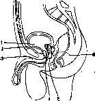
Figure 15. Approaches to the prostate: 1—transvesical; 2—between the bladder and the symphysis; 3—below the symphysis with stripping of the root of the penis; 4—lateral perineal; 5—straight perineal; 6—ischiorectal.
—~<- into the cavity of the bladder. The mucosa of the internal opening of the urethra is opened around a catheter introduced beforehand into the bladder, and by a blunt method (Fig. 16) hypertrophied masses are enucleated from the prostate, while the gland itself remains in place. The resulting cavity is tamponed. Bleeding in this case is usually small if the glandular masses of the tumor are removed entirely within the bounds of the so-called surgical capsule. With more significant bleeding, the wound should always be subjected to careful revision, because usually the bleeding is caused by incomplete removal of the hypertrophied tumor nodules. Individual authors recommend abundant irrigation of the wound with hot water for the purpose of hemostasis, however, the majority do not resort to what is extremely incorrect and yields significant
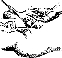
Figure 16. Enucleation of the prostate by the transvesical route.
method of late bleedings, but contents itself with tamponade of the prostatic bed. Drainage is inserted into the bladder, and wound healing proceeds by granulation. In the postoperative period, some authors do not introduce a catheter into the urethra at all (Martynov), others introduce a catheter during the operation and tamponade the bed around the catheter (Illyes, Fronshtein), and finally, a third group (Freyer, Kholsort) introduces it on the 4th-5th day. Investigations by Vishnevsky have shown that the regeneration of the urethral mucosa, removed together with the hypertrophied masses of the prostate, and the fusion of the urethral segments occur even without the introduction of a catheter, yet with a catheter the postoperative period proceeds better and shorter. Patients (elderly people) must be mobilized as soon as possible (hypostatic pneumonia, bedsores), and in the postoperative period special attention must be paid to the heart and lungs (inhalation of carbon dioxide). While yielding a good functional success, prostatectomy is far from being an indifferent intervention for patients. This explains the fact that there is no unity of opinion in setting indications for surgical intervention—some authors (Martin, Thompson, Ilyinsky, Chaika) recommend operating in all cases of diagnosed hypertrophy of the prostate gland, believing that early intervention provides a better postoperative period and less danger, while others (Suter, Casper, Fronshtein) believe that in the 1st stage one should operate only in the presence of a tongue-shaped lobe, and in the 2nd and 3rd stages prostatectomy is indicated when conservative treatment for some time does not produce an effect (residual urine and intoxication phenomena do not disappear, and retentions recur). Conservative operations also include the operation of ligation of the vas deferens (see Vasoligature). Experimental work (Przhevalsky, Borman) has shown that after it, atrophic changes are observed in the normal prostate. In the hypertrophied prostate gland, reverse development does not occur, but only a slowing of its growth is observed. In addition, clinically, a decrease in dysuric phenomena is observed as a result of improved blood circulation in the prostate gland due to the elimination of the reflex influence originating from the testicles and causing spasm of the sphincter. Dunaevsky and Temkin indicate that in the first stage of the disease, vasoligature is directly indicated as a prophylactic method that suspends the development of the pathological process. The failure of conservative operations, on the one hand, and the high risk of prostatectomy, on the other hand, have long made it necessary to seek safer methods of operation that would destroy the mechanical obstacle in the bladder neck to the outflow of urine. Such a method was proposed long ago by Bottini (see Bottini's operation), but was abandoned because the operation was performed blindly. Recently, Luys and Caulk proposed using an electrocoagulator for this purpose, performing burning under visual control (cystoscope or urethroscope). In the hands of these authors, the operation yields good results. X-ray and radiotherapy do not yield good therapeutic results in hypertrophy of the prostate gland. Prostate massage, which was previously widely used in hypertrophy of the prostate gland, must now be abandoned, since by massaging the gland we improve its blood supply and can cause an increase in its growth, and furthermore one can never be certain that the hypertrophied gland does not contain the beginnings of a malignant neoplasm. Among malignant tumors of the prostate, cancer and sarcoma are observed. The accumulation of clinical material indicates that among diseases of the prostate gland, its cancerous lesion is a far from rare occurrence. Cancerous neoplasm is especially frequent in hypertrophied prostate glands. According to Albarran's statistics, out of 306 prostate hypertrophies there were 28 clinically diagnosed cancers, whereas anatomical studies of 100 surgically removed hypertrophied prostates showed that 14% had cancerous degeneration. In total cancer mortality (Moscow, 1923-1927), according to autopsy data, prostate cancer is encountered in only 0.89%. In cancer material, it is necessary to note histologically special forms, which are expressed by the appearance of separate areas of atypical epithelial proliferation against the background of a banal picture of prostatic adenoma. Albarran and Halle classify these cases as cancers, yet Rothschild, Kaufmann, Frisch, Fronshtein, and others indicate that such epithelial proliferation can also be encountered in healthy, non-hypertrophied prostates of elderly people and in inflamed glands of young people, and therefore cannot be recognized as indisputable proof of the transition of hypertrophy into a malignant neoplasm. Secondary cancers of the prostate gland are encountered significantly less frequently than primary ones and are usually diagnosed only on the autopsy table. Most often they represent the result of tumor growth into the prostate from surrounding organs (rectum, urinary bladder). While with respect to the rectum it is possible in most cases to establish with certainty which organ (rectum or prostate) is primarily affected, this is significantly more difficult with respect to the prostate and bladder. Metastases of malignant tumors from other organs to the prostate are extremely rare. Kaufmann considers them altogether doubtful. According to Posner's statistics, out of 100 cases of prostate cancer, the age of 45-50 years accounts for 5 cases, 50-60 years for 20 cases, 60-70 years for 40 cases, 70-80 years for 30 cases, and 80-85 years for 5 cases. Chronic inflammatory processes in the prostate gland, like any irritating factor, apparently do not remain without influence on the frequency of occurrence of malignant neoplasms of the prostate gland. Taking into account, however, the frequency of gonorrhea and constipation—the two main factors of chronic prostatitis—it should be considered that chronic prostatitis in the anamnesis does not yet imply that it is a causative factor of malignant degeneration of the prostate gland. Individual authors (Oppenheimer) indicate that work in aniline production can serve as a predisposing factor to malignant degeneration of the prostate. Prostatic cancers in their histological structure belong to adenocarcinomas, more rarely to carcinoma solidum or colloid cancer, and cancroid is observed very rarely. Macroscopically, three anatomical forms should be distinguished: 1) a limited cancer node in a normal or hypertrophied prostate gland (Frisch's cancerous gland), 2) cancerous degeneration of the entire prostate gland, 3) diffuse infiltrating cancer (Guyon's carcinose prostato-pelvienne). This macroscopic difference is by no means a symptom of the stage or duration of the disease, but depends on the morphology of individual forms of cancer, which in some cases develops slowly, and in others exhibits a sharp capacity for growth, infiltration, and involvement of surrounding tissues and organs in the process. In the first form, the dimensions of the prostate gland may be increased very little or even remain unchanged, while in the second, the tumor grows into the pelvic tissues and reaches enormous sizes. The prostatic capsule in prostate cancers is involved in the neoplastic process very rapidly, especially in cases of infiltrating cancer. Prostate cancer has a pronounced tendency both to metastases in distant organs and to transition to surrounding tissues. The seminal vesicles are most frequently affected (in 4/5 of all prostate cancers according to Posner), with the seminal vesicles being involved in the process per continuitatem. According to Motz, in 75% of all prostate cancers, the urinary bladder is also affected. Ingrowth of the intestinal wall is observed, conversely, rarely (10-12%), but at the same time the tumor compresses its lumen from the outside and can be a cause of defecation disorders. Involvement of lymph glands in prostate cancers is observed in far-advanced cases as a rule, but can also be in an early stage when the prostate is still very small. The first to be affected are the glands located along the course of the vessels, the anterior and lateral vesical, then the iliac and hypogastric. The growth of the lymph glands usually proceeds very quickly; they fuse with each other into a dense package that can fill the entire lesser pelvis and compress the organs located here. Spreading along the course of the vessels, the glands can reach the level of the diaphragmatic crura, forming a dense infiltrate running parallel to the spine. Inguinal glands as a rule are not affected. Characteristic of prostate cancer is the frequency of metastases to the bone system (Kaufmann, Recklinghausen); Mayo's statistics are indicative in this regard: out of 539 patients with prostate cancer, X-rays detected bone metastases in 246. Most often the pelvic bones are affected (Mayo—123 times), then the spine (107 cases), mainly its lower thoracic and lumbar sections, followed in descending order by the femur bones, ribs, humerus, sternum, parietal bones, tibia, scapula, clavicle, fibula, and forearm bones. Located in the form of a diffuse infiltrate at the ends or in the middle of long bones, the cancerous elements cause the formation of dense bone masses in the area of spongy tissue and rarefaction (osteoporosis) of the compact layers.
Sometimes, in cancerous changes in the bone skeleton, nothing can be detected either clinically or radiologically on the part of the prostate gland, and only a histological examination of the prostate obtained at autopsy reveals foci of atypical epithelial proliferation in it. The symptoms of prostate cancer are extremely uncharacteristic; according to Fenwick's statistics, only in 60% of cases does the disease begin with local phenomena, and in most of these cases the clinical picture is the same as in prostate hypertrophy. In the remaining 40%, primary symptoms appear from the organs affected secondarily—the skeleton and the lymphatic system. The most frequent symptoms are pain and hematuria. According to Young, pain occurs in 30% of all cases. Appearing at the beginning of the disease only during urination or defecation, it later becomes constant and is localized in the perineum and rectum. Hematuria in cancerous lesions of the prostate gland occurs, according to Posner, in 10% of cases, according to Mayo in 13.7%, and according to other authors in 30-46% of all cases of the disease. Depending on the nature of the lesion, it varies in intensity and duration. Dense cancers of the prostate gland give prolonged and microscopic hematuria, while soft forms cause profuse, suddenly appearing and suddenly ceasing hemorrhages. In 60% of all malignant neoplasms according to Fenwick and in 90% according to Temkin, disorders of the act of urination are observed. The latter becomes more frequent, especially at nights, the urine stream becomes thin, and patients have to strain in order to empty the bladder. Subsequently, as a rule, complete urine retention develops, forcing recourse to catheterization. Catheterization does not present great technical difficulties, but in contrast to prostate hypertrophy, upon emptying the bladder the patient usually not only does not feel relief, but, on the contrary, severe spasmodic pains occur, often requiring morphine injections. Along with urination disorders, disorders of the rectum may also be observed in the form of difficulty and pain during defecation. At the same time, depending on the direction in which the tumor grows—anteriorly or posteriorly, toward the bladder or toward the rectum—the phenomena from one or the other organ prevail. In any case, it must be emphasized that defecation disorders are observed much less frequently than urination disorders. The general condition of the patient usually changes for the worse very early, even before the appearance of metastases. Loss of weight and strength by patients, general emaciation are often the first objective symptoms of the disease. Since a purulent infection of the urinary tract with a remitting temperature usually quickly joins the cancerous intoxication, cachectic phenomena also usually develop at a very rapid pace.
Figure 17. Perineal prostatectomy. Release of the m. recto-urethralis.
Recognition of early forms of malignant neoplasms of the prostate gland presents very great difficulties. The main sign is an enlargement of the prostate gland upon palpation. In the initial stage, this enlargement is due to one of the lateral lobes in the form of the formation of a tubercle in it. In later cases, the entire gland is enlarged, nodular, and very dense. The boundaries of the tumor in the initial stages are sharply expressed; later, when the neoplasm grows through the capsule and pelvic fiber, the lateral boundaries cannot be determined. The entire prostate appears immovable upon palpation, and sometimes long after it the patient feels significant pain in the pelvic cavity. Temkin's observations indicate that in cancerous lesions of the prostate gland there is usually a discrepancy between the patient's subjective complaints and the data of palpation examination. A significant number of local complaints in the presence of a slightly enlarged, dense prostate almost unmistakably indicates a malignant neoplasm in it. Young points out that cancer usually develops in the posterior lobe of the prostate, whereas simple prostate hypertrophy affects the lateral and middle lobes. Due to this circumstance, upon examination of the bladder relief of the prostate affected by cancer, the spherical protrusions characteristic of hypertrophy, separated by deep grooves, are not visible, but only a forward bulging of the entire mucosa of the anterior part of the Lieutaud triangle is noticeable. Burckhardt, Fronstein, and others attach great importance during cystoscopy to the unevenness and nodularity of the lateral edges of the prostate gland protruding into the bladder cavity. With simple hypertrophy, there is no such nodularity. The diagnosis is especially difficult in cases where a malignant disease already develops in a hypertrophied prostate gland. The relative frequency of this fact forces a number of surgeons to be very radical in the indications for surgical intervention in hypertrophy. When deciding the question of whether it is generally necessary to operate in a malignant lesion of the prostate, one must proceed from the stage of the disease. If one is dealing with an early stage when it is possible to hope to shell out the tumor from the capsule in the same way as is done with ordinary hypertrophy of the prostate gland, then one should insist on operation in order to subsequently subject the operated person to radiation treatment. At the same time, however, surgical intervention should consist not in enucleation of the prostate, but in its radical removal together with the capsule. With a large, clearly contoured tumor, one should also resort to surgical intervention in the form of removal of the prostate, unless the cystoscopic picture indicates germination of the tumor into the bladder and there are no metastases. With germination of the capsule and bladder by the tumor, treatment can only be symptomatic and consists in prescribing internal preparations of morphine and bladder washings. In individual cases, frequent urges and agonizing tenesmus force recourse to a symptomatic operation—the imposition of a suprapubic bladder fistula. The technique of surgical intervention was developed by Young and consists of the following: an incision on the perineum at an acute angle with the apex facing the scrotum (Fig. 17), exposure of the prostate as in perineal prostatectomy. Transverse section of the membranous urethra (Fig. 18), after which a special two-bladed tractor is inserted into the bladder (Fig. 19). Pulling on the latter, the prostate together with the capsule and seminal vesicles is dissected out from the surrounding tissues from the posterior and lateral surfaces. The tumor is brought downwards, the anterior wall of the bladder is incised (Figs. 20 and 21), the incision continues along the mucous membrane of the bladder in front of the ureteral orifices, the mucosa is dissected from the tumor, which remains hanging on the vas deferens. Transection of the latter makes it possible to remove in one piece the prostate with the capsule, prostatic urethra (bladder neck), seminal vesicles, and part of the vas deferens. The peripheral segment of the urethra is sutured to the bladder stump, an indwelling catheter is inserted. Voelcker somewhat modified Young's operation in the sense of the approach to the tumor; he makes a linear incision in the left
Figure 18.
ischiorectal region (Fig. 22). This incision gives the operator great scope since it allows easy mobilization of the rectum, however, this method has not received widespread distribution because the trauma inflicted on the patient is significantly greater than with Young's method. Surgical intervention is extremely large and can therefore be offered only to strong patients presenting no symptoms of cachexia. By removing the neck of the urinary bladder, the integrity of its locking apparatus is violated, so that patients must be prepared for the fact that in the postoperative period, and often even after complete healing of the wound, they may have urinary incontinence. The magnitude of the surgical intervention forces a number of authors to sharply limit the indications for it; thus, for example, Blum points out that out of 30 consecutive cases of prostate cancer he decided to operate only 2 times. Bumpus (Mayo Clinic) operates on prostate cancers only if there is no undoubted evidence of malignancy (i.e., early cases), Willan and Freyer do not go further than applying a fistula. Statistics of postoperative results are generally not very encouraging, especially if not "early" cases were operated on. According to Verhoogen's summary statistics, the immediate operative mortality is 17%, recurrences are observed in 30%, and complete recovery—from a year and a half to two years—is only in 29%. The prognosis in prostate cancers is generally very bad: 40% of patients died within the first 7 months after diagnosis; according to Young's statistics, 70% of patients die within the first year. Such a high mortality rate, not observed in cancers of other organs,
Figure 19. Young's tractor.
Figure 20. Young's operation. Incisions of the capsule.
ischiorectal. Figure 19. Young's tractor.
Figure 21. Enucleation of the prostate according to Young.
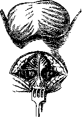
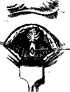

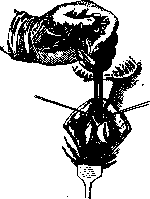
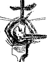
is explained by the location of the prostate, its relationship to other organs, and finally the tendency of this disease to metastasize. Cases with the presence of metastases, cachexia, cases accompanied by involvement of the lymphatic glands, and cases of disseminated prostate cancer with involvement of the pelvic cellular tissue are absolutely contraindicated for radical surgical treatment. X-ray therapy is quite widespread in the treatment of cancers of the prostate gland. While not a radical method, it causes a reduction in the volume of the malignant tumor and, as a rule, acts symptomatically, bringing about a reduction in pain. The treatment of prostate cancers with radium is widely used in America. Bumpus reports the results of treating 1,000 cases. Radium was used either perineally or by the endovesical route. In the first method, capsules containing radium were introduced for a certain period into the bladder through a cystoscope or a previously established fistula; in the second, radium needles were inserted into the prostate gland through the thickness of the perineum or after preliminary exposure of it through perineal or suprapubic fistulas. No one has seen long-term recovery from radiotherapy for prostate cancer, and the best American statisticians speak of prolonging the life of patients on average to a maximum of 22 months. Sarcomas of the prostate gland are observed less frequently than cancers: according to the anatomical statistics of Gebele, 2 sarcomas occur for every 29 cancers. In contrast to cancers, sarcoma of the prostate gland is usually observed in childhood, sometimes even in infancy (6-9 months) of age. According to Poschas statistics, 50% of all described cases belong to the age under 10 years, 25% to the age from 10 to 30 years, 5% to 30-50 years, and 20% over 60 years. All histological varieties of sarcomas are found in the prostate gland, but round-cell and spindle-cell sarcomas take first place, followed by myxosarcomas and angiosarcomas. The course of prostate sarcoma is initially asymptomatic, and this explains the circumstance that in the described cases the tumor occupied the entire gland. The size of the tumor ranges from a walnut to a child's head (Paschkis). At the beginning of the disease, the contours of the gland are clearly defined, but as a result of the extremely rapid growth of the tumor, the surrounding tissues are involved in the process, and the urethra, bladder, and bowel are compressed and displaced. These factors are the immediate cause of the onset of urinary disorders in the form of frequent urination and urinary retention, and defecation disorders in the form of stubborn constipation. The invasion of the lesser pelvis cellular tissue by the tumor causes the immobility of the neoplasm, which is often palpable above the pubis, as if filling the entire pelvic cavity and making it impossible to orient oneself in the details and contours of the organs. In contrast to cancers of the prostate gland, its sarcomas give

Fig. 22. Removal of the prostate by literal incision: metastases are extremely rare. In lymphosarcomas, the lymph glands are affected: pelvic, retroperitoneal, mesenteric, and lumbar. At the beginning of the disease, the process is asymptomatic; only with the growth of the tumor and the compression of the bladder and rectum do disorders of urination and defecation appear. Pain and hematuria are usually absent. Cachexia comes to the fore. The younger the patient (Holtzov), the faster the tumor growth. On average, the duration of the disease does not exceed two years. Diagnosis is based on rectal examination data. The presence of a large prostate gland at a young age always makes one think of its sarcomatous involvement. The consistency of the gland is soft, moderately dense, and never reaches such hardness as in cancer. Sometimes the tumor masses fluctuate and can be mistaken for an abscess. The prognosis is grim. Surgical treatment is technically similar to the treatment of prostate cancers. All authors reject surgery in childhood, since the operative mortality is very high, and children who undergo surgery quickly die from metastases. In adulthood, the results of surgical intervention are also not very favorable; the best results were obtained by Mac Gowan—4 years without recurrence after surgery. Almost all authors recommend roentgenotherapy by irradiation of the perineum, alone or in combination with radium, which is introduced to the prostate gland through an opened bladder. The experience of the Mayo Clinic indicates a patient survival time of up to 7 years with this treatment method. Prostate cysts are rare: according to Wesson's statistics (1925), 33 cases have been described. True, retention, and echinococcal cysts should be distinguished. Retention acquired cysts arise as a result of the compression and closure of the excretory ducts of the prostate gland by developing connective tissue; congenital ones are the consequence of abnormal development of the lower end of the Müllerian ducts. The size of the cyst varies widely—from 5 liters in Schmidt to 2 cm3 in Semyanyako. The contents of the cyst are grayish-yellow in color, rich in protein, and contain epithelial cells and detritus under a microscope. In the hypertrophied prostate gland, cystic cavities are observed quite often and are the result of the disease. Symptoms are expressed in frequent urination and premature, sometimes painful ejaculation. Cysts that do not cause complaints require no therapy. For large cyst sizes and sharply expressed subjective complaints, opening the cyst or enucleating it through a perineal incision is indicated. Prostate atony is a concept first introduced by Porosz. As a result of various pathological factors, the muscular tissue of the prostate loses the tone inherent in healthy individuals and relaxes, resulting in the stretching of individual alveoli by prostate juice and stagnation of the prostatic secretion in them. The etiology of this condition is not always clear—one can think that it is in direct relationship with venous stasis in the lesser pelvis, prolonged urinary retention, sexual excesses, coitus interruptus, mastonin, prolonged unsatisfied excitations, a sedentary lifestyle, and finally, constant jolting riding (drivers' disease). However, with the most careful questioning of patients, it is sometimes impossible to establish any of the listed moments, and one can think (Holtzov) that certain individuals have a constitutional predisposition to the disease. Both the entire gland and part of it can be atonic. Histological examination reveals dilation of individual alveoli, their overfilling with juice, and atrophy of muscle elements. Upon palpation through the rectum, the prostate appears as a flabby bag of doughy consistency, poorly configured and difficult to distinguish from surrounding tissues. The sensitivity of the prostate is not impaired in this process. When pressing on the gland into the urethra, an abundant amount of whitish, turbid liquid is released, containing under the microscope elements of prostate juice, with an insignificant number of leukocytes, the absence of microorganisms, and a significant number of lecithin bodies. Prolonged retention of the secretion in the atonic prostate gland can lead to secondary irritation of its tissue by the secretion and cause inflammatory phenomena in it—aseptic or stagnant prostatitis. In such cases, the abundantly secreted juice contains a greater or lesser number of leukocytes. The differential diagnosis between chronic prostatitis and aseptic prostatitis should be based on the data of microscopic examination of the juice. In the former, the number of lecithin bodies is reduced, whereas in aseptic prostatitis, on the contrary, local leukocytosis is caused by chemical irritation of the prostate gland by lecithin bodies that have not been released outside, a significant amount of which is found in the prostate juice. Furthermore, leukocytes in chronic prostatitis disappear from the juice extremely slowly; in aseptic prostatitis, on the contrary, one or two massages are enough for them to disappear. Local complaints of patients boil down to a feeling of heaviness in the anus and the discharge of a whitish fluid from the urethra during urination and defecation (prostatorrhea). Very often there are simultaneous frequent urges to urinate, accompanied by an intermittent stream. The main complaints, however, come down to general nervous disorders and are often fixed in organs that would seem to have no direct relation to the sex glands. This includes a feeling of constant fatigue, irritability, a decrease in mental balance, insomnia, gastrointestinal disorders, palpitations, pain in the spine and sacral region, i.e., what was previously described under the name of sexual neurasthenia. At the same time, patients complain of a decrease and sometimes even a complete disappearance of erections, and this is often preceded by increased excitability. We find the explanation for the occurrence of all these complaints in the increased absorption into the general bloodstream of the secretion retained in the prostate, which can cause the same symptoms upon its artificial introduction into the organism. The diagnosis of atony is extremely simple and is hindered only by the fact that in the general examination of a patient who does not make complaints of a local nature, the examination of the prostate is usually neglected. At the same time, it is undoubtedly that palpation of the prostate could provide a key to explaining a number of symptoms of a seemingly purely 'nervous' nature. The prognosis of the disease is dubious in the sense of a complete cure. It is not always easy to restore the tone of the prostate muscles, and patients are often doomed to prolonged treatment and relapses of the disease. However, the prognosis is inextricably linked with the living and occupational conditions of the patient, on the one hand, and with the constitution and duration of the disease, on the other. Therapy should consist in removing the secretion accumulated in the prostate in order to prevent its enhanced absorption and in restoring the tone of the muscular apparatus. For this purpose, gland massage, faradization, and water procedures in all forms are prescribed, and internally—preparations of arsenic and strychnine. Prostate stones are divided into true ones, i.e., formed in the tissue of the gland itself, and false ones, i.e., located in the prostatic urethra and from there penetrating into the prostate by layering and pressure. True stones are surrounded by prostate tissue on all sides and have no direct contact with urine. False stones are constantly washed by the latter, protrude into the lumen of the urethra, where they can be easily detected with a urethroscope and removed endourethrally. Infectious processes of the prostate play an undeniable role in the etiology of stones (true ones). Furthermore, the latter are quite frequently found in hypertrophy of the prostate. The stone-forming process should be conceived as follows: a chronic inflammatory process causes partial necrosis of the glandular epithelium, which desquamates in the form of a shapeless mass, fills individual follicles of the prostate, undergoes amyloid degeneration, and forms the core of the future stone, onto which phosphate and lime salts are layered. Prostate stones can be single or multiple (584 in the case of Pavlov-Silvansky) and can be located in one lobe or fill the entire gland. Single or multiple small stones of a purely organic composition do not produce any clinical symptoms and can be discovered only on the operating table as an incidental finding. Large or infected stones cause pain during urination and urinary disorders in the form of frequency or retention of urine. The pains spread to the region of the anus and intensify with pressure on the perineum (sitting on a hard surface). During sexual intercourse, the pains sharply intensify, an admixture of blood is observed in the semen (see Hemospermia), and patients complain of frequent painful pollutions. The diagnosis is based on palpation data. The prostate containing stones is enlarged in size, dense, bumpy, and gives a sensation of crepitation. In the urine after massage, one can often detect a number of calculi that have fallen out of the prostate. X-ray examination gives a positive answer in cases of encrusted primary stones, making it possible to judge their size, shape, and quantity. Small organic prostate stones that do not produce clinical symptoms do not require treatment. Large multiple inorganic stones are subject to removal by prerectal prostatotomy.
Individual authors (Holtsov, Pavlov-Silvansky) recommend performing intracapsular prostatectomy to avoid recurrences.
Related articles
Mentioned in
Cite this page
“Prostate Gland.” Soviet Medical Encyclopedia. English translation of Bolshaya Meditsinskaya Entsiklopediya, 1st ed. (Moscow, 1928–1936), ed. N. A. Semashko. https://sovietmedicalencyclopedia.pages.dev/article/prostate-gland/