Prosencephalon
Historical document, translated for reference. It reflects medical knowledge of the 1920s–30s and is not medical advice.
Summary
This entry defines the prosencephalon as the forebrain, detailing its anatomical subdivisions into the telencephalon and diencephalon. It also includes a significant portion of the entry for leprosy, covering its historical, etiological, and epidemiological aspects as understood in the early 20th century.
Encyclopedia article (1928–1936)
PROSENCEPHALON, forebrain; together with the mesencephalon it is called the cerebrum; together with the mesencephalon and rhombencephalon it forms the brain. In turn, it is subdivided into 1) telencephalon, or endbrain (hemispheres and pars optica hypothalami) and 2) diencephalon—interbrain (thalamus opticus, metathalamus, epithalamus). P. develops from the primary forebrain vesicle, which subsequently differentiates into the intermediate vesicle or diencephalon and two lateral vesicles of the hemispheres (telencephalon). Weakly developed in lower vertebrates, it reaches its highest development in man. Lit.—see Brain. LEPROSY. Contents: History... 251 Etiology... 252 Epidemiology... 257 Statistics and geographical distribution... 259 Pathological anatomy... 261 Clinical picture... 264 Prophylaxis... 273. Leprosy (lepra). With the word lepra, the 70 translators of the Bible (Septuagint) translated the Hebrew term Zaraath, denoting the disease described in chapter 13 of the 3rd book of Moses (Leviticus). Regarding the question of what content is hidden behind this term, why the 'LXX' translated it with the word lepra, and whether both these terms correspond to the modern concept of leprosy, there exists a rich literature. Most authors hold the opinion that by the word Zaraath one must understand heterogeneous dermatoses, including, probably, leprosy, which undoubtedly existed among the Jews of that time. According to other authors, this term refers to the so-called white leprosy, which supposedly existed at one time and then disappeared. Others are convinced (Minch) that under the name Zaraath a classic description of disseminated vitiligo is given, the so-called pesi, which is very common even now in Central Asia. There are even authors who argue that syphilis (Pogorelsky) is hidden under this name, etc. Leprosy is a chronic disease, distinguished like tuberculosis by a prolonged course. It is caused by a specific microbe and must be classified as a contagious disease. The history of leprosy is hidden in deep antiquity. There are indications (Sauton) that leprosy was known in Egypt many centuries before our era, and Brugsch, the famous Egyptologist, found an indication in a papyrus he discovered that leprosy existed in Egypt at least 2,400 years before our era. Herodotus (5th century B.C.) has a passage where he speaks of the fact that in Persia there exists a disease which he calls lepra or leuke, and reports that those possessed by this disease, as 'having sinned against the sun, dared neither to enter the city nor to meet with other persons.' However, Hippocrates, a contemporary of Herodotus, calls quite diverse diseases by the names lepra and leuke, into which one can only with difficulty include some forms of leprosy. A little later, Aristotle (4th century B.C.) already described leprosy more accurately as a disease common in Greece, calling it satyriasis or leontia, and the physician Aretaeus shortly after this first introduced the term leontiasis, which is used even today to denote certain stages of the course of leprosy (Babes). Later, this disease in Greece received the name elephantiasis, under which it was known for a long time. Under the same name, it was first indisputably described by the Roman physician Aulus Cornelius Celsus, who lived in the time of Augustus: 'completely unknown in Italy,' says Celsus, 'but very common in some countries, is the disease quem elephantiasin Graeci vocant.' This continued until one of the major Christian writers, Origen (3rd century A.D.), 'explained' that by the Hebrew name 'Zaraath,' which the '70 translators' translated with the term lepra, one should understand precisely leprosy. Since then, the name 'lepra' for leprosy has become the generally accepted term. Along with Egypt, leprosy existed long ago, even in prehistoric times, in Phoenicia, from where, thanks to trade relations, it could spread to other, even distant countries, where it received the name 'morbus phoenicicus' accepted by some ancient authors. Leprosy also appeared long ago in India, China, and Japan, for which there is a series of evidence in the most ancient writings of these countries. It is considered generally accepted that Europeans brought leprosy to America, and leprosy owes its further spread to the importation of Negro slaves from Africa, whom Europeans used in abundance as a labor force. It is believed that leprosy penetrated into Europe mainly through Greece and Italy, where it could have been brought partly from Egypt, partly from Persia or from Asia Minor. From these two centers (Greece and Italy), leprosy spread throughout Western Europe. The great migrations of peoples that occurred at the turn of the transition from ancient times to the Middle Ages played no small role in such a spread of leprosy, and the Crusades played a particularly sad role in the spread of this disease, when over the course of 2 centuries the popular wave was thrown back and forth 7 times, from Western and Southern Europe to Palestine through Egypt and Asia Minor, i.e., countries heavily affected by leprosy. Having received an impetus for strong spread in European countries since the time of the Crusades, leprosy reached its maximum by the end of the 12th century and especially in the 13th century, calling into existence a huge number of leprosaria, of which there were up to 2,000 in France alone. From the 14th and 15th centuries, leprosy began to decline and by the beginning of the 19th century had decreased so much that both doctors and society completely forgot about it. And only in the 40s of the 19th century was attention drawn to it again by the famous studies of Danielssen and Boeck, and in the 70s by the no less famous bacteriological investigations of Hansen and Neisser. Interest in the disease increased even more after three international conferences (Berlin in 1897, Bergen in 1909, and Strasbourg in 1923), thanks to which the question of leprosy again took an honorable place in world scientific thought. Etiology. Hansen, investigating leprotic nodules in a fresh (unstained) state in 1871, first discovered bacilli in them. It was only possible to stain these bacilli for Neisser in 1879. Since then, bacilli are easily detected in all nodular forms of leprosy. In macular-nerve forms, bacilli were not found for a long time, and therefore it was believed that the very changes in the skin were caused here by local neuritis, apart from the participation of bacilli [Unna's neuroleprids, Arning's 'leprids']. At the present time, there is no doubt that leprotic bacilli are found here too, but for the most part only in meager quantities; sometimes there are so few of them that it is not easy to find them. Furthermore, it turned out that not all leprotic bacilli have the typical appearance of an acid-fast rod, but that, on the contrary, they are also found in an acid-sensitive form and, moreover, often in the form of grains. To detect these forms, Much proposed a modified method of prolonged Gram staining; it turned out that in those cases of macular leprosy where acid-fast bacilli were not detected, they were discovered by staining according to Much. The causative agent of leprosy (according to the terminology of recent years - Mycobacterium leprae) belongs to the group of acid-fast microbes and can be detected in smears and tissues by any of the methods proposed for staining this extensive group (see separate table, figs. 4 and 6). In its morphological properties and structure, it is very close to the causative agent of tuberculosis and has the appearance of a thin rod, the width of which is determined at 0.2–0.5 µ, and the length is subject to significant fluctuations (1.5–6 µ and more). The rod is straight or slightly curved. In a stained state, it either has a uniform appearance and seems homogeneous or consists as if of separate small grains arranged along its length [Lutz's Coccothrix]. The latter form is apparently observed more often in old foci and infiltrates and is regarded by some as a form of degeneration. Many look at these grains as the well-known Babes-Ernst grains, which are encountered, among other things, in all bacteria of the diphtheria series. In the rod, one can sometimes see a series of free, remaining unstained spaces (Neisser's vacuoles), which were previously completely incorrectly taken for spores. Sometimes the ends of the rod are pointed, sometimes, on the contrary, they are slightly thickened and flask-like swollen. Besides such typical forms, less regular forms are encountered in leprotic material, fully corresponding to those 'fragments' of bacilli that were noted by Spengler in tuberculosis. In the same material, one can also encounter freely lying small coccoid grains, the diameter of which is equal to the width of the bacilli. These grains, arranged singly or in short chains or small clusters, are sometimes encountered where bacilli are absent. When treated with osmic acid, small bodies are detected in the rods, staining black, which is attributed to the content of fat or lipoids (Unna). Rods released from leprotic tissues do not possess their own movement and are devoid of cilia. Leprotic bacilli absorb coloring substances more easily than tubercular ones and therefore can be stained with relatively weak solutions of aniline dyes. Many, especially in the past, thought to use this property to distinguish them from tubercular ones.
Of all the methods proposed for this purpose, the method of Baumgarten is the most popular; it is based on the rapid staining of leprosy bacilli with a very weak solution of fuchsin, by which tuberculosis bacilli are not stained. Subsequently, however, it turned out that all these methods, not excluding Baumgarten's, prove to be more or less 'reliable' only in those cases where recognition is already easy and presents no difficulties, namely in cases of nodular leprosy, where there are many bacilli and they are arranged in typical clusters. In all other cases, these methods proved to be of little practical use. Cultivation of leprosy bacilli. All works to date on the isolation of leprosy cultures can be divided into the following groups: 1. From leprous material, cultures of acid-fast bacilli were isolated, which in morphological respects fully correspond to genuine leprosy bacilli, but proved inactive when inoculated into animals. Such cultures were first obtained by Bordoni-Uffreduzzi (1889) from the bone marrow of a leprous corpse on glycerinated coagulated serum. Similar cultures were obtained by Clegg (1911) and his colleagues (Brinckerhoff, Currie, and Hollmann), as well as by Duval (who later, however, renounced his cultures) on liquid nutrient media in which amoebae or other protozoa or even some bacteria (biophytes or saprophytes) were simultaneously or previously grown. From this, leprosy bacilli in pure form were easily obtained. The cultures were pigmented yellow or orange. 2. From leprous tissues, cultures of bacilli were isolated that were completely similar to leprosy bacilli and fully retained their acid-fast properties; however, the bacilli themselves proved poorly adapted to the artificial conditions of their existence and could not be isolated even in the second generation. Such cultures were obtained by Emile Weil in the yolk of a chicken egg and on some solid media containing an admixture of pure yolk. Similar cultures were obtained by Ch. Nicolle on coagulated rabbit serum, brain agar, and yolk agar. Recently (1928-1930), similar cultures were obtained by G. S. Kulesh when inoculating pieces of leprous tissue (skin, spleen) into ordinary nutrient broth, with the growth of bacilli occurring either only in the pieces themselves or in the form of a very thin brownish film on the surface of the medium, while the broth remained transparent. 3. From leprous nodules, a filamentous microbe was obtained, belonging to the group of streptothrix or actinomycetes (Barannikov, Kedrovsky, V. Williams, Rost, Deycke, Bayon, Reenstierna, and recently de Araujo and Bulkin). 4. From leprous tissues, cultures of bacilli were isolated that were similar to true leprosy bacilli, but unlike them, they were either immediately devoid of acid-fast properties or retained them only in the first generations and lost them in subsequent passages. Acid-fastness was already reduced from the very beginning or was preserved only in the granules contained within the bacilli, while the bacilli themselves took on a counterstain. The latter category is the most numerous. This fact, significant in itself, acquires special importance in connection with the data noted above that in leprous tissues, along with acid-fast bacilli, one can always find a sometimes significant admixture of acid-sensitive forms. The weak point of all these observations was considered to be the lack of direct evidence that the isolated bacteria were indeed true leprosy bacilli. This gap was filled by the observations of Kedrovsky, who, by passing the culture of acid-sensitive bacilli he had isolated through animals, converted it into an acid-fast variety and caused a number of leprosy-like changes in the internal organs of rabbits and mice with the latter. A detailed study of the cultures isolated in leprosy, just like the study of tuberculosis cultures, leaves no doubt that both leprosy bacilli and tuberculosis bacilli, under certain conditions, can pass into a more complex state of a filamentous fungus of the Streptothrix or Actinomyces type. Apparently, such a transition (a kind of 'mutation') is generally possible in that group of diphtheroids to which all microbes of the diphtheria series should be assigned, including the group of acid-fast bacilli. Thus, the difference in the conclusions of the authors who obtained, on the one hand, cultures of acid-fast or acid-sensitive bacilli and, on the other, fungal cultures of the streptothrix type, completely disappears—especially since both old works and especially recent works prove the possibility of the transition of streptothrix filaments into diphtheroid bacilli both in vitro and in vivo (Williams, Bayon, Kedrovsky, Trius and Politova, Bulkin). Regarding immunity in leprosy, information is insufficient. Since the time of the work of the 'English Commission in India' (1890), it has been known that in families affected by leprosy, far from all members contract this disease, even if they lived and were in completely identical conditions with the closest contact with each other. A sick spouse transmits their disease to a healthy one in only 20% of cases. Children born to sick parents and living with them in one family do not all contract the disease by any means. On the other hand, patients are very often puzzled as to where and from whom they could have contracted leprosy, since they had no cases of prolonged contact with patients before the disease. All these 'paradoxes' of infection are well known, but nothing has yet been provided to explain them. It has been established that the child's organism is significantly more susceptible to infection than other ages. The fact that children get sick more often if the mother is sick in the family than if the father is sick is easily explained by the closer and more intimate contact of young children with the mother. Men get sick somewhat more often than women, which is obviously connected with the more 'mobile' nature of their life and work and has nothing to do with questions of immunity. Racial differences in the incidence of leprosy apparently play no role. If it is noted that out of 2-3 nationalities inhabiting one or another endemic leprosy area, one becomes infected more often and more than others, this most likely depends on the difference in social and living conditions rather than on special properties of 'racial constitution'. Eitner was the first to apply the complement fixation reaction in leprosy. As an antigen, he took an aqueous extract from lepromas. From subsequent works, it turned out that the serum of patients with the nodular form reacts positively with the specific antigen from lepromas in the majority of cases (50-80%), while the serum of patients with the macular form gives significantly less constant results. The method of preparing the antigen proved to be very important. Aqueous extracts from lepromas mostly gave a negative result even in nodular forms. Nishiura obtained inhibition of hemolysis with an aqueous extract from skin nodules and leprous liver in the nodular form in 89% of cases, and in the neural form in 48%. Bieler and Eliasberg successfully used an antigen prepared from leprosy bacilli after dissolving lepromas with antiformin. Using stronger solutions of antiformin to dissolve the bacteria along with the tissue, Bulkin prepared an antigen that was sufficiently sensitive and completely specific for leprosy. An antigen prepared from a leprous testicle containing a huge number of bacilli proved to be particularly sensitive. In leprosy, one often has to observe a positive Wassermann reaction (70-75% for tuberous forms and 35-40% for macular-neural forms). The serum of leprosy patients often reacts positively if tuberculin is used as an antigen. The Bordet-Gengou reaction was also used to prove the identity of the bacilli isolated from leprous tissues with true leprosy bacilli (Duval—in relation to his culture, Bayon, as well as Birger and Krichevsky—in relation to the cultures of Duval and Kedrovsky). Mesinescu obtained inhibition of hemolysis with the serum of lepers by using as an antigen an extract from leprosy-like nodules taken from rats that spontaneously contracted a process in many respects similar to human leprosy. Cultures of the leprosy pathogen are also used to prepare an antigen which, possessing specificity, could serve for diagnostic purposes, especially for early forms of the disease (Bulkin, Lewis, Blanc, Pangalos et Joannides). The question of agglutination in leprosy is poorly developed. As an antigen, an emulsion of bacilli from macerated lepromas (Gaucher, Abrami, Sugai), leprosy cultures (Spronk, Birger, Krichevsky, et al.), and bacilli of rat 'leprosy' (Dean) were taken. The first attempts to inoculate leprosy into a rabbit through the anterior chamber of the eye were discredited by the objection that the local changes obtained were caused by the microbes introduced into the tissue, which were contained in the inoculum itself, without their multiplication having occurred. Melcher and Ortmann, by inoculating leprous material in the same way, caused a number of changes in the internal organs, lymph nodes, and lymph follicles of the cecum and its appendix (sacculus rotundus) in rabbits. The scientific authorities of that time (1885-86) who saw these changes expressed their opinions—some for leprosy (Koch), others for tuberculosis (Hansen, Neisser). Experiments by V. V.
Ivanov's experiments on guinea pigs seem to suggest the possibility of inoculating leprosy into these animals. Sugai successfully inoculated leprosy into a special breed of Japanese mice (dancing mice) by injecting leprous material into their testicles. The inoculation of leprous material into monkeys (subcutaneously) led to the formation of nodules at the site of inoculation that grew gradually and then underwent regression, the 'epithelioid' cells of which contained a large number of leprosy bacilli. In numerous experiments by Reenstierna, the growth of the nodules was accompanied by hair loss and, correspondingly, loss of eyebrows. The nodules, as a rule, resolved after some time. Passaging from one animal to another was unsuccessful. In the internal organs of some monkeys, cheesy nodules developed, which contained a large number of leprosy bacilli in their 'epithelioid', sometimes vacuolated, and often multinucleated cells. de Araujo (1931) inoculated leprosy into white rats and white mice, in which he observed the formation of small and large nodules containing a huge number of bacilli in their arrangement typical for leprosy (cigar bundles, globi). However, de Araujo obtained a picture identical to this in the same animals by introducing leprous material that had been previously subjected to prolonged exposure to methyl alcohol or formalin, and even prolonged heating at 120°.

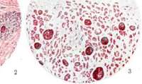


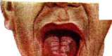
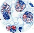
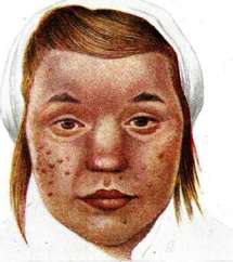
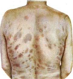
Fig. 1. Brown leprosy cells. Fig. 3. Tuberculoid leprosy. Fig. 3. Leprous cells. Fig. 4. Encephalitic membrane of a foam-like cell (schematic). Fig. 5. Leprous cell. Fig. 6. Smear from the nose. Fig. 7. Leprous cells. Fig. 8. Leprous infiltration of the tongue. Fig. 9. Tuberculoid leprosy. Fig. 10. Spotted-tuberculoid leprosy. (Figs. 1 and 5—stained with hematoxylin-eosin. Figs. 2-7—stained according to Ziehl-Neelsen). To the article Leprosy. Epidemiology. It is necessary to acknowledge that leprosy is an unconditionally contagious disease, and therefore the entire complicated question of the paths of transmission of this disease reduces to the question of how the infectious agent is transmitted: by congenital means, as some think, by direct transfer from the sick to the healthy, as others think, or whether some intermediate factors take part in the transmission. That leprosy can be transmitted by congenital means, there can be no doubt. Some researchers have observed leprosy in newborn children or in children of such an early period of life that the origin of the disease in them could be explained only by intrauterine infection (Reshetillo). In practical terms, however, such cases cannot have significance, since all such children with early manifestations of leprosy turn out to be non-viable and soon perish. Children who have acquired leprosy at a later period of life, in the vast majority of cases, do not develop properly in the sexual sense and are deprived of the ability to produce offspring. The well-known fact that leprosy patients in the majority of cases early lose the ability to procreate, that this ability is generally lowered in them to a greater or lesser degree, and that consequently, if leprosy were transmitted only by inheritance, it would soon disappear spontaneously, does not speak in favor of the congenital path. Thus, a number of indirect indications force us to admit that congenital transmission cannot play the exclusive role in the spread of leprosy that was recently attributed to it. Therefore, the latest researchers see the immediate cause of the spread of leprosy in the ability of its pathogen to be transmitted directly from the sick to the healthy. This doctrine rests on the following data: 1. Leprosy is spread mainly among the poor population living in filth and overcrowding, in the absence of the most elementary rules of hygiene. With the improvement of hygienic conditions, with the raising of the level of culture and well-being, the disease also disappears. The example of Norway, Iceland, and the Hawaiian Islands shows that if patients are placed in proper conditions of care and made safe for those around them, then cases of leprosy begin to fall rapidly. 2. Leprosy is transmitted not only to children from sick parents but also to outsiders who are in close contact with the sick. 3. In countries affected by leprosy, outsiders are sometimes infected. True, such infection is not observed so often, but this only speaks to the fact that the transmission of leprosy is possible only under particularly favorable circumstances, among which poor social, economic, and hygienic conditions and the closest communication with the sick stand in the foreground. The same considerations explain why Europeans who have become infected in leprous countries and returned to their homeland serve as a source of disease for those around them only in rare cases. However, such cases are still observed. 4. The contagiousness of leprosy is also confirmed by those individual cases where the disease occurred on the basis of direct infection, for example, cases of the disease in doctors or nursing staff who became infected from patients as a result of injury during an operation. 5. Finally, the experiments on inoculation of animals, which were reported above, also speak in favor of the contagiousness of leprosy. The recognition of the contagiousness of leprosy is hindered not a little by complete ignorance regarding the primary foci of the lesion. On the basis of personal research, Sticker came to the conclusion that the mucous membrane of the nose serves as the path for the penetration of the infection: out of 153 lepers, he did not find bacilli in the nose in only 4 cases, and in 9 patients, bacilli were found in the absence of any changes in the mucous membrane. However, later researchers partly could not detect bacilli in the nose with such constancy, and partly attributed their presence to a secondary lesion of the mucosa. Just as little is known regarding the method of transmission of the infection. The view that animals (rats) play a role in the spread of the infection, in which diseases have been discovered that are in a pathologico-anatomical respect to a significant degree similar to human leprosy, is still poorly substantiated, since in notoriously leprous countries such leper-like diseases are encountered far from with the constancy that one would expect in the event that this factor actually had such great significance. Even fewer data can be cited in favor of the view that one or another insect plays a role in the transmission of the disease. Likewise, there are as yet no solid grounds to assume, as some admit, that the
Leprosy here is to a significant extent prevalent both in the north (Egypt, Tunisia, especially Abyssinia) and in the south of Africa (Union of South Africa), but to an even greater degree, the entire eastern coast of the continent with its adjacent islands is affected, among which Madagascar occupies the first place. The infection is particularly severe in the central part of the continent, mainly in that vast part of its western coast where the colonies of France, Belgium, and England are concentrated, and according to local physicians, the degree of infection increases as one moves deeper into the interior of the continent. Rogers cites a huge percentage of infection for all these colonies, up to 40, even up to 60-70 (Ivory Coast) per 1,000 population; for the southern part of Cameroon, 130 per 1,000; and for the eastern outskirts of the Belgian Congo, he gives a staggering, even seemingly improbable figure of 200 per 1,000, i.e., 20%. However, for the 'Kissi' region in French Guinea, the same Rogers gives an even higher figure—32%. In North America, leprosy is weakly distributed. Mexico with its islands is more significantly affected (according to Rogers, on the island of Cuba the number of patients reaches 1,500 = 0.73 per mille; on the island of Trinidad, 526 = 1.70 per mille), but the infection reaches particularly significant strength in the northern part of South America, where in some countries it reaches 19.1 and even 25 per 1,000 population (French and Dutch Guiana), and in Brazil the number of patients according to the latest data is 12,000-15,000 (approximately 0.72 per 1,000). In the southern states of South America, leprosy is encountered less frequently (in Argentina, according to Rogers, 750 = 0.13 per mille; in others, even less; while in Bolivia, Peru, and Chile, there are only isolated cases). Almost on all islands belonging to Oceania or Polynesia, leprosy is developed to an extreme degree. The Hawaiian Islands are particularly affected, where the number of patients according to data from 1896 reached approximately 5,000 out of 58,000 population, i.e., exceeded 8.5%. By 1909, the number of patients, due to the high mortality among them, fell to 6%, and at the present time, it apparently should not exceed 400-500, since Rogers, citing Hoffmann, counts only 611 patients in 1919, which constitutes 2.32 per 1,000 population. Leprosy is distinguished here by particular malignancy. The Philippine Islands must also be classified as heavily affected. On the island of Culion, the largest leprosarium in the world is located, housing over 5,000 patients. The total number of patients here reaches no less than 15,000. Australia can be considered almost free of leprosy. In Queensland (northeastern coast) in 1921, according to Rogers, there were 50 patients (0.07%). In New South Wales in 1920, there were 20 patients. The total number of patients in Western Europe at the present time is small. J. E. Abbot, who personally visited a number of countries in 1923 and 1926, gives the following figures: France—up to 1,500 patients, Norway—130, Sweden—32, Finland—75, Estonia—226, Latvia—210, Lithuania—21, Germany—8 (all imported), Yugoslavia—84, Italy—226, Switzerland—25, Iceland—67, Holland—13, Spain—578, Portugal—200, England—100. In Poland, Austria, Czechoslovakia, and Hungary, no leprosy patients have been established. In Belgium, Holland, and Denmark, there are only imported cases. In 1926, 13 such accidental patients were interned in Dutch hospitals. Incomplete data: Crete—400 (according to Rogers up to 1,000), Cyprus—250, Malta—247, Romania—338. Regarding Turkey, Greece (the mainland), and Bulgaria, no information is provided. According to Rogers, who cites Düring, there are 500-600 patients in Turkey.
In the USSR, as of January 1, 1931, there were 1,431 leprosy patients, of whom 1,032 were isolated in leprosaria. Out of the total sum of 1,431, the share of the RSFSR accounted for 1,289, and the share of the union republics for 142 patients. The patients in the USSR were distributed as follows: A. Isolated in leprosaria of the RSFSR: Krutye Ruchyi (Leningrad Region)......
128 Astrakhan (Lower Volga Territory) ....
131 Karachay-Cherkess I North Caucasian Territory
11 Total............. 399 Pathological anatomy. Changes in the skin in the tuberous form consist of the formation of granulomas. A characteristic feature of such 'lepromas' are the so-called lepromatous cells, often—though hardly correctly—called Virchow cells, since Danielssen had already drawn attention to these cells for the first time, considering them characteristic of leprosy. Hansen also drew attention to these same pigmented cells while searching for the specific microbe, calling them 'brown bodies' [see color plate (255-256), fig. 1]. These cells are characterized by the presence of vacuoles, sometimes in such a large quantity that they appear as if foamy (see color plate, fig. 3, 5-7). Virchow's merit lies in the fact that he was the first to note the presence of fat in these vacuoles, to the study of the chemical structure of which increased attention has been paid only recently (Cedercreutz, Herxheimer, Stein). In former times, these cells were considered epithelioid and were derived, as in other granulomas, from fixed connective tissue cells and vascular endothelium. At the present time, however, after the works of Aschoff and Anichkov, they are inclined to be considered histiocytic elements. Lepromatous cells are characterized by the content of lipoids, vacuolization, and the presence of bacilli. The very process of their formation is conceived as follows: due to the absorption of bacilli and their gradual multiplication inside the protoplasm, the cell increases more and more in volume; under the influence of the bacilli, the deposition of lipoids occurs within it, in which vacuoles of various sizes are then formed. Herxheimer views the accumulation of lipoids in the cell as lipoid degeneration, while he explains the formation of vacuoles by the splitting and emulsification of fats. Stein, on the contrary, explains the accumulation of lipoids in lepromatous cells as an infiltrative process, perhaps standing in connection with the administration to patients of significant quantities of lipoids as a therapeutic agent; he is inclined to explain the origin of the vacuoles by the disintegration and dissolution of microbes under the influence of the lipoids themselves or perhaps under the influence of lipases induced by them. Cedercreutz (1921) classifies the lipoids partly as ordinary isotropic, partly as anisotropic fats (cholesterol and its esters). The content of bacilli in lepromatous cells is not uniform—sometimes there are few of them and they lie scattered in the protoplasm, then their quantity increases more and more and finally reaches a huge magnitude, with the bacilli, arranging themselves in parallel rows, either forming clusters resembling a 'bundle of cigars' (Babes) or producing round-shaped colonies, permeated by regularly outlined vacuoles of various sizes (Neisser's globi). In these colonies, the bacilli for the most part adhere so closely to each other that they give the impression of tightly fused heaps, in which individual specimens are distinguishable only perhaps at the periphery. The bacilli lie in the lipoid inclusions of the cells around the vacuoles, sometimes arranging themselves around them in a radius or wreath-like (Herxheimer, Stein). The vacuoles themselves either remain free or, according to Stein, include 'remnants of disintegrating bacilli'. In Unna's opinion, the bacilli in such compact heaps are glued together by a special mucus-like substance—apparently a product of the secretion of the bacilli themselves, to which he gave the name gloea. The primary multiplication of cells during the formation of a cutaneous granuloma occurs around small vessels, nerve endings, and those skin appendages which are intertwined with a rich network of blood capillaries (hair follicles, sweat and sebaceous glands). The settling here and multiplication of microbes occurs immediately in many places, or, having started in one place, it soon spreads to neighboring areas, so that upon examination of a lepromatous nodule or infiltrate, one usually finds several nodules at once, separated from each other partly by old, partly by newly proliferated connective tissue, which sometimes forms a kind of capsule around the nodules themselves, consisting of 2-3 layers of collagen fibers. The nodules occupy the entire thickness of the skin, sometimes passing into the underlying subcutaneous tissue, and only very rarely reach the epidermis, for the most part being separated from it by a thin strip of connective tissue of the papillary layer. Calling this boundary layer 'protective' or 'precautionary' (Lie) can only be done conditionally, since in chronic cases the granulomatous process can reach the epidermis itself and, by destroying it, lead to ulceration. Lepromatous nodules and infiltrates are characterized by a richness of vessels and the stability of the tissue of which they consist. True, in lepromatous cells one can note phenomena of pyknosis, wrinkling of nuclei, and sometimes their disintegration, but it only reaches the formation of even small foci of coagulation necrosis in exceptional cases. Likewise, the nodules only rarely undergo softening and liquefaction with the formation of a cavity filled with a mucopurulent mass, rich in polymorphonuclear leukocytes with lepromatous bacilli enclosed within them (the so-called lepromatous abscess). On the contrary, such nodules and entire infiltrates very often undergo 'fibrous transformation,' whereby the development of connective tissue sometimes leads to the almost complete destruction of the lepromatous tissue or even to the complete disappearance of the leproma through the formation of an intradermal scar. Changes in the skin in the macular or macular-nervous form are less typical. The formation of granulomas is not observed here, and all changes are reduced to the formation of a chronic inflammatory infiltrate, proceeding mainly along vessels, nerve branches, and skin appendages. As with any chronic inflammation, in this infiltrate one finds round, plasma, spindle-shaped, and epithelioid cells, but all these elements in their totality do not produce those dense, more or less distinctly expressed clusters that are inherent in the nodular forms. Typical lepromatous cells are usually absent here. By positioning itself around peripheral nerve endings, the infiltrate compresses and destroys them. Sometimes, especially in large nerves, the infiltrative process penetrates between individual nerve fibers, which leads to a thickening of the nerve trunk, often so significant that the nerve is easily palpable through the skin. The fate of such an infiltrate is not uniform. Sometimes it scleroses, turning into connective tissue; in other cases, becoming enriched with cells, it reorganizes into the form of genuine lepromatous granulation, and then one speaks of a 'leproma of the nerves'; finally, in a third series of cases, the infiltrate, undergoing coagulation necrosis, leads to the formation of extensive cheesy foci, giving a picture completely similar to the changes in tuberculosis (tuberculoid changes of nerve trunks). Very often in macular and macular-nervous leprosy, one finds changes, the nature of which has only begun to be clarified recently. These changes consist of the appearance of small areas scattered throughout the thickness of the skin, built of 'epithelioid' cells with an admixture of lymphoid elements (lying for the most part at the periphery), with coagulation necrosis beginning in the center and the formation of typical Langhans cells. In view of the exceptional similarity of these changes to tuberculous ones, such cases are called tuberculoid leprosy [see color plate (p. 255-256), fig. 2] (Jadassohn, Klingmüller, Kedrowsky, and recently Darier and Pautrier and Boez). Changes in internal organs are well studied only in the tuberous form. The most affected organs here are the lymph nodes, liver, spleen, and (in men) the gonads. In lymph nodes during fresh lesions, the formation of small granulomas occurs, consisting of plasma and 'foamy' lepromatous cells and immersed in the preserved tissue of follicles and sinuses. In older lesions, both the follicular apparatus and the sinuses can be completely filled with massive clusters of lepromatous cells, arranged without any particular order among the remnants of the preserved tissue. Individual cells, confined to the trabeculae and walls of capillaries, can be scattered everywhere quite randomly' (Stein). The process is accompanied by a thickening of the trabeculae and reticulum and, in general, by the proliferation of connective tissue. A similar picture is observed in the spleen. Small granulomas, sometimes consisting almost entirely of lepromatous cells, confined mainly to the central arteries and to the walls of venous sinuses, are also encountered here. Individual lepromatous cells, sometimes reaching large sizes, can be seen scattered throughout the tissue of the organ. In the liver, the process is expressed by the formation of small granulomas in the periportal tissue, partly by even smaller clusters of lepromatous cells between the trabeculae near the capillaries and sometimes the central veins, or even in the lumen of the vessels themselves. Besides these cells, bacilli are encountered here in the endothelium of the intertrabecular capillaries, in individual Kupffer cells, often swollen and vacuolated, and even, it is said, in the liver cells themselves. The lesion of the testicles consists of the formation of multiple granulomas in the intertubular connective tissue. Granulomas and the entire inflammatory process in general can reach significant development here, due to which the interstitial tissue proliferates to a high degree and, by compressing the seminiferous tubules, leads to their atrophy and disappearance.
The membrana propria of the tubules becomes hyalinized, swells, and thickens; the cellular layers gradually disappear, so that in the final stages, the testicular tissue is found to be unusually sharply altered: the walls of the tubules are excessively thickened, partly due to the proliferation of fibrous tissue, partly due to the swollen, hyalin-degenerated membrana propria; the lumen of the tubules is narrowed—sometimes almost to complete obliteration—and is lined by only a single row of strongly flattened cells, and in the lumen itself, one can often encounter foamy cells bearing all the signs of leprosy and containing typical "globes" of leprosy bacilli (Babes, later Stein). Leprosy of the lungs is observed extremely rarely. The lesion proceeds either very slowly or more acutely. In the first case, the process consists of inflammatory infiltration of the interalveolar and peribronchial tissue, which leads to compression and collapse of the small bronchi and alveoli. The process resembles tuberculous peribronchitis and is accompanied by the formation of cirrhotic foci, either localized or more diffuse (Babes, Bonome). Leprosy-containing bacilli cells in the septa or alveoli are encountered in small numbers. In cases proceeding more acutely, sometimes even with the expectoration of bloody sputum containing leprosy cells and leprosy bacilli (Doutrelepont, Wolters), large accumulations of actual leprosy cells packed with bacilli are found in the infiltrated septa and alveoli. In the central nervous system, leprosy (granulomatous) changes have not been found to date. Sudakevich, Babes, Lie, and others have seen accumulations of bacilli, sometimes in large numbers, in the ganglion cells of the spinal cord, Gasserian, cervical sympathetic, and intervertebral ganglia. Sudakevich saw them in the terminal nerve branches and tactile corpuscles of the skin. In some cases, they were also found in the endothelium of the capillaries of the brain (Babes, Stahlberg, and others). All these observations concern mainly the nodular forms, in which a huge number of bacilli are scattered in the tissues. In macular-nerve forms, such findings are noted less frequently (Babes, Vnukov, Lie). Bacilli have not been detected in the cerebrospinal fluid. Lesions of the mucous membranes (mainly of the upper respiratory tract, very rarely of the intestine) consist of their leprosy infiltration, which leads to their thickening (local) and later to ulceration [see color plate (col. 255-256), fig. 8]. The skeletal system is affected either in the form of osteitis or periostitis, which leads to uneven nodular thickening of the bones and to the formation of tuberosities along their periphery (in cutaneous leprosy), or in the form of atrophy and resorption, sometimes so complete that, for example, the phalanges of the fingers, and even the hands and feet, disappear (in nerve leprosy).
V. Kedrovsky. Clinical features. The primary affect in leprosy is usually impossible to find. Even during the long incubation period (on average 3-5 years), characteristic signs of the beginning disease are not noticed. Rarely can prodromal phenomena be established: rheumatoid and neuralgic pains, itching, a sensation of crawling ants, numbness in the limbs at night, vesicular rash similar to pemphigus, nasal congestion from dry rhinitis, and nosebleeds. The former classification of leprosy into 1) nodular and 2) anesthetic (Danielssen and Boeck), or maculo-anesthetic (Hansen), or nervous (Virchow and Neisser) forms does not make it possible to draw a definite line between these forms, which is why ambiguity and confusion were created. Figure 1. Tubercular leprosy (mild form). Figure 2. Maculo-anesthetic leprosy. Figure 3. Macular leprosy. Figure 4. Sinking of the nose in leprosy. Figure 5. "Lion face". Figure 6. Tubercular leprosy—before treatment (patient from the Tokyo leprosarium). Figure 7. The same—after 1 year of treatment with Hydnocarpus anthelmin-tica oil. Figure 8. Tubercular leprosy (severe form). The International Conference of Leprologists-Experts (Manila, 1931) proposed distinguishing 2 forms of leprosy: 1) cutaneous and 2) nervous—with precise assignment of certain manifestations to one or another definite form. In the following presentation, the latter classification is adopted. 1. Cutaneous leprosy (Lepra cutanea) includes the forms: macular, maculo-anesthetic, tubercular or nodular. Cutaneous leprosy usually presents as a polymorphic rash. Macular erythema (Lepra maculosa) appears predominantly on the face and on the extensor surfaces of the limbs. This rash can have a temporary character, or in the future leads to the formation of rings and garlands with the disappearance of pigment in the central parts (morphaea alba) or with its increase (morphaea nigra). When palpating the spots with a finger, one can easily be convinced of some density in them, especially at the periphery. In the area of the spots, sometimes weakly, sometimes sharply expressed disorders of sensitivity are found (hyperesthesia or paresthesia, and then anesthesia)—Lepra maculo-anaesthetica. In other cases, the spots form well-defined, dense plaques rising above the surface of the skin; often simultaneously with them or later, hill-like small nodules, tubercles, and nodes arise, protruding above the surface of the skin—Lepra tuberosa or Lepra nodosa. All these elements have a reddish-violet or brownish-bronze color, are dense and painless. Their number is unequal: in some cases, there may be only one or two nodes, in others almost the entire skin is dotted with them. They are located on any area, but only rarely affect the Scarpa triangle, the popliteal and elbow flexures, and the scalp. A favorite place is the face, where the lesion is often expressed in the form of solid, diffuse disfiguring infiltrates, separated by deep furrows. Such nodes and diffuse infiltrates, located on the forehead, superciliary arches, wings of the nose, cheeks, and chin, often give the face a stern expression resembling the face of a beast—facies leonina. The color of the skin takes on an earthy or dark violet color. On the surface of the infiltrates, one can observe an abundant number of telangiectasias. These changes are pathognomonic for leprosy. The eyebrows fall out, and in the case of the localization of tubercles on the eyelids, the eyelashes also fall out. The hair of the beard and mustache is affected insignificantly. On the head, small or larger bald patches are encountered in the form of alopecia areata. Due to infiltrates and edema, the auricles increase in size (1 1/2-2 times), their lobes are sharply infiltrated. [See separate table, figs. 1-8 and color table (pp. 255-256), figs. 9 and 10.] On the trunk, the nodes are located on the abdomen, back, and to a lesser extent along the axillary line and along the line of the spinal column, not reaching large sizes here. On the trunk, diffuse erythemas or sharply defined plaques are found. Rarely are small lichenoid nodules observed, sometimes clustered, sometimes diffuse. The nodes on the upper and lower limbs reach the size of a cherry and larger. Upon their fusion, extensive indurations of cartilaginous consistency are obtained, without inflammatory phenomena around the periphery, painless or slightly painful. These lesions are accompanied by a cyanotic color and dense edema of the skin of the forearms and hands (cushion-like indurations), shins, and ankle joints. The fingers of the hands can acquire a "bottle-like" shape. The edematous swelling of the limbs can turn into a compact, densely plastic infiltrated mass with the proliferation of connective tissue. These changes lead to elephantine thickenings, which gave grounds to call leprosy elephantiasis graecorum. On the soles, a clearly expressed picture of hyperkeratosis is present. On the palms and soles, tubercular eruptions are encountered, not sharply protruding above the surface of the skin in the form of limited dark violet indurations. Extensive infiltrates and nodes can lead to ulcerative processes, necrosis, and even gangrene, due to which partial rejection of the phalanges of the hands and feet (mutilation) is observed. The lesion of the mucous membrane of the mouth and nose reduces to the formation of small grayish-white tubercles on the hyperemic nasal septum and larger infiltrates on the soft and hard palate, tongue, uvula, and arches. On the nasal mucosa, the tubercles disintegrate quite quickly, giving erosions and ulcers. The lesion of the nose is accompanied by bleeding, mucopurulent discharge, the formation of crusts and scars, entailing the closure of the nasal passages and severe respiratory disturbances. The shape of the nose can change sharply both due to the destruction of the cartilaginous part of the septum (sinking of the nose) and due to infiltrates of the bridge of the nose or wings. The lesion of the lips (tubercles, infiltrates) often leads to a kind of leprous elephantiasis. The tongue most often presents phenomena of sclerotic glossitis with limited or merging infiltrates covered with mucus and desquamated epithelium (of the type of syphilitic mucous plaques). Taste is preserved for a long time. The tonsils, mucous membrane of the cheeks, and gums are affected relatively rarely. Often, sharp deformation or complete destruction of the uvula and arches is observed. Perforation of the hard palate belongs to rarities. The process often spreads to the larynx. Changes in it are characterized by disfigurement of the epiglottis by infiltrates, ulcers, and scars, its curling into a tube when replaced by scar tissue, infiltrates and ulcers in the region of the arytenoid cartilages, false and true vocal cords, the interarytenoid and subglottic space. All the above-mentioned changes cause one or another degree of narrowing of the larynx and a condition of the voice called vox rauca leprosorum. In cutaneous leprosy, eye involvement is often encountered (up to 85%). The most common form of the disease is episcleritis, to which keratitis and iritis can be added, which, however, can also develop independently. Leprous iritis often leads to blindness. Diseases of the posterior part of the eyeball are extremely rare. Lymph nodes in cutaneous leprosy increase. They are painless, fused together, and mobile. The genital organs are affected much more often in men than in women. The testicles and epididymides become bumpy, dense, and painless (orchitis or orcho-epididymitis). There may be a slight discharge from the urethra, sometimes with leprous bacilli (leprous urethritis). In advanced forms, there occurs, for the most part, complete atrophy and desolation of the testicles, leading to azoospermia and infertility. In women, menstrual disorders are observed—irregularity in timing and duration, cessation. Very often, in the cutaneous form, thickening of the nerve trunks is observed (especially nn. ulnaris, radialis, peronaei, auricularis, etc.). 2. The nervous form of leprosy (Lepra nervorum, s. nervosa) is caused by the involvement of peripheral nerves and the central nervous system. This form can present extremely complex and variegated pictures in the form of sensitivity disorders, trophic disturbances, or paralyses and their subsequent results: atrophy, contractures, ulcers. The nervous form is not accompanied by leprous skin changes. Anesthesia is the dominant sign here. It is located symmetrically on the limbs, starting most often from the outer surfaces as a narrow band, gradually widening and capturing the entire surface of the limbs. In other cases, one can observe the development of anesthesia in segments throughout the body. Due to the loss of sensitivity (mainly temperature and pain) and trophic disturbances, blisters form—pemphigus leprosus, ulcers (mal perforant), especially on the feet and hands, and necrosis, which lead to the rejection of the phalanges of the hands and feet (Lepra mutilans). The muscles of the face, upper and lower limbs, and sometimes the trunk undergo atrophy. Amyotrophies of the face can result in the inability to close the eyelids and create absolute immobility of the facial features (Antonin's mask). The lower eyelid and lower lip sag; "hare" eye leads to clouding and ulceration of the cornea, which can entail complete loss of vision.
Progressive atrophy of the muscles of the thenar eminence and the interosseous muscles leads, in accordance with the distribution of the paralysis, to the formation of a contracture of the Aran-Duchenne type in the form of a 'claw hand'. Thickening of the nerve trunks begins mainly with the ulnar nerve and develops in the following order: auditory (great), tibial behind the great condyle, supraorbital, peroneal. Abscesses of the nerves (ulnar) are encountered, accompanied by severe pain. The thickening of the nerves is uniform, but not beaded. Sharp contractures are often observed. Vasomotor disorders manifest as a sensation of cold, cyanosis of the hands and feet. Painless panaritiums of the Morvan type are also encountered, which end in necrosis and the falling off of one or two phalanges or the entire finger. On the part of the psychic sphere, depression is sometimes observed, while specific psychoses of the Korsakoff type are noted as an exceptional rarity. Lesions of the meninges (leprotic meningitis) are also rarely encountered. The course of P. is usually chronic in the form of separate bouts. Remissions can last for months and even years. According to the severity of the course, the disease can be divided into 3 groups: 1) mild form—there are small areas with impaired sensitivity and insignificant trophic disorders (neural-1) or there are one or several leprotic spots and a few limited infiltrates or nodules (cutaneous-1); 2) moderate form—when there are significant disturbances of sensitivity, paralysis, atrophy, blisters, etc. (neural-2), or there is a large number of spots, or nodules, or infiltrates, and lesions of the mucous membranes (cutaneous-2); and 3) severe form—extensive anesthetic areas, significant motor disorders and trophic changes, paralysis, atrophy, contractures, trophic ulcers and mutilations (neural-3); extensive lesions of the skin and mucous membranes (extensive infiltrates, necrosis, ulcers) in various stages of development (cutaneous-3). Kuznetsov distinguishes 4 periods in the course of the disease: I—latent (prodromal), II—period of flowering (benign and malignant), III—period of stability, and IV—relapse-free period. In the cutaneous form, the patient usually feels satisfactory and complains only of rheumatoid and neuralgic pains. The cutaneous elements for the most part do not bother the patient, and only under the influence of trauma, forced treatment, or exacerbation can they undergo inflammation, suppuration, and disintegration. In other cases, the course of P. is distinguished by great severity: lepromas disintegrate and ulcerate, necrosis and gangrene occur on the extremities, complicated by secondary infection, and the patient complains of sharp pains in the joints and along the nerve trunks. Stenotic narrowing of the larynx is often present, requiring immediate assistance (lazaret form). In neural P., the process proceeds slowly and sluggishly and reminds of itself with pains, paralysis, atrophy, and contractures, which lead the patient to complete disability. In the course of P., exacerbations are often observed, occurring spontaneously or after various unfavorable factors. This is the so-called 'leprotic reaction' or 'leprotic fever' (of English authors). It influences the course of the process sometimes beneficially, and sometimes, conversely, sharply worsens it. In the course of P., one can observe the simultaneous existence of cutaneous and neural manifestations. Active leprotic cutaneous changes can disappear, leaving only neural changes. Such forms have the name of secondary neural. Diagnosis. In developed forms, the clinical diagnosis presents no difficulties. All anamnestic material with an analysis of the epidemiological characteristics of the patient's place of residence, his connection with those around him, the onset and development of the disease, and at least an approximate clarification of the causes often suggest the thought of P. if the patient had relatives and acquaintances with P. Therefore, here morphological signs in many cases give way to etiological factors. Further clarification of the diagnosis can be based on the characteristic signs of cutaneous and neural lesions. These include spots, tubercles, nodules and infiltrates, lesions of the mucous membrane of the nose and mouth, thickening of the nerve trunks, enlargement of the lymph glands, disturbance of sensitivity, and trophoneurotic disorders. It is especially easy to recognize the nature of cutaneous lesions if one pays attention to the absence of pain in them, their cartilaginous consistency, the different age of the elements (leprids), their bronze-brown or violet-red color, the change in the coloration of the superciliary arches with the loss of eyebrows, more often from the outer side, and puffiness and change in the coloration of the face. Since in practice cutaneous forms in the form of tuberous or spotted varieties are most often encountered, while neural forms without cutaneous lesions are observed extremely rarely, the diagnosis of the disease is in most cases not difficult. The pure neural form of P. presents great diagnostic difficulties. In individual cases, errors are observed. Thus, a case is described where one of the experts in this field, Lere, presented his observation to Berlin neuropathologists, who confirmed the diagnosis of syringomyelia. The same case was shown in Berlin at a congress on P. and was recognized by dermatologists as a typical case of P. When diagnosing neural P., sensitivity (temperature, pain, and tactile), trophic, and motor disorders are examined. In the early stages, pains similar to rheumatoid or neuralgic ones are noted, or widespread hyperalgesia is observed. Anesthesia is preceded by hyperesthesia with the same localization and distribution as the anesthesia, so that one can observe changes in sensitivity sometimes in the form of separate scattered islands, sometimes in the form of bands, sometimes in the form of a glove on the hands, etc. A characteristic sign for neural P. is the systematic nature of the anesthesia. Disorders of sensitivity are predominantly observed on the extremities; they begin on the hands and feet and then spread further upward. In view of the fact that neuritis of specific nerves lies at the basis of neural P., one can speak of a classical localization of anesthesias. These areas correspond on the arms to the sphere of the ulnar nerve, and on the legs to the sphere of the peroneal nerve. Muscle atrophy is observed mainly on the extremities again. Paralysis of the peroneal nerve is often observed, and the gait is difficult. An ataxic gait is not encountered; the Romberg symptom is absent. Tendon reflexes on the extremities can be completely normal. Among trophic disorders, one can note the formation of pemphigoid blisters, changes in the nails (softening, destruction), and a sharp decrease or cessation of sebum secretion. In other cases—especially in early diseases—cutaneous lesions are expressed so 'faintly' that a diagnosis is impossible without bacterioscopic examination. Such an examination is subjected to: 1) discharge from the mucous membrane of the nose, 2) punctate from lymph glands, 3) tissue fluid of cutaneous elements. When examining mucous membranes, discharge from ulcers is used. In the absence of ulcers, a scraping is examined. Deycke recommends examining for bacilli the tissue fluid or serous fluid exuding from the superficial layers of the mucous membrane upon light friction with some disinfected object, best of all the burnt end of an ordinary match. The tissue fluid of lepromas is most conveniently obtained by 'scooping': with a sterile scalpel, one penetrates deep into the tissue of the element and, before blood appears, turns the knife, tilting the handle to the right, and quickly scoops out a part of the tissue with the blade, which is then smeared on a glass slide. For the purpose of 'enrichment', it is sometimes useful to apply the method of Greenbaum and Schamberg: a small amount of sterile physiological solution is drawn into a sterile syringe and, after piercing the skin in the area of the element, the physiological solution is first introduced into the tissue, and then, by repeated suction, a small amount of material is pumped out, which is then examined for bacilli. Lowe, considering the earlobes to be the most frequent and earliest site of localization of lesions in P., recommends cutting off a small piece of the lobe for diagnostic purposes and smearing a glass slide with the cut surface. In all cases, no matter how clearly they are expressed, it is recommended to resort to histological examination of biopsied pieces. In the case of the early appearance of rashes and, in general, in cases unclear in clinical terms, such examinations become mandatory. Of particular attention in diagnostic terms are those cases of spotted-neural P. where, upon microscopic examination, a picture of tuberculosis-like changes is found. To exclude the tubercular nature of the lesion, one has to resort to experiments on animals here. For staining bacilli, the method of Ziehl-Neelsen or Ehrlich is usually used. The method of Baumgarten also gives good results. Sometimes it is necessary to apply the Gram-Much method as well. Immunity reactions have not yet acquired practical significance for the diagnosis of P. The Bargehr reaction did not justify the hopes placed on it. The Rubino reaction in the hands of various authors gave very mixed results. The Kulesh reaction (with leprin) is in the development stage.
The complement fixation reaction has not yet been sufficiently developed for leprosy; leprosy is most often confused with syphilis. The deformity of the nose somewhat resembles syphilitic changes, but unlike syphilis, in leprosy it is not the bony part of the septum that is destroyed, but the cartilaginous part, and the nose collapses below the bony part with the formation of a transverse dense ridge. Among skin tumors, leprosy resembles neurofibromatosis, multiple leiomyomas, idiopathic Kaposi's sarcoma, and leukemic skin tumors. The absence of cardinal signs of leprosy (anesthesia and bacilli) allows for their exclusion. Various forms of ringworm may resemble the spots in leprosy, but the presence of fungus and the absence of bacilli and anesthesia easily exclude leprosy. Erythema nodosum differs from cutaneous leprosy by the suddenness of its appearance, the tenderness of the nodes, and an acute but short-lived course. Tuberculous skin lesions differ from the manifestations of leprosy by the absence of anesthesia, color, consistency, and the nature of the arrangement of the elements. Vitiligo, as well as pellagra, are not accompanied by sensory disturbances, and the lesions in these conditions differ sharply from the eruptions in leprosy. Syringomyelia has a great similarity to neural leprosy. Unlike leprosy, the facial muscles are never affected, and the hair of the eyebrows does not suffer. Prognosis. The course of leprosy, if left to itself, is unfavorable. Cases of spontaneous recovery are extremely rare. The question of curability can be considered almost resolved. Early forms of cutaneous and neural leprosy respond to therapeutic intervention to such an extent that patients become visibly healthy. Developed forms are more difficult to treat, but they also regress significantly under the influence of anti-leprosy agents and the improvement of sanitary and hygienic conditions. In the Culion leprosarium (Sinope), from January 1, 1922, to October 31, 1929, 1,355 patients out of 6,800 regularly treated during this period were discharged as practically healthy (so-called negative), i.e., 19.6%, with treatment lasting from 4 months to 8 years. In view of the possibility of the disease returning later (1931), it has been proposed to designate such cases as "quiescent" or "arrested." In cases that do not respond to improvement, the average duration of the disease is 6-8 years for the cutaneous form and 10-15 years for the neural form. Mortality in leprosy is caused mainly by pulmonary tuberculosis (46.4%), nephritis (18.2%), amyloidosis, and arteriosclerosis; leprosy itself is the primary cause of death in only 2.8% of cases. Treatment. There is still no specific remedy for the treatment of leprosy. Among the agents used for leprosy, Oleum Chaulmoograe (chaulmoogra oil) and its derivatives have become widely used. Oleum Chaulmoograe is obtained from the seeds of Taraktogenos Kurzii (King). Opinions differ regarding the pharmacodynamic action of the oil. Some believe that Oleum Chaulmoograe preparations possess a bactericidal effect due to the content of cyclic unsaturated fatty acids in them, while others believe that they only mobilize the body's defensive forces. Mercado sees the essence of the action of the oil and its derivatives in leukocytosis and increased phagocytosis. Rogers asserts that Oleum Chaulmoograe acts by increasing lipase and lipolytic enzymes in the blood, with subsequent dissolution of the fat-wax-like envelope of the microbe. In 1920, Walker and Sweeney showed that chaulmoogric acid and gynocardic acid are 100 times more active than phenol and, in a 1:100,000 solution, kill acid-fast bacilli. If one admits that therapeutic activity is determined by the content of unsaturated fatty acids (most authors point to gynocardic acid), then of all the oils used so far, the first place should be occupied by oil from Taraktogenos Kurzii (Oleum Chaulmoograe) and from Hydnocarpus wightiana (Oleum wightiana). Oleum Chaulmoograe was previously used exclusively internally. It is recommended to prescribe it in ascending doses of 5-30 drops 3 times a day in milk. The negative side of internal administration is dyspeptic phenomena. Since 1888, subcutaneous injection of the oil in pure form or mixed with guaiacol, camphor, creosote, thymol, etc., has been used. All these mixtures are injected under the skin or, more often, intramuscularly at 0.5-10 cm3 two to three times a decade, depending on the reaction. A course of 30-35 injections with a break of 1-1.5 months is recommended. Ghosh (1916) isolated 5-6 acids from the oil, the sodium salts of which were named gynocardates. They are used intravenously in a 3% aqueous solution with 0.5% phenol and 0.5% sodium citrate, starting from 0.5 to 15 cm3. A negative property of these salts is the obliteration of veins at the injection site. The sodium salt of gynocardic acid with a lower melting point, under the name "Alepol," is devoid of this side effect. Dean and Hollman (1918) isolated 4 ethyl esters from Oleum Chaulmoograe. The group of such esters includes Mugrol and Antileprol (Engel-Bey, 1907), which are used in the USSR. They are administered under the skin or intramuscularly at 0.5-5 cm3 two to three times a decade. To avoid irritation and pain, 0.5% crystalline iodine is added to them. Great success is noted from the intradermal application of ethyl esters of Oleum wightiana with 0.5% crystalline iodine in a dose of 0.05-3 cm3 directly into the skin lesions. In 1919, Rogers prepared a salt from cod liver oil and named it Natr. morrhuatum. It is a mixture of glycerides of palmitic, stearic, and some other acids. It is used under the skin, intramuscularly, or intravenously in doses of 0.5-5 cm3. A similar preparation was manufactured in the USSR by Prof. Slovtsov and Astanin. Over the last decades, ethyl esters of castor and perilla oils, soluble preparations of olive, soybean, and linseed oils, as well as a mixture of copper salts with fatty acids, have been tested. Among other chemotherapeutic agents, preparations of antimony, arsenic, gold, and mercury are used. In 1920, Cawston especially recommended an antimony preparation—Oscol stibium. Of the synthetic gold preparations (Triphal, Krysolgan, Solganal, Sanocrysin), krysolgan has a good effect in leprous eye disease. For a combination of syphilis and leprosy, the mercury preparation "Atacol" has been proposed, containing 26.7% mercury, 67% iodine, and 5.4% calcium. Good results have been noted from a mixture of Hg with Oleum Chaulmoograe and Leprol (leprol—sodium sulfonate salt of fatty acids contained in Oleum Chaulmoograe). Muir successfully treated leprous patients with potassium iodide in doses of 0.06 to 14.4. Palldrock (1922) proposed freezing leprous nodes with CO2 snow for 3-4 seconds with light pressure for the cutaneous form. Do not freeze more than 10 areas at once and move on to other areas only every 3-4 weeks. Treatment continues for about 2 years with 4-month breaks. After this, the author uses additional treatment with Solganal, which is injected into a vein at 0.1-0.5 twice a week for 5 months. Andruson successfully used light cauterization of the nodes with a Paquelin cautery. Cauterization is done quickly, no more than once a week, first on the periphery of the nodes; as the leproma decreases, they move toward the center. Among biological preparations, extracts, vaccines, autolysates, etc., have been used. This group includes: 1) Nastin—a lipoid-neutral extract obtained by Deycke from his own culture of Streptothrix leproides; 2) leprin—an emulsion from leprous nodes in glycerin broth (Babes); 3) leprolin; 4) leprolysin and other vaccines prepared from various leprous cultures (Rost, Kedrovsky, etc.); 5) autolysate of tubercle bacilli prepared by Row. For lesions of the mucous membrane of the mouth and nose, swabbing and painting with a 5% solution of chromic acid or a 50% solution of lactic acid and alkaline rinses are used. For leprous reactions and severe neuralgic pains, a good result is achieved with the help of lacto- and autohemotherapy or intramuscular injections of Ephedrin at 0.05-0.1 or a solution of adrenaline (1:1,000)—3 drops in 30 drops of physiological saline. The choice of one or another method of treatment must be coordinated in each case with the general somatic condition of the patient and the stage of the leprous process. Treatment should not be forced. By alternating courses of treatment with periods of rest and tonic treatment (arsenic, iron, strychnine, and other tonic agents), as well as by keeping the patient in sanitary and hygienic conditions and having them perform dosed labor processes, it is possible in most early and even in developed forms of leprosy to obtain significant success, up to a clinically visible cure.
N. Pavlov. Prophylaxis. The starting point for modern measures to combat leprosy must be: 1) recognition of the absolute contagiousness of the disease; 2) recognition of the fact that leprosy is one of the most severe diseases both for the patient himself and for those around him. Only the indisputability of these two facts, taken together and complementing one another, can serve as a justification for those "severe" measures which we consider possible to apply to leprosy patients. These measures consist in removing the sick persons as a source of further infections from society and in this way "neutralizing" them. All the above-mentioned (three) international conferences on the study of leprosy came to the conclusion: to recognize the isolation of leprosy patients as the best means against its spread and to "recommend" for its implementation the system adopted in Norway. But, reviewing the work of the conferences and their conclusions in chronological order, one must come to the conclusion that both the views of doctors and the resolutions of the conferences have changed significantly towards a softer attitude to the question of isolation. While the Berlin Conference (1897) insistently recommended the system of "compulsory notification, supervision, and isolation" used in Norway for all nations with autonomous local government, the Strasbourg Conference (1923) recognizes the "necessity" of isolation only for countries where leprosy is endemic; for all other countries where leprosy is not widespread, the conference only "recommends" isolation according to the Norwegian system "in a hospital or at home," while adding: "if possible." In general, the conference concludes, "legislative prescriptions must differ depending on the country where they are applied." The isolation system introduced in Norway in 1856 was voluntary. From 1885 it became compulsory. One of the most important moments of this isolation is the compulsory registration of patients. Since the leprosaria existing at that time could not immediately accommodate all the discovered patients, isolation at home was permitted along with isolation in leprosaria, which was allowed each time at the discretion of the chief inspector in charge of all leprosy affairs in the country. The included diagrams (Fig. 1 and 2) characterize the movement of patients over the entire period of the introduced and still operating system. It can be seen from them that the number of patients during this period (1856-1927) decreased from 2,858 to the modest figure of 90. If one also takes into account that in 1926 and 1927 there were no new cases of the disease in Norway, then together with Chief Inspector Lie, one must come to the conclusion that the leprosy endemic in the country is on the eve of its cessation. Despite the compulsion, isolation in Norway was never complete, however, leprosy began to decline rapidly 15 years after the enactment of the isolation law. The chief medical inspector for leprosy affairs in the country, Lie, explains this circumstance by the fact that along with isolation, the establishment in the country (1857) of so-called "people's committees" for sanitary education played a huge role, the duties of which included familiarizing the population with general sanitary-hygienic requirements regarding housing, clothing, and nutrition, as well as with the manifestation and course of diseases, mainly infectious ones, including leprosy. However, one cannot limit the idea of "education" to narrow
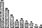
Figure 1. Number of new leprosy cases in Norway from 1857 to 1925 by five-year periods. ...frames of sanitation. Raising the general cultural development of the population, its social and living standards, and economic well-being is still the most important link in the chain of measures to combat infectious diseases in general. It is precisely this factor—the raising of cultural... 25C0 \ Total number of patients ----------------patients in districts ----------------hospitalized patients
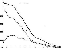
Figure 2. The movement of leprosy in Norway from 1856 to 1927. [The general] level of the population, and not isolation—many authors attribute decisive importance to [it] in the relatively rapid disappearance of leprosy in the countries of Western Europe. In a prominent place, one must put the widespread introduction among physicians and paramedical personnel of knowledge about leprosy—about its clinical picture, spread, epidemiology, bacteriology, and treatment. Children fall ill with leprosy more often than others. According to data from the English Leprosy Commission, which worked in 1890–1891 in India, out of 2,371 leprosy patients, 264 family cases were discovered, of which 130 fell to the share of children, i.e., half of all discovered cases. According to Lee's observations, children with a sick father fall ill in 10.27% of cases, with a sick mother—in 16.36%, with both sick parents—in 39.19%. Therefore, one of the most essential measures in the fight against leprosy should be considered the earliest possible removal of children from their parents. Such children are either given into the care of relatives or separate 'shelters' are created for them. In Russia, until the 1880s, government bodies viewed leprosy as a non-contagious disease. In the early 1880s, G. N. Minkh first spoke out insistently with the opinion about the 'contagiousness of leprosy and the necessary measures against it in Russia.' Soon he was supported by E. Wahl, K. K. Dehio, and O. V. Petersen. A convinced and decisive opponent of theirs was A. G. Polotebnov. The dispute between the two groups of the most prominent representatives of the clinical science of that time contributed much to the revitalization of the very question of leprosy, and it was from this very moment that a period of ever-increasing attention to patients and the disease began for us. Thanks to the efforts of Prof. Petersen and the Zemstvo physician P. N. Prokhorov, the first of the currently existing colonies for leprosy patients in the European part of the USSR was founded in 1895 (Krutye Ruch'i, Leningrad Oblast, near the town of Kingisepp). Then leprosaria began to arise: Astrakhan (1896), Nikolayevsky on the Amur (1896, transferred to Vladivostok in 1925), Tersk (1897), Kholm (1905), Karachay-Cherkess (1914), Irkutsk (1926), and two in Yakutia: Vilyuysk (1892) and Sredne-Kolymsk (1907). In the very recent time, one leprosarium each has been opened in Uzbekistan (Samarkand), Kazakhstan (Kzyl-Orda), and Turkmenistan (Ashkhabad). Leprosaria must be organized in such a way that patients go to them 'voluntarily and willingly' (Strasbourg Conference). For this, it is necessary to give them such an appearance that patients, upon entering them, feel the change in their situation the least acutely. In the USSR, the main mass of patients falls on agricultural elements. Therefore, the idea that our leprosaria should be primarily medical-labor colonies of an agricultural type does not meet with objections from anyone. Labor itself in the fresh open air, connected with muscular tension, correctly set up and dosed in proportion to the strength of the patients, is recognized by Soviet leprologists as one of the powerful factors in the treatment regimen. Besides field and garden work, special craft workshops with special instruction should be organized in leprosaria. Educational work (cinema, clubs, theaters, talks, library, etc.) should be widely established. Isolation at home comes down to a separate room, the absence of close and prolonged communication with those around, the use of one's own household inventory, especially linen and bedding, and medical control. Patients should absolutely be forbidden from occupations connected with supplying the population with food, clothing, and other consumer goods, as well as occupations requiring close communication with healthy people (barbers, laundresses, private and public service, etc.). Visiting public places and institutions (baths, places of entertainment, public gatherings, etc.) should also be prohibited. Legislation. In every country, there is its own legislation regarding leprosy, not everywhere, however, sufficiently developed. France still has no legislation on leprosy, and the 300 patients in Paris itself can freely reside in the city. England, with its solidified cultural and everyday forms, also relates very skeptically to any restrictive and coercive measures in relation to lepers, both in the metropolis and in the colonies. On the other hand, Estonia and Latvia require the mandatory removal of patients to a leprosarium, while Norway, Sweden, and Finland allow isolation at home. In Russia, the first step of government bodies in the leprosy matter can be considered the circular of the Medical Department of 18/IV 1895, which established the mandatory registration of leprosy patients by a card system. The next step was the circular of 2/IV 1902, which outlined in general terms those main lines along which measures to fight leprosy subsequently proceeded. After the October Revolution, an important stage in the development of regulations on protecting the population from leprosy is the issuance by the People's Commissariat of Health of the RSFSR of two circulars of 4/IV 1923 (Nos. 74 and 75), by which were given: 'Instruction on the fight against leprosy' and 'Regulation on leprosaria.' Then on 10/VII 1923 followed a decree of the Council of People's Commissars of the USSR, by which were entrusted: 1) to the People's Commissariat of Health—registration of all lepers in the Union; mandatory isolation of them in leprosaria or at home; provision in budgets of funds for the maintenance of leprosaria; 2) to the People's Commissariats of Agriculture—provision of leprosaria with land plots; 3) to the People's Commissariats of Social Security—provision for persons placed in leprosaria and their dependents; 4) to the People's Commissariat of Transport and the People's Commissariat of Health of the RSFSR—issuance of instructions on the preferential transport of leprosy patients by rail and water routes; 5) to the People's Commissariat of Foreign Affairs together with the People's Commissariat of Health and the People's Commissariat of Internal Affairs—conclusion of agreements with foreign states on prohibiting the entry of leprosy patients from abroad. At the present time, a draft of a new law on the fight against leprosy is being developed.
V. Kedrovsky. Lit.: Kulesha G. and Malinin I., Materials on the study of leprosy in the Kuban, Rostov-on-Don, 1928; Minkh G., Leprosy in the south of Russia, Vol. I–II, Kiev, 1884–94; Prospects for the fight against leprosy, collection edited by A. Vladimirov, V. Kedrovsky, I. Kovalev, and A. Miskinov, M.-L., 1929; Pospelov V., Index of works and articles on leprosy in the Russian language, Russkiy vestnik dermatologii, Vol. VIII, No. 2, 1930; Leprosy and the fight against it in the USSR, collection edited by V. Kedrovsky, I. Kovalev, and A. Miskinov, M.-L., 1927; Reshetillo D., Leprosy, SPB, 1904 (lit.); Aoki T. u. Aoki Y., Untersuchungen uber die Friihdiagnose u. Therapie der Lepra, Tokyo, 1930; Cook V., The epidemiology of leprosy in Australia, Melbourne, 1927; Deycke G., Die Lepra (Spez. Pathologie u. Therapie innerer Krankheiten, edited by F. Kraus u. Th. Brugsch, Vol. II, Part 1, B.-Wien, 1919, lit.; Russian ed.—F. Kraus u. Th. Brugsch, Infectious diseases, Vol. I, issue 2, P., 1916); Gron K., Lepra in Literatur und Kunst (Hndb. d. Haut- u. Geschlechtskrankheiten, edited by J. Jadassohn, Vol. X, Part 2, B., 1931); Jadassohn J., Lepra (Hndb. d. pathogenen Mikroorganismen, edited by W. Kolle, R. Kraus u. P. Uhlenhuth, Vol. V, Part 2, Jena-B.-Wien, 1928, lit.); Klingmuller V., Die Lepra (Hndb. d. Haut-u. Geschlechtskrankheiten, edited by J. Jadassohn, Vol. X, Part 2, B., 1931, lit.); Kobayashi W., Uber die viscerale Lepra, Kyoto, 1929; Kupffer A., Uber die Verbreitungswege der Lepra-Beobachtungen aus Estland, Arch. f. Schiffs-u. Tropen-Hygiene, Vol. XXXVI, Beihelt 3, Lpz., 1932; Lewandowsky F., Lepra (Hndb. d. inn. Med., edited by G. Bergmann u. R. Staehelin, Vol. I, Part 2, B., 1925, lit.); Marras A., La terapie deila lepra i resultati ottenuti coi moderni trattemento, Sassari, 1929; Rogers L. a. Muir E., Leprosy, L., 1925; Ruge R., Muhlens P. u. zur Verth M., Krankheiten und Hygiene der warmen Lander, Lpz., 1930; p. 381-396; The principles of the prophylaxis of leprosy, League of Nations, Health organisation, Geneve, 1931. Periodical publication.—International journal of leprosy, Manila, since 1933. 277
Related articles
Mentioned in
Cite this page
“Prosencephalon.” Soviet Medical Encyclopedia. English translation of Bolshaya Meditsinskaya Entsiklopediya, 1st ed. (Moscow, 1928–1936), ed. N. A. Semashko. https://sovietmedicalencyclopedia.pages.dev/article/prosencephalon/