Lichen
Historical document, translated for reference. It reflects medical knowledge of the 1920s–30s and is not medical advice.
Summary
Lichen is a term historically used for various skin diseases, with Hebra clarifying that true lichens are characterized by nodular eruptions maintaining their typical appearance throughout the disease course. Lichen ruber planus is the most typical form, characterized by itchy, small, shiny, polygonal papules that tend to merge, with various theories about its etiology including nervous, toxic, infectious, and hereditary factors.
Encyclopedia article (1928–1936)
LICHEN (from Greek leichen-lichen), a name inherited from Hippocrates and Galen and used in dermatological literature of the 18th and 19th centuries to denote the most diverse skin diseases, but mainly in the sense given to it by Willan, to denote any itchy nodular eruptions regardless of their etiological essence. To this period belong the names: L. tropicus, L. urticatus, L. pilaris, L. agrius, L. circumscripus, L. simplex, L. herpeticus, L. arthriticus and others. Only F. Hebra in the second half of the 19th century brought clarity to this one of the most confusing questions in dermatology, proposing to call lichens only those skin diseases in which nodular eruptions form that retain their typical appearance throughout the entire chronic course of the disease and undergo no changes in the eruptive elements of a higher order, i.e., vesicles or pustules. Only L. ruber and L. scrofulosorum meet this requirement, with the latter only partially, since with it, nodules often further transform into vesicles and pustules. All other lichens were classified as various other skin diseases, although it should be noted that to the present day many dermatoses that are not true lichens in Hebra's sense continue to be called lichens by tradition, e.g. L. pilaris, L. urticatus, L. syphiliticus and others. The only typical lichen is L. ruber, two forms of which—L. ruber planus and L. ruber acuminatus (see Pityriasis)—most consider as independent nosological units. Lichen ruber planus (red flat lichen, syn. L. planus, L. Wilson, lichen vrai) was first described in detail in 1869 by Wilson (E. Wilson); a relatively common chronic skin disease characterized in its typical form by eruptions of itchy, small, shiny, polygonal papules tending to merge. The question of the etiology of L. ruber planus has not yet been resolved; there exist nervous, toxic, infectious, hereditary theories and hypotheses of special skin predisposition closely related to the latter. The nervous theory, adhered to by Nikolsky and others, is based on the not infrequent cases of coincidence of L. ruber planus with organic or functional nervous diseases, and in some of these cases, with improvement of the nervous condition, L. ruber planus also recedes. The nervous theory is also supported by cases of L. ruber planus appearing strictly on one half of the body or along the course of nerve trunks, as well as favorable results from treating the area of spinal nerve roots with X-rays (see below—treatment). Du Bois sees in L. ruber planus a reaction of the skin to intoxication coming from the gastrointestinal tract. The school of Jadassohn until recently adhered to the infectious theory, while at the same time attributing a certain role to the 'soil'. In favor of this theory are cases of L. ruber planus in spouses or in two members of the same family living together; a relatively small number of such cases have been described in the literature; thus, up to 1924 Spitzer collected only 7 cases of marital L. ruber planus; in favor of the infectious theory is the frequently observed cyclic course of L. ruber planus, its occurrence after trauma, sometimes enlargement of lymph glands, etc. Darier believes that L. ruber planus is caused by some filterable virus similar to that of herpes zoster; according to him, this hypothesis could explain the not too rare, but pathogenetically still completely unclear cases of the occurrence of typical L. ruber planus after the administration of salvarsan preparations to the body (see separate table, figure 4). An invisible parasite is activated as a result of intoxication, trauma, infection, etc. due to humoral or even moral shock. This view as yet has no solid evidence. In recent years, hypotheses treating of special skin predisposition as the main cause of the occurrence of L. ruber planus1 have attracted the main attention. Buschke and Sklarz speak of lichenoid predisposition, Samberger—of hyperkeratotic. Both these hypotheses, differing little from each other, cannot be considered separately from the hereditary theory of L. ruber planus, since according to them this dermatosis is a definite clinical type developing under the influence of one or another harmful factor in individuals with corresponding, probably hereditary, skin predisposition. The skin of such individuals always responds with a homogeneous eruption of the L. ruber planus type to any (exogenous or endogenous) sufficiently strong irritation, an example of which can be the reaction of Pirquet in patients with L. ruber planus: typical eruptions of this dermatosis usually develop at the sites of scratches. According to Buschke and Sklarz, the irritation is transmitted to the skin through the autonomic nervous system. The hereditary theory is based on corresponding observations, and in this regard, particularly convincing are cases of L. ruber planus in blood relatives living separately, as well as the only case so far in the literature of this disease in 3 generations: in a grandmother, son and grandson (Joseph). Spitzer up to 1924 collected 39 cases of L. ruber planus in blood relatives. One must agree with Siemens that for the occurrence of L. ruber planus an idiotype predisposition is probably necessary, i.e. L. ruber planus should be classified among the so-called idiopathic diseases. In different countries L. ruber planus is distributed differently, constituting in general from 0.1% (Copenhagen) to 1.47% (London) of all skin diseases; in cities of the USSR L. ruber planus constitutes about 0.5% of all skin diseases. According to Sabouraud, L. ruber planus is histologically the most 'autonomous' skin disease. From the side of the epidermis are noted: hyperkeratosis, parakeratosis, slight acanthosis, phenomena of swelling and edema of intercellular spaces; dilated follicles are usually filled with horn. The border of the epidermis with the dermis is usually indistinct due to the dense infiltrate closely adjacent to the epidermis, individual cells of which penetrate it. In the upper part of the true skin around the dilated vessels there is a limited infiltrate consisting of lymphocytes, polynuclearg
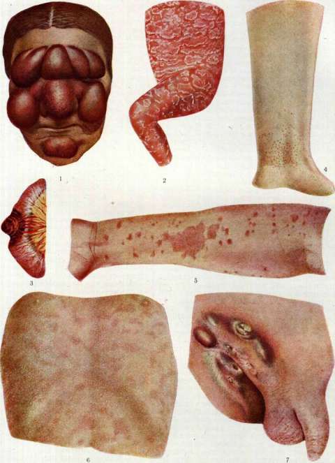
Figure 1. Leukemic nodes of the skin. Figure 2. Levers' disease. Figure 3. Richter's hernia,
Fig. 4. Lichen ruber planus (after the illustration of Neosalvarsan). Figure 5. Lichen ruber planus. Figure 6. Lichen trichophyticus. Figure 7. Lymphogranuloma inguinale. (Figures 4, 5 and 6 - according to models from the State Venereal Institute, fig. 1 - according to Riecke, fig. 7 - according to Frey-Hoffmann). To the article Lichen, Rehn's disease, Lymphogranuloma, fibroblasts and a small amount of mast cells; characterized by a sharp demarcation of the infiltrate from below. Course of the disease and symptomatology. The clinical picture is characterized by small, flat, shiny, skin-colored, pink and pink-red, polygonal, intensely itchy papular eruptions, having a clear tendency to merge and form dark red and violet, scaling, shagreen-like plaques (see separate table, fig. 5). The size of individual eruptions ranges from a pinhead to a millet grain; initial eruptions are often in the form of pink dots. The nodules gradually reach the size of a hemp seed and a lentil and often acquire a lilac hue; in their central parts, a navel-like depression appears. On developed nodules, the so-called network sign (Wickham's sign) is observed, consisting of opalescent white or grayish stripes and dots, forming a network on the surface of the papules, which becomes especially noticeable if the nodules are moistened with water or petroleum jelly, which makes the horny layer transparent. Fresh papules hardly scale, developing ones are usually covered with one or two thin silvery scales; strong scaling occurs in acute or severely irritated forms, as well as in L. ruber corneus. Both sexes apparently get sick equally often, although there are indications of a predominance of men; the predominant age of patients is from 20 to 55 years; the disease also occurs in older children, but in infants it is very rare. Data on the involvement of the oral mucosa among different authors are contradictory: from 17% and less to 75% of all cases; more often the cheeks, edges and back of the tongue and the base of the oral cavity are affected. Clinically, L. ruber planus of the mucosa manifests either as a diffuse white leukoplakic thickening of the mucosa, or as opalescent porcelain-like spots, or represents a delicate network of light white superficial lines, resembling that on skin eruptions. L. ruber planus of the oral mucosa does not cause any subjective sensations. On the skin, it is always accompanied by greater (L. r. pruriginosus) or lesser itching. Sometimes the nails are affected (clouding, pits, grooving, etc.). Complication with pyogenic infection in L. r. planus is almost never observed; in some cases, secondary erythroderma arises, spontaneously or as a result of irritating drug therapy (see Dermatitis, Dermatitis exfoliativa secundaria).- According to the course, acute, subacute and chronic forms of L. ruber planus are distinguished; according to spread - diffuse and localized. Acute eruptions more often have a diffuse character and are often accompanied by general phenomena; the skin suddenly diffusely reddens, swells and scales, with only typical elements of L. ruber planus visible in places. This form (L. ruber planus acutus, neuroticus or scarlatiniformis) either relatively quickly passes spontaneously or gradually transitions into a chronic form. Eruptions of L. ruber planus are often symmetrical, located mainly on the flexor surfaces of the forearms, especially in the wrist joint area, and on the legs, often on the lower back, genitals, lateral surfaces of the trunk, palms, soles and oral mucosa; rarely is the rash on the face and on the hairy part of the head. On the palms and soles, L. ruber planus can be in the form of callous thickenings surrounded by a purple border.- Varieties by location of the rash: papules of L. ruber planus often take the form of rings (L. r. planus annulatus, s. circinatus); in other cases, the eruptions group in the form of figures, semicircles, etc. (L. ruber planus marginatus, or serpiginosus), sometimes they are arranged more or less in regular stripes and lines (L. ruber planus linearis, striatus), in rare cases - along the course of a nerve (L. ruber planus zosteriformis). Wilson also distinguished L. r. planus aggregatus and L. r. planus discretus. Atypical forms of L. ruber planus. Sometimes the nodules of L. ruber planus deform, turning in their central parts into vesicles (L. ruber planus pemphigoides); in rare cases, L. ruber planus proceeds in the form of a persistent red rash without papules [mainly on the trunk and limbs - L. ruber planus erythematosus (Crocker)].- With reverse development, L. ruber planus usually leaves only slight pigmentation (more intense in cases treated with arsenic); in some cases the pigmentation is unusually strong, obscuring the main disease (L. ruber planus pigmentosus), in other cases the nodules after their disappearance leave skin atrophy in the form of white, pearly, irregularly outlined spots or in the form of true, sometimes even keloid-like scars; this form has many names: L. planus atrophicus of Hallopeau, L. albus of Zumbusch, L. planus sclerosus, L. morphoeicus (Crocker), L. keloidiformis (Pavlov). Patho-anatomically in this form, sclerosed areas of tissue are found between the epidermis and the infiltrate. When localized on the legs, less often on other sites, the eruptions of L. ruber planus are sometimes covered with abundant, dense, firmly adherent grayish-ash horny layers resembling asbestos: L. ruber planus hyperkeratoticus, or corneus; in other cases (also more often on the legs) the rash consists of flat warty, as if pricked with a pin, porous (with a cellular surface) pink-red elevations, also covered with a small amount of horny masses - L. r. planus verrucosus, or hypertrophicus; histologically in this form - thickening of the epidermis and papillomatosis. The name L. obtusus (from Latin obtuse - blunt) is given to itchy rashes consisting of hemispherical truncated papules. Three forms are distinguished: 1) L. planus obtusus (Unna), 2) L. ruber monileformis (Kaposi) and 3) L. obtusus corneus or vulgaris. The first two forms are only special varieties of lichen planus, while the latter should be classified with nodular prurigo. L. planus obtusus consists of relatively little itching, up to pea-sized, dry, 'flattened', non-scaling shiny papules, from bluish-red to brownish-red in color, often with a slight depression in the center; often a typical L. ruber planus nodule can be found nearby. Remaining as a rule limited, L. planus obtusus sometimes spreads to almost the entire skin. L. ruber monileformis (from Latin monile - necklace), coral lichen, a very rare form of lichen planus, consisting of large, up to cherry pit-sized, flattened papules, arranged like beads and alternating with small nodules and pigmentations. The differential diagnosis in ordinary cases presents no difficulties. Ring-shaped L. ruber planus when localized on the genitals can be mistaken for ring-shaped papular syphilid; the correct diagnosis in such cases is helped by the absence of a special infiltrate and weeping, the color of the nodules, itching, etc. From limited neurodermatitis, L. ruber planus differs in the smaller size of individual nodules, their polygonality, the absence of marked lichenification, the secondary nature of itching, etc. From psoriasis, L. ruber planus differs in a completely different nature of scaling, the absence of the phenomena of the stearin spot and pinpoint bleeding, itching, localization, etc. Usually the diagnosis is difficult in cases of acute L. ruber planus with secondary erythroderma, when individual typical nodules are absent; they become noticeable only after the acute phenomena have passed.- The prognosis is favorable, although one can never guarantee patients against relapses.- Treatment is currently only empirical-symptomatic in nature. Of internal remedies, the best is arsenic, which needs to be given for a long time and in relatively large doses (often - up to several thousand Asian pills); when treating with arsenic, one must always keep in mind the possibility of very intense pigmentation at the site of the eruptions. In acute cases, it is better to use remedies that calm the nervous system (bromine, etc.) instead of arsenic. The French school, in contrast to the German, uses arsenic much less in the treatment of L. ruber planus, replacing it mainly with hydrotherapy in the form of warm water showers and electrotherapy (static shower, d'Arsonval currents). Mercury treatment (internally or injections) as well as bismuth (injections of biochinol) often brings success. Among the new methods of treatment, it is necessary to note the indirect X-ray therapy on the spine area, proposed by French authors and used in various modifications. Hufschmitt and Pautrier use filtered oblique rays in order to act on the spinal roots. Gouin and Bienvenue use unfiltered rays in order to act on the sympathetic nodes. The results of this method are often good. The lumbar puncture recommended by Ravaut also sometimes brings success, mainly in relation to itching.
Local treatment consists in the application of antipruritic agents: 1/2-2% carbolic alcohol, Unna's mercuric-carbolic ointment (Hydr. biehlor. corrosivi 0.05-0.25, Ac. carbolici 2.0, Ung. Wilsoni 50.0), and others. For warty, hyperkeratotic, and generally chronic forms, salicylic, tar, and chrysarobin ointments are used. In stubborn cases with limited spread of the process, X-rays have a good effect. L. ruber planus of the mucous membranes is almost unresponsive to treatment. Lichen nitidus (Latin nitidus-shining) is a rare skin disease, first described in 1907 by Pinkus, characterized by the appearance of very small, the size of a pinhead, smooth, pearl-like shiny, flat, sharply delimited, slightly elevated nodules of round or polygonal shape, the color of normal skin, sometimes with a depression in the center. Despite their dense arrangement, the nodules never fuse, and there is no itching. L. nitidus is localized mainly on the skin of the penis, and less frequently on other areas. In the cases of Arndt, Kyrle, and Mc Donagh, L. nitidus was universal; only the face, palms, and soles were free from the rash. The mucous membranes are not affected. Men are more frequently affected. The course is prolonged. The nodules of L. nitidus do not scale and upon regression leave no trace. Patho-anatomically-a sharply delimited, tubercle-like granuloma in the papillary and subpapillary layers of the true skin; the infiltrate consists of lymphocytes, epithelioid cells, fibroblasts, and giant cells; in the center of the granuloma are dilated vessels, and in old elements-necrotic foci. Inoculation of affected tissue to guinea pigs is without result. Most researchers (Pinkus, Lewandowsky, Kyrle, etc.) see the cause of L. nitidus in tuberculous intoxication and consider it a tuberculid. The basis for this view, in addition to the frequent simultaneous presence of tuberculosis in patients, is the histological picture and usually positive tuberculin reactions. Darier objects to the tuberculous origin of L. nitidus; he sees it as 'an atypical variety of flat L. with a special histological structure.'-In differential diagnosis, it is necessary to take into account the miliary form of L. ruber planus (in L. nitidus there is no tendency to fusion and grouping, a different color of the rash, no itching, a completely different histological picture, etc.) and L. scrofulosorum.-The prognosis is favorable. Treatment in most known cases remained unsuccessful. Fox (H. Fox) achieved a cure with X-rays in one case. General strengthening measures are recommended. Lichen syphiliticus - syphilitic lichen (syn. Syphilis papulosa miliaris, syphilide papulo-granuleuse, lichenoid syphilide, micropapular syphilide), a skin rash occurring in the secondary period of syphilis, more often as a recurrent, much less often as a fresh primary rash. L. syphiliticus occurs approximately 10 times less frequently than the ordinary lenticular papular syphilide (Mulzer), and develops mainly in syphilitics who also have tuberculosis. Patho-anatomically-in the papillary layer there is an infiltration of lymphocytes, plasma cells, and sometimes giant cells, with the papillae surrounding the follicles playing the main role in the process; in the upper layers of the papillary layer around the capillaries and in the epidermis, the pale spirochete is found. L. syphiliticus develops either on completely unchanged skin or on spots of a previous roseola. Upon regression, the nodules of L. syphiliticus scale off, leaving no trace or leaving brown pigment spots; in rare cases, flattening over time, pinpoint, depressed scars remain. From the ordinary lenticular papular syphilide, L. syphiliticus differs in smaller size (nodules from poppy seed to pinhead) and convex (resembling shot) papules, significantly greater density of the rash, the connection of most elements with follicles, and some resistance to specific treatment; in addition, individual nodules often have a small pustule on top, which then forms a crust. The grouping of the rash is very characteristic for recurrent L. syphiliticus. Subjectively-sometimes slight itching. L. syphiliticus is localized more often on the trunk, less frequently on the limbs and face. In differential diagnosis, it is necessary to take into account L. scrofulosorum and L. ruber acuminatus (see Pityriasis rubra pilaris). The smaller infiltrate, paler, brownish, rather than brown-red color of the nodules, their frequent fusion and the absence of any signs of syphilis distinguish L. scrofulosorum from L. syphiliticus. In terms of prognosis, L. syphiliticus, according to a number of syphilologists, predicts a more severe course of syphilis in the future. Lichen trichophyticus, the most common variety of trichophytids (first noted in 1911 by Jadasson), a more or less widespread rash appearing on the hypersensitive (allergic) skin of patients with trichophytosis, mainly deep trichophytosis. For the development of L. trichophyticus, as well as other trichophytids, besides the presence of a trichophytosis focus, a state of special increased skin sensitivity to trichophytosis fungi and their products is necessary; the immediate cause of L. trichophyticus is usually irritation of the primary focus from which the fungi or their toxins are carried by the blood to the skin, causing a rash there and quickly dying due to the intense protective reaction of the allergic skin; therefore, fungi are very rarely found in the rashes of L. trichophyticus. The hematogenous origin of L. trichophyticus, as well as other trichophytids, is evidenced by the presence of fungi in the blood and lymph glands, the sudden onset and course of the process, the symmetry of the rashes, etc. At present, both hematogenous-microbial and hematogenous-toxic pathogenesis of trichophytids is accepted (for details, see Trichophytosis): Patho-anatomically in L. trichophyticus, hyperkeratosis, acanthosis, and in places complete destruction of the follicle walls and formation of cavities are found in the epidermis; in the true skin-around the vessels and sebaceous-hair follicles-an inflammatory infiltrate, sometimes with an admixture of plasma, epithelioid, and giant cells. Symptomatology and course: A few days or 1-2 weeks after irritation or injury to the primary focus, usually deep trichophytosis (kerion Celsi) (X-ray therapy, intradermal injections of trichophytin, its injection into the focus of lesion, incisions of the latter, pyrogallic therapy, etc., strongly irritating agents and methods), a more or less widespread symmetrical rash suddenly appears, consisting of small, the size from the tip to the head of a pin, conical or flat, shiny, somewhat dense nodules. The color of the latter varies between pink, brownish-red, and red. Most rashes are associated with follicles. In the very first days of their existence, the nodules become covered with scales; sometimes a blister or pustule forms on the top of one or another nodule, which is soon replaced by a crust [see separate table (pp. 247-248), fig. 6]. The rash of L. trichophyticus is often accompanied by transient general phenomena: malaise, fever, headache, etc. Sometimes there is intense itching. According to the spread, disseminated and limited L. trichophyticus are distinguished; in some cases, the latter is localized exclusively regionally, i.e., near the primary focus of trichophytosis. L. trichophyticus has no favorite localization, more often located on the trunk and limbs. Besides the typical form of L. trichophyticus, it often runs according to the type of L. spinulosus (Jadasson, Lewandowsky, etc.) and according to the type of L. ruber planus. Initially typical, L. trichophyticus sometimes after some time takes on an eczematous appearance. Often simultaneously with L. trichophyticus there is another morphological variety of trichophytid of scarlatiniform or exudative character.-The course of typical L. trichophyticus is usually rapid: 2-3 weeks; L. trichophyticus spinulosus lasts longer. In differential diagnosis, it is necessary to take into account the possibility of confusion with L. scrofulosorum and with L. syphiliticus. The latter differs in greater infiltration, copper-red color, characteristic grouping of the rash and the presence of other signs of syphilis. Chronic course, pale-yellowish or brownish color of the rashes and their frequent fusion allow to distinguish L. scrofulosorum. The main thing in diagnosis is the presence of the primary focus of trichophytosis, as well as positive skin reactions to trichophytin.-Prevention: careful observation of the entire skin cover of trichophytosis patients during treatment; at the first suspicion of the beginning of a trichophytid, it is necessary to stop the irritating therapy of the primary focus.-Treatment: indifferent pastes, mixtures for shaking and powders. Similarly to L. trichophyticus in trichophytosis, in microsporia, both deep and superficial, L. microsporicus occurs. The conditions of development, pathogenesis, symptomatology, path. anatomy and treatment are the same as in Lichen trichophyticus. L. simplex chronicus-see Vidal's lichen. L. haemorrhagicus-see Purpura. L. pyodermicus-see Pyoderma.
Lit.: Bobovich A., Case of lichen trichophytieus, Venereol. i dermat., 1924, № 2; Brauder., Etiology and pathogenesis of trichophytids, ibid., 1926, № 3 and 4; Brauder R. and Per M., Lichen nitidus (Pin-&us), ibid., 1926, № 1; Gorbuliev S., Red flat lichen according to data from the G. V. I. polyclinic, ibid., 1929, № 1; Darrie J., Foundations of dermatology, M.-L., 1930; Iordan A., 80 cases of red flat lichen, Russ. vestn. dermatologii, 1927, № 2; Fournier A., Doctrine of syphilis, vol. 2-Secondary period, M., 1909; Arndt G., Contributions to the knowledge of Lichen nitidus, Dermatol. Ztschr., B. XVI, 1909; Bloch W., Zur Pathogenese d. Trichophytide, Arch. f. Derm. u. Syphil., B. GXXIX, 1921; Bueler P., Uber Lichen obtusus, ibid., B. CXXXVI, 1921; G alewsky E., Contributions to the Aetiology of Lichen ruber, familiarer Lichen ruber, Lichen ruber under spouses, Lichen ruber and irritation, ibid., B. CXXIX, 1921; Guth A., Uber Lichenoide (kleinpapulose, spinulo-se), Trychophytie, ibid., B. CXVIII, 1914; Hallopeau H., Du lichen plan et particulierement de sa forme atrophique, Union med., v. XLIII, 1887; Kaposi M., Lichen ruber, monileformis-korallen-schnurartiger Lichen ruber, Vierteljahresschr. f. Dermat., B. XIII, 1886; Mietscher G., Die Trichophytien (Handb. d. Haut- u. Geschlechtskrankh., hrsg. v. J. Jadassohn, B. XI, B., 1928, lit.); Pinkus F., Uber eine neue Kn6tchenformige Hauteruption-Lichen nitidus, Arch. f. Derm. u. Siphilis, B. LXXXV, 1907; Riecke E., Lichen ruber (Handbuch d. Hautkrankheiten, herausgegeben v. F. Pick, B. II, Wien, 1905, Literatur); Spitzer R., Uber familiaren Lichen ruber planus, Arch. f. Derm. u. Syph., B. CXLVI, 1923-24.
Related articles
Mentioned in
Cite this page
“Lichen.” Soviet Medical Encyclopedia. English translation of Bolshaya Meditsinskaya Entsiklopediya, 1st ed. (Moscow, 1928–1936), ed. N. A. Semashko. https://sovietmedicalencyclopedia.pages.dev/article/lichen/