Thalamus Opticus
Historical document, translated for reference. It reflects medical knowledge of the 1920s–30s and is not medical advice.
Summary
This article from the 1928–1936 Soviet medical encyclopedia describes the anatomy, phylogenetic development, and structural organization of the thalamus opticus. It details the various nuclei of the thalamus and their connections to other brain structures, including the visual pathways and the cerebral cortex.
Encyclopedia article (1928–1936)
THALAMUS OPTICUS, the optic thalamus, is the most voluminous and structurally complex of the basal ganglia (see); it represents a collection of gray matter, permeated by fibers and separated from the same formation on the other side by a ventricle. The thalamus opticus develops from the diencephalic part of the prosencephalon. Its phylogenetically oldest medio-ventral section, the palaeothalamus, directly connected with the extrapyramidal system, is, as it were, a remnant of that higher receptor apparatus which was characteristic of animals not yet possessing a cortex. In all lower vertebrates, the palaeothalamus is represented as a central node, closely connected with the brainstem parts and the striatum. In mammals, morphologically differentiating more and more, it enters into connection with the neoencephalic parts of the brain, assuming at this stage the structural forms characteristic of the neothalamus. Its further development therefore proceeds to some extent in parallel with the development of the cerebral cortex, with which it is connected both centripetally and centrifugally. Comparative anatomical study of intrathalamic nuclei in the series of animals appears to be still far from complete. Its anterior nucleus is distinguished by great stability in the series of mammals, which is explained by its phylogenetically very old connections with the olfactory system. The external nucleus develops in parallel with the course of development of the medial lemniscus and cerebellar pathways to the thalamus, as well as partly also thalamostriatal and striatothalamic connections. Figure 1. Structure of the thalamus opticus: 1-nucleus ant. thal.; 2-g. lam. med. ant.; 3-zona reticularis; 4-lam. medul. int.; 5-lam. medul. ext.;
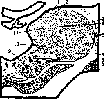
6-lam. medul. med.; 7-fibr. rubro-conjunctivo-thalamica;
Regarding the structure of the internal nucleus, well developed in all mammals, it is still little studied phylogenetically. The pulvinar is related to the evolution of the visual pathway system. At lower stages of phylogenesis, this nucleus is relatively poorly developed, and the lateral geniculate body is still a constituent part of the thalamus opticus—the metathalamus. In lower vertebrates, the role of the terminal station of the visual pathways is played by the tectum of the superior colliculus, called the tectum opticum. In mammals, in contrast to the diencephalic and mesencephalic parts, the significance of the occipital cortex for the visual act is already clearly outlined. As one ascends the ladder of development, the lateral geniculate body becomes more and more separated from the thalamus opticus and in humans clearly appears as an independent formation, although connected with the pulvinar, which reaches its maximum development here. The dorsal and medial surfaces of the thalamus opticus, opening into the third ventricle, are almost completely free; in the ventral and lateral directions, it is closely connected with the adjacent parts (Fig. 1). Its dorsal, slightly convex surface is covered with ependyma and a layer of fibers—the stratum zonale—of cortical origin, descending through various parts of the internal capsule and running above the thalamus in the sagittal direction. Between the dorsal and medial surfaces lies a white lamina (stria medullaris, s. taenia thalami), which serves as a boundary between them. A significant part of the fibers of this lamina begins at the base of the brain, in an area closely connected with the olfactory system. Posteriorly, it expands into a triangular formation (see Habenula). The upper surface of the thalamus is separated from the caudate nucleus by a groove, along which runs a white horn or boundary strip (stria cornea, s. terminalis), called boundary because it separates the brainstem from the hemispheres. The medial surface of the thalamus is located almost vertically. With its anterior two-thirds, it forms the outer wall of the third ventricle. On the medial side, both optic thalami are connected to each other by a powerful gray commissure (commissura media, s. mollis, s. massa intermedia) lying almost in the middle, forming the mesothalamus. The lateral, or outer surface of the thalamus opticus corresponds to the internal capsule, especially its posterior limb. With its ventral, or lower part, the thalamus opticus is in contact with the subthalamic region—regio subthalamica (see), s. hypothalamus. Structurally, the thalamus opticus is not a homogeneous formation, but represents a collection of a whole series of nuclei, although not always clearly delimited from each other (Fig. 2). Upon histological examination, three medullary laminae are found in the optic thalamus. The external one (lamina medullaris externa), running along its outer edge, separates the external nucleus from the reticular layer (zona reticularis thalami), which is directly adjacent to the internal capsule. Inward from it runs the internal lamina (lamina medullaris interna), separating the internal nucleus from the external one; in the anterior-upper section, it bifurcates, forming the lamina medullaris anterior, enclosing the nucleus of the thalamus of the same name, located in its anterior pole, known as the tuberculum anterius. Thus, with the help of these layers, the thalamus opticus is divided into three main nuclei: nucl. anterior, nucl. externus, s. lateralis, and nucl. internus, s. medialis. Adjacent to the latter two without sharp boundaries is the nucl. posterior, s. pulvinar thalami. The anterior nucleus (nucl. anterior) is the most sharply contoured of the formations included in the thalamus opticus. According to Edinger, this nucleus is of paleoencephalic nature. Cytoarchitectonically, it is constructed uniformly—from multipolar cells, thereby resembling the anterior part of the adjacent external nucleus. The external, or lateral nucleus (nucl. externus, s. lateralis) is phylogenetically a very old section of the optic thalamus; it is well developed already in fish and reptiles. According to Edinger, it is not homogeneous and consists of a group of smaller nuclei, occupying the upper and lateral part of the thalamus and running along almost its entire length. Cytoarchitectonically, like the anterior nucleus, it is constructed quite uniformly—from multipolar cells of different sizes with well-developed dendrites. In the ventral part of the external nucleus, there is a special inclusion, more compact in its structure than the main mass, the semilunar nucleus of Flechsig—nucl. semilunaris, s. arcuatus, or the cup-shaped nucleus (Chizh). The ventral section of the external nucleus, rich in lemniscal fibers, is often designated as a special ventral or ventrolateral nucleus (nucl. ventralis or nucl. ventro-lateralis). The internal, or medial nucleus (nucl. internus, s. medialis) is much more complex in its structure than the external one. Its cells are polymorphic—sometimes polygonal, sometimes quadrangular, with weakly developed dendrites. Nissl granulation in them appears poorly differentiated. In its lower-outer part, at the junction with the external nucleus, a special conglomerate of cells appears in the form of the so-called centre médian de Luys, differing in its structure from the main nucleus by cells of a stellate or triangular shape. This nucleus, rich in fibers and surrounded by its capsule, is related to...
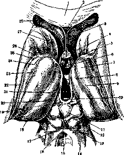
Figure 2. Diencephalon. Thalamus opticus, its relation to the third ventricle and surrounding formations: 1-genu corporis callosi; 2-cavum septi pellucidi; 3-septum pellucidum; 4-columnae fornicis; 5-recessus triangularia; 6-commissura alba ant.; 7-tuberculum ant. thalami; 8-massa intermedia; 9-thalamus opticus; 10-commissura alba post.; 11-glandula pineale; 12-corp. quadrigem. sup.; 13-corp. quadrig. inf.; 14-brachium conjunctivum; 15-lingula cerebelli; 16-frenulum veli medul. ant.; 17-n. trochlearis; 18-pulvinar; 19-habenula; 20-trigonum habenulae; 21-stria medullaris; 22-cauda nucl. caudati; 23-taenia chorioidea; 24-lamina affixa; 25-stria terminalis; 26-v. terminalis; 27-caput nucl. caudati; 28-cornu ant.
to the loops of the lemniscus. The posterior nucleus of the thalamus (nucl. posterior, s. pulvinar thalami) in humans is very voluminous and is in direct contact with the internal and external nuclei. In its internal part, it is homogeneous, whereas in its external part, its cellular groups are arranged in the form of columns separated by fibers of the optic radiation. In the median part, connecting both thalami (mesothalamus, s. commissura intermedia, s. mollis), there is a significant accumulation of gray matter, known by the name nucl. intracommissuralis, the cells of which pass partly into the main mass of the Thalamus Opticus. This central gray mass, together with other cellular accumulations located near the III ventricle and connected with the hypothalamic region, is recognized by its structure and connections as the central apparatus of the sympathicus (Edinger), the sympathetic nuclei of the thalami optici. The Thalamus Opticus is a connecting link between the phylogenetically lower brainstem parts and the cerebral cortex. Its extensive connections with the latter form the corona radiata thalami above it. The greater part of it consists of thalamofugal (fibrae thalamo-corticales), and a smaller part of thalamopetal fibers (fibrae cortico-thalamicae). Individual rays of the corona, consisting of bundles of fibers that are more closely connected at the edges of the thalamus than as they approach the cortex, have long been called the peduncles of the Thalamus Opticus. One distinguishes (purely schematically, of course) the anterior, superior, inferior, and posterior peduncles. The anterior connects the anterior and partially the internal and external nuclei of the thalamus with the frontal lobe. The superior, or middle, peduncle—the widest—connects the internal and external nuclei with the posterior parts of the frontal and parietal lobes. The posterior part consists mainly of fibers of the optic radiation of Gratiolet and connects the thalamus with the occipital lobe and partly with the parietal. The inferior peduncle represents a bundle of fibers connecting the thalamus with the temporal lobe and the insula. Thus, the Thalamus Opticus is the main subcortical basin into which impulses of the most diverse origin flow and from which, in turn, they go to the cerebral cortex, to the subcortical nodes, and to the brainstem parts. Most caudally, into its ventral section, enter the proprioceptive systems—tract. bulbo-thalamicus (fibers from the nucleus of the Goll's tract) and, somewhat more laterally, fibers from the nucleus of the Burdach's tract. More orally from them terminate the fibers of the anterior cerebellar peduncle with the rubro-thalamic tracts (radiatio rubro-conjunctivo-thalamica). As for the exteroceptive system, the second neurons of the tract. spino-thalamici enter the most lateral part of the ventral section of the thalamus. The fibers of the trigeminal nerve, according to Ramón y Cajal, enter the semilunar nucleus. In the centre médian de Luys terminate the afferent pathways of the nn. vagi and glosso-pharyngei, and in the central gray matter of the mesothalami—the remaining fibers of visceral sensitivity. Into the pulvinar thalami enter a part of the fibers of the tract. optici, and from it go through the Wernicke's zone to the fissura calcarina the fibers of the tract. thalamo-occipitalis, which are part of the optic radiation of Gratiolet (see Visual pathways, centers). From the above-mentioned ventral section of the thalamus begins the third sensory neuron (tract. thalamo-corticalis), which goes through the internal capsule to the psychomotor region and terminates near the 3rd and 4th layers of the cortex of the posterior central and superior parietal gyrus, and also partly the anterior central. Along with these corticopetal pathways, the Thalamus Opticus is connected with the cortex also corticofugally through the mediation of the cortico-thalamic system (tract. cortico-thalamicus). The Thalamus Opticus is supplied with these latter connections not only from the central gyri but also from other sections of the cortex. Minkowski showed the presence in the thalamus of fibers from almost all gyri of the convex surface of the brain. Especially significant are the connections connecting the cortex of the frontal lobes with the thalami in the form of the tract. fronto-thalamicus. The corticopetal part of this system arises mainly in the anterior nucleus and in the marginal zone of the external nucleus, and terminates in the 3rd layer of the cortex; its corticofugal part goes from the 5th layer of the cortex to the anterior section of the internal nucleus, and also to the ventral nucleus. The main connections of the Thalamus Opticus with the extrapyramidal system are carried out primarily through the mediation of the lenticular loop—fibers connecting it with the striatum in a bidirectional direction (fasc. thalamicus Forel and fibrae strio-thalamicae) (see Extrapyramidal system). Further, it should be noted the transition of a part of the fibers of the medial lemniscus into the corp. mamillare, which carries out, through the bulbo-thalamo-mamillary pathways, the connection of the sacral segments with the hypothalamic region. From the middle nucleus of the corp. mamillaris, from its dorsal section, departs the Vicq d'Azyr's bundle (fasc. mamillaris princeps), which soon divides into two branches, of which one (tract. mamillo-thalamicus) goes upward, crossing the hypothalamus, and terminates in the anterior nucleus of the Thalamus Opticus. This pathway also contains fibers of the reverse direction (tract. thalamo-mamillaris). A whole series of connections of the Thalamus Opticus with the brainstem parts is known. From its posterior-internal section originate mainly the descending pathways. A part of them terminates in the red nucleus—the aforementioned fasc. thalamicus Forel, which also contains ascending fibers. Another part of them consists of pathways to the superior colliculus, then the fibrae thalamo-reticulares, which go, according to Bekhterev, to the nucleus of the reticular formation of the pons, as well as the pathway traced by Flechsig and Bekhterev to the inferior olive (tract. thalamo-rubro-olivaris). The majority of descending thalamic pathways are interrupted in the indicated mesencephalic stations. The most important of them goes from the red nucleus to the spinal cord—the tract. rubro-spinalis (Monakow) or the fasc. praepyramidalis Thomas corresponding to it in its greater part—a group of fibers located in front of the pyramidal tract. However, Meynert and Kölliker described thalamofugal pathways, tracing them to the spinal cord. Wallenberg, after destruction of the dorso-medial section of the thalamus in cats, noted degeneration in the anterior-lateral columns. Bekhterev also pointed to the presence of direct thalamo-spinal pathways (tract. thalamo-spinalis). The Thalamus Opticus is thus an intermediate switching station to neo-encephalic neurons, but at the same time, it is also a place for processing the impulses flowing into it and for closing reflex arcs that are phylogenetically older and shorter than the cortical ones. As for thalamic excitations, they arise from three sources. Firstly, this includes all proprioceptive and exteroceptive impulses related to general and special sensitivity. The second part of thalamic excitations consists of impulses either arising in the diencephalon itself or brought here through the blood and liquor in the form of chemical and hormonal factors. The third category of its excitations consists of impulses going in a descending direction from the cerebral cortex. Until recently, the cerebral cortex was considered the sole source of the origin of sensations; however, through the works of a whole series of authors, and especially Head, it was proven that a prominent role in the mechanism of sensations also belongs to the thalami. This is proven by the fact that in cases of destruction of the sensory centers of the cortex, a complete loss of sensitivity does not occur if the named subcortical sensory centers are preserved. On the question of the significance of the cortico-thalamic pathways connecting these two central apparatuses of sensitivity, there is not yet unanimity. While Head and Holmes, as well as Förster, consider them inhibitory pathways regulating the excitability of subcortical centers, Wallenberg, on the contrary, considers them as sensitizing these centers, making them more receptive to stimuli. According to Minkowski, they serve to activate receptor mechanisms. Already purely anatomical data force one to acknowledge the existence of a closely closed circle and interaction between the paleo-encephalic and neo-encephalic apparatuses of sensitivity. And indeed, the role of the Thalamus Opticus in the realization of sensory functions is not limited to the simple transmission of received impulses to the cortical apparatus. It primarily takes an active part in the processing of the received excitations. The product of this thalamic activity, according to Head, is the affective tone of sensations. At the same time, one of the main tasks of the Thalamus Opticus is the setting of the mechanisms of sensitivity in a strictly defined direction—either for the correct evaluation of received stimuli in the cortical analyzers and their auxiliary apparatuses, or for their utilization by transmission to the motor elements of the extrapyramidal system. In doing so, it regulates not only the direction of the processes but also their intensity, maintaining the flow of reactions at a certain level. In this respect, it functions, apparently, in close contact with the system of the striatum. As is known, the Thalamus Opticus not only sends its impulses to the latter but also receives them from it—this is evidenced by the early-established thalamopetal pathways. Here we see the same closed circle of influences and the same interaction that we encounter when considering the relationships between the cortex and the Thalamus Opticus.
Being at the center of all nervous and humoral influences and possessing the ability to sensitize received excitations, the Thalamus Opticus plays a most prominent role in emotional-affective life. A whole series of mimico-somatic reactions and expressive movements is built on the connection of the Thalamus Opticus with the extrapyramidal system and the hypothalamus (see Subcortical functions). It has long been established by Nothnagel, Bechterew, and others that with an isolated lesion of the Thalamus Opticus on the side opposite to the focus, "psycho-reflexes" are destroyed, and "mimic paralysis" occurs—this side of the face does not participate in laughing, crying, and other expressive movements, although it retains the ability for voluntary movements. In contrast to this, with the loss of voluntary innervation along the pyramidal tracts, especially if bilateral, increased affectivity is noted (Head-Holmes symptom). The clinical pathology of the Thalamus Opticus has been developed, although not yet fully, nevertheless quite widely, and at the present time one can speak not of just one thalamic syndrome, but of a whole series of them. The classic thalamic syndrome, or more accurately the syndrome of the posterior-inferior part of the external nucleus of the Thalamus Opticus, is outlined quite clearly by Dejerine and his students (Roussy, Egger, Thomas). Clinically, it is characterized by: 1) a decrease in superficial and especially deep sensitivity on the opposite side of the body; 2) hemialgia with a severe character of pain, almost unresponsive to therapy; 3) hemiataxia; 4) weakly expressed hemiplegia, or rather hemiparesis, usually without contractures; the Babinski symptom is for the most part absent; 5) choreoathetosis. The most characteristic feature of the syndrome in its pure form is hyperpathia—a specific pain sensation with diffuse localization in the entire opposite half of the body. Also typical for this syndrome is the diffuseness of pain during peripheral irritation, a long sensation of it after the cessation of irritation, and the inaccuracy of the localization of the latter. The symptom of increased affective reactions during relatively insignificant physical irritations, noted earlier by Head and Holmes, also takes place here. Of the various types of sensitivity, deep sensitivity suffers the most, especially in the upper limb. The loss of proprioceptors to a significant degree causes the peculiar position of the hand in this syndrome ("thalamic" hand). Often there is complete astereognosis. Homonymous hemianopsia is also observed. Of the five symptoms mentioned above, Roussy considers only the first three to be characteristic of the Thalamus Opticus, attributing the latter to the lesion of neighboring parts. On this basis, Roussy and Cornil distinguished from the classic thalamic syndrome the so-called thalamo-striatal type, encountered in cases where the lesion goes beyond the limits of the Thalamus Opticus and captures the posterior part of the lenticular nucleus or, as Thomas thinks, the connections of the latter with the thalamus, and clinically manifests itself by the presence of choreoathetosis. The main vessel nourishing the Thalamus Opticus is the posterior cerebral artery with its branches. Hillemand, in connection with this, attempted to distinguish several thalamic syndromes. The form described by Dejerine-Roussy depends, in his opinion, on the softening of that region of the Thalamus Opticus which is supplied by a branch of the named artery—the ramus thalamo-geniculatus. Another syndrome, according to Hillemand, is encountered with the occlusion of the ramus thalamo-perforati. Clinically, it corresponds to the so-called rubro-thalamic syndrome described by Chiari, Foix, and Nicolesco. Here, motor cerebellar symptoms come to the fore, whereas sensory disturbances are weakly expressed. In 1926, Foix, Chavany, and Bascourret distinguished yet another variety from the classic syndrome of the Thalamus Opticus—the vegetative syndrome. The authors see at its base the presence of two kinds of phenomena—thalamic as such and vegetative. The latter consist of hemihyperhidrosis on the affected side, especially on the face, in ocular symptoms with the character of irritation of the sympathicus on the same side, and in an increase of the pilomotor reflex, predominantly on the face and neck on the side of the hemiparesis. The indicated phenomena in combination with a slight hemiparesis and hemialgia constitute this variety of the thalamic syndrome. The authors link the described form with the localization of the process in the internal nucleus of the Thalamus Opticus, especially in its periventricular elements. The indicated variety of the thalamic syndrome has also been noted in Russian literature.
Related articles
Mentioned in
- Basal Ganglia
- Brain (551 General diagnostics of brain diseases)
- Brain (cerebrum, a comprehensive term for the entire)
- Brown-Séquard Tract
- Capsula Externa
- Commissural Fibers
- Corpora Geniculata
- Faure Jean-Louis
- Fennel (medicinal or Florentine, fennel, Foeniculum)
- Fever (a complex of phenomena in the organism,)
- Habenula
- Hemialgia
- Hemiataxia
- Hypothalamus
- Infundibulum
- Internal Capsule
- Konstantin von Monakow
- Projection Fibers
- Prosencephalon
- Substantia Nigra
Cite this page
“Thalamus Opticus.” Soviet Medical Encyclopedia. English translation of Bolshaya Meditsinskaya Entsiklopediya, 1st ed. (Moscow, 1928–1936), ed. N. A. Semashko. https://sovietmedicalencyclopedia.pages.dev/article/thalamus-opticus/