Commissural Fibers
Historical document, translated for reference. It reflects medical knowledge of the 1920s–30s and is not medical advice.
Summary
Commissural fibers are nerve fibers in the central nervous system that connect the two halves of a given structure. They are found in the brain, spinal cord, and cerebellum, forming connections between corresponding areas of the cerebral hemispheres.
Encyclopedia article (1928–1936)
COMMISSURAL FIBERS (adhesive fibers), are located in the central nervous system and serve to connect both halves of a given structure. Commissural fibers are present in the brain, spinal cord, and cerebellum. In the brain, they form a double connection between the two hemispheres; they connect not only symmetrical areas of the hemispheres, but also gyri belonging to different lobes. The commissural fibers include: corpus callosum (see), commissura alba anterior, commissura alba posterior in the brain and spinal cord, psalterium, or commissura fornicis, commissura grisea, Ganser's commissure, Gudden's commissure, Meynert's commissure, commissura habenularum, commissura media. Commissura alba ant. (anterior cerebral commissure) is a bundle of myelinated fibers arranged in the shape of a horseshoe with the concavity directed forward (Fig. 1 and 2). The middle part of this bundle enters into the composition of the anterior wall of the third ventricle together with the anterior pillars of the fornix; the posterior surface of commissura albae ant. is covered with ependyma; on cross-section it has the shape of an oval measuring 5x4 mm. On the sides, the anterior commissure divides into two parts - pars olfactoria and pars temporalis (hemisphaerica). Pars olfactoria is poorly developed in humans and consists of two thin bundles, which, separating from the middle part, proceed to the substantia perforata anterior, and then to the trigonum olfactorium and to the bulbus olfactorius; this part includes fibers connecting both olfactory lobes and forming a kind of chiasma olfactorium, as well as fibers connecting the olfactory lobe with the temporal. Pars temporalis, on the contrary, consists of two thick bundles, going
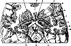
Figure 1. Fronto-vertical section of the brain: 1-commissura alba ant.; 2-genu corp. callosi; 3-chiasma opt.; 4-caput nuclei caudati; 5-capsula int.; 6-nucl. lenticul.; 7-claustrum; 8-insula Reili; 9-tr. opt.; 10-corpus mammillare; 11-peduncul. cerebri; 12-aquaeduct. Sylvii; 13-fossa interpeduncularis.
under the corpus striatum; externally from nucl. amygdal. they reach the temporal lobe; the beginning and end of this part have not yet been finally clarified; it is assumed that it ends not only in gyrus hippocampi, but also in gyri lingualis and fusiformis. The function of commissura albae anter. consists in connecting the primary and cortical olfactory centers of both hemispheres. Commissura alba post, (posterior cerebral commissure) is located in the diencephalon near the epiphysis, at the site of the bend of its lower plate (see Fig.); into it enters the anterior end of the fissure separating the epiphysis from the quadrigemina, and divides it into upper and lower parts. Under commiss. alba post. III ventricle passes into aquaeductus Sylvii. The question of the origin and termination of the fibers of the posterior commissure remains controversial.
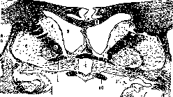
Figure 2. Frontal section of the cerebral hemisphere: 1 - commissura alba ant.; 2-corpus callosum; 3-lateral ventricle; 4-nucl. caudat.; 5-capsula int.; 6-nucl. lentic; 7-claustrum; 8-capsula ext.; 9-capsula extrema; 10-n. opticus.
the nucleus of Darkshevich, corpus quadrigeminum ant., nucleus interstitialis or thalamus opticus (see Darkshevich's nucleus). By its function, commissura alba post, is classified by many authors as belonging to the pathways of light reflexes. Commissura media, s. mollis, s. massa intermedia connects both thalamus opticus (see). Psalterium, or commissura fornicis connects the posterior pillars of the fornix (see Fornix). Commissura Ganser (Ganser) is also located in the diencephalon, is also known under the name of the anterior hypothalamic commissure (commissura hypothalamica anterior) or commissura Foreli; it consists of thin fibers going from the area lying in front of the red nucleus to the nucleus lenticularis of the opposite side. Posterior hypothalamic commissure, s. commissura hypothalamica poster., or Forel's commissure, consists mainly of fibers connecting both corpora Luysi; to a small extent, fibers of the fornix enter into it. Commissura Gudden (Gudden) is included in the composition of tractus optici (see Gudden's commissure). Commissura Meynert (Meynert) passes over the chiasma and over the tractus opticus, but has no relation to the visual fibers; its significance is not fully clarified; it is assumed that it connects 1) corpus Luysi with the nucleus lenticularis of the opposite side, 2) lemniscus medialis with the opposite nucleus lenticularis, and finally it has been suggested that it has a relation to the ganglion opticum basale. Commissura habenulorum-: see Habenula. Commissural fibers in the cerebellum connect the cortex of different lobules of one hemisphere with the other. In the spinal cord, commissural fibers are also present - commissura alba anterior et posterior, commissura grisea.
Related articles
Mentioned in
Cite this page
“Commissural Fibers.” Soviet Medical Encyclopedia. English translation of Bolshaya Meditsinskaya Entsiklopediya, 1st ed. (Moscow, 1928–1936), ed. N. A. Semashko. https://sovietmedicalencyclopedia.pages.dev/article/commissural-fibers/