Common Carotid Artery
Historical document, translated for reference. It reflects medical knowledge of the 1920s–30s and is not medical advice.
Summary
This article describes the surgical anatomy and procedures for ligating and suturing the common carotid artery, including approaches, complications, and treatment of aneurysms. It covers both historical techniques and clinical outcomes from the 1930s Soviet medical perspective.
Encyclopedia article (1928–1936)
COMMON CAROTID ARTERY at the level of the same cartilage; then the anterior edge of the sternocleidomastoid muscle lies in the middle of the length of the incision. With a knife, 1) the skin, 2) subcutaneous tissue, 3) the superficial muscle (platysma) are incised, under which the branches of the external jugular vein are ligated and tied; the anterior edge of the sternocleidomastoid muscle and the flat, thin omohyoid muscle extending obliquely downward and backward from the hyoid bone are exposed. With a grooved probe, the outer sheath is incised (Figure 25. Ligation of aa. carotis communis, internae et externae). Along the artery runs the ramus descendens n. hypoglossi; behind it externally is the v. jugularis int.; between it and a. carotis communis is the n. vagus; in the lower angle of the wound laterally lies the m. sternocleidomastoideus; medially is the m. omohyoideus. (After Bier-Braun-Kummell.)
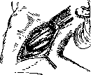
bJ"
sternocleidomastoid muscle, the muscle is retracted laterally with a blunt hook, then the deep layer of the same sheath and the common sheath of the vascular-nerve bundle are incised. Here, with an anatomical forceps, the proper sheath of the artery is ruptured, and from without to within between the v. jugularis int. and a. carotis communis, very carefully handling the vein (which tears easily) and avoiding catching the n. vagus (behind the vessels) and the ramus descendens n. hypoglossi (in front of the artery), a Deschamp needle with silk is passed and the artery is ligated 2-3 cm below its bifurcation. Damage to the sympathetic trunk of the neck, located behind the vascular bundle and separated from it by a layer of deep aponeurosis, can be easily avoided. The wound is sutured in layers. - Ligation of a. carotis communis below the omohyoid muscle in the sterno-mastoideo-tracheal fossa (according to Pirogov) is somewhat more complex than the described method, since in addition to the already known layers, it is necessary to expose the thyroid gland and the muscles that lower the larynx. All layers are incised just as in the previous method, with the only difference that here the sternocleidomastoid muscle overlaps the artery more, and if it is significantly tense, it sometimes has to be cut. After incising the deep layer of the sheath of the sternocleidomastoid muscle, the thyroid gland with the muscles covering it (m. sternohyoideus and m. sternothyreoideus) is retracted medially. Ligation of the vessel with the same precautions. Layered suture of the wound. When locating a. carotis communis by operative means, it is necessary to keep in mind all the anomalies of its origin and branching described above. - Ligation of a. carotis communis between the branches (in spatio inter-glosso-pharyngeo according to Pirogov). According to Zang-Sedillot, the position of the patient is the same as in the above methods. The incision (4-6 cm) is made upward and begins at the joint to reach the artery between the branches of the sternocleidomastoid muscle. The reason for exposing and ligating or suturing a. carotis communis are wounds to the artery by stab or gunshot, less frequently - transverse cuts in suicide attempts, malignant tumors, destruction of the arterial wall by suppuration and aneurysms, passing a thread or temporary ligature during operations on the neck, face or head. When ligating a. carotis communis, in addition to the danger of insufficient blood supply to the brain (softening), there is the threat of damage to the intima of the artery with subsequent thrombosis (Perthes). Therefore, the artery should not be tied too tightly; as for temporary ligature, besides not tying it too tightly, it is very good to use material in the form of a strip rather than a thread; even better is to compress it simply with a finger or with special vessel clamps by Hopfner; rubber tubes are placed on the latter's jaws. - Suture of a. carotis communis (see Vascular suture). The number of cases of suture on a. carotis communis is small. Moskalenko cites the statistics of Goodman, where out of 24 ligations of a. carotis communis there were 5 deaths and in 9 cases of suture - 0 deaths. The figures speak in favor of suture, but they are small and therefore not very conclusive. Aneurysms of a. carotis communis and their treatment. Aneurysms of this artery are more common than aneurysms of the external or internal carotid artery: 45% of all aneurysms fall on the common carotid artery (Crisp, according to Dyakonov and Lysenkov). Most often, the upper end of the artery is affected, the area of the bulbus arteriosus. Symptomatology and clinical picture - see Aneurysm, traumatic aneurysms. Arteriovenous aneurysms of a. carotis communis are formed after puncture, incised and gunshot wounds. They are rare. Surgical diseases and injuries of a. carotis ext. and int. and their treatment. The external and internal carotid arteries can be wounded from the outside (stabs, gunshot wounds) as well as from the side of the pharynx. Gunshot wounds of a. carotis ext. occur in 2½% (approximately) of all vascular wounds (Punin). The clinical picture and prognosis are described above. The best method of treatment of injuries to a. carotis ext. is ligation (with a mortality of only 1¼% according to Lipps, according to Tikho). Mortality is directly dependent on thrombosis that has spread to the internal carotid artery: to avoid thrombosis, it is necessary (according to Wieting) to ligate the artery above the origin of a. thyr. sup. Due to the relative safety of ligation of this artery, it was proposed as a therapeutic procedure in tumors of the head and face; it is assumed that the reduced blood flow will cause atrophic changes in the tumor. Ligation is also performed as a preliminary step in resection of the upper jaw. Aneurysms of a. carotis ext. This variety is not often encountered in practice. Clinically, the aneurysm is characterized by the presence of a pulsating tumor in unison with the heartbeats under the angle of the lower jaw, symptoms of compression of the hypoglossal and lingual nerves; sometimes tonsillar protrusion is observed. The prognosis is very favorable due to the great ease of access to the vessel. Treatment consists of ligation of the afferent trunk of a. carotis ext. or a. carotis communis. According to Walter's data, ligation of a. carotis communis (below the inter) was applied in 17 cases with three fatal outcomes; ligation of a. carotis externa (above the inter) - 2 times with one death (according to Le Fort); finger compression was successfully applied - 1 case (R. Delbet), enucleation of the sac - 1 case (Philagrius) and incision of the aneurysm - 1 case (according to Tikho). The frequency of injuries to a. carotis int. is expressed as 1.2% of all vascular wounds. The prognosis for injury to this artery is more serious than for injury to a. carotis ext., since ligation of the first leads to softening of the brain, paralysis and even death in 25%; therefore, where possible, vascular suture should be used (Moskalenko). Aneurysms of a. carotis int. are also not often encountered; here we are talking about the extracranial part of the vessel, since the intracranial part often serves as the site of development
c
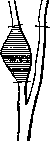
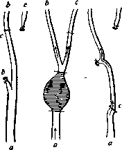
Figure 26. Replacement of a. carotis int. in its aneurysm with the trunk of a. carotis ext. according to Rehn's method (according to Schjering); a-a. carotis commun.; b-a. carotis int.; c-a. carotis ext.
Figure 27. Operation on aneurysm of a. carotis communis according to Hoffmann's method: a - a. carotis communis; b-a. carotis int.; c-a. carotis ext.
The exposure of a. carotis int. et ext. is indicated in injuries, aneurysms, hemorrhages, in cases of erosion by a purulent process and other causes, in tumors of the areas supplied by these arteries, and in resection of the upper jaw. The position of the patient is the same as in ligation of a. carotis communis. The incision from the angle of the lower jaw downward for 5 cm along the anterior edge of the sternocleidomastoid muscle. With a knife, the skin and subcutaneous tissue are incised; along
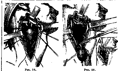
Figure 28. Resection of the bifurcation of the common carotid artery for an aneurysm of it. The facial vein and digastric muscle have been divided between two ligatures. Ligatures have been applied to the internal and external carotid arteries directly from the sac. Above, Moure's clamps have been applied. Below the aneurysm, after opening its own sheath, a provisional suture has been placed on the common carotid artery. (After Petit-Dutaillis.) Figure 29. Resection of an aneurysm of the bifurcation of the common carotid artery with subsequent end-to-end anastomosis between the external and internal carotid arteries. Left drawing - anastomosis completed; removal of the aneurysmal sac from top to bottom; one must remember to spare the vagus nerve and sympathetic nerve (the latter is in the depth of the wound). At the bottom right of the drawing, the sac has already been removed, suture of the digastric muscle. (After Petit-Dutaillis.) To a grooved probe - the subcutaneous muscle, under which the external jugular vein is encountered; if necessary, the latter is divided between two ligatures; further, the cervical aponeurosis is cut, under it the vascular sheath. In the lower corner of the wound passes the common facial vein - it is deflected downward; in the upper corner of the wound - the posterior belly of the digastric muscle and the hypoglossal nerve. (When applying a ligature under the site where the artery crosses the digastric muscle, one must remember the superior laryngeal nerve passing behind the artery.) Both vessels are opened; they must be differentiated: the external carotid artery lies in front of and medial to the internal carotid artery; tracing the vessels upward, on one of them we will encounter branches
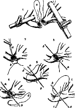
Figure 30. End-to-end anastomosis of the external and internal carotid arteries. Different moments of the anastomosis: 1 and 2 (a) - situational suture on the upper semicircle of the arteries; 3 - application of a situational suture (c) on the posterior semicircle of the arteries after rotating them on their axis; a - upper situational suture (after rotation it turned out to be below); b - lower situational suture, now upper; 4 - application of a situational suture (d) on the anterior semicircle of the arteries; 5 - application of a continuous suture between situational ones on the anterior semicircle; 6 - application of a continuous suture on the posterior semicircle. (After Petit-Dutaillis.)
this will be the external carotid artery. Another way to determine which artery is anterior is to press it and see if the pulsation in the superficial temporal artery has ceased; if there is no pulsation in it, then again the external carotid artery has been pressed. - Resection of the internal carotid artery according to Renu (reported by Moskalenko). Indications: aneurysms. The artery is exposed as described above. The aneurysm is excised according to Filagrius. The central end of the internal carotid artery is ligated tightly, the peripheral end remains in a clamp; the external carotid artery is divided, at the level of the upper segment of the internal carotid artery a ligature is placed on the peripheral end and a clamp on the central end of the external carotid artery. The latter is then brought to the peripheral end of the internal carotid artery and sutured end-to-end with a circular vascular suture. As a result of this operation, blood is directed from the common carotid artery through a segment of the external carotid artery into the internal carotid artery (fig. 26). The wound is closed tightly. Hoffmann proposed after removing the sac to divide the external carotid artery and bend it downward, suture it with a circular suture with the central segment of the common carotid artery. The external carotid artery will now serve as a link between the common carotid artery and the internal carotid artery (fig. 27). In case of an aneurysm located at the bifurcation of the common carotid artery, Quemi proposed connecting the peripheral ends of the external and internal carotid arteries to each other with an end-to-end vascular suture. After applying ligatures to the external and internal carotid arteries at the sac, the arteries are divided above the site of ligation and clamped, after which they proceed to suture them together (fig. 28, 29 and 30).
A. Siritkin. CARPA ON OK PACK A (Carpano), modification of Gram's staining for tissues. Sections are first stained with eosin (Eosin B. A. aquosa 1%) for 1 min., quickly washed in 95% alcohol, stained for 5 min. with a heated solution of crystal violet (Krystallviolett 0.1; Alcohol 95°-2.0, carbolic water 2%-50.0), placed for 1 min. in Lugol's solution (0.1 I + 0.2 KJ + 40.0 Aq. dest.), differentiated under the microscope in 95% alcohol. Mounted in Canadian balsam (see also Gram's method).
Related articles
Mentioned in
Cite this page
“Common Carotid Artery.” Soviet Medical Encyclopedia. English translation of Bolshaya Meditsinskaya Entsiklopediya, 1st ed. (Moscow, 1928–1936), ed. N. A. Semashko. https://sovietmedicalencyclopedia.pages.dev/article/common-carotid-artery/