Carotid Artery
Historical document, translated for reference. It reflects medical knowledge of the 1920s–30s and is not medical advice.
Summary
The article describes the anatomy and embryology of the carotid artery, including its origin, course, branches, and relationship with surrounding structures in the neck. It details the differences between the right and left common carotid arteries and their topographical relationships.
Encyclopedia article (1928–1936)
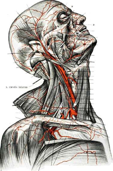
CAROTID ARTERY. Contents: Anatomy and embryology............382 Pathological anatomy............419 Clinical ...................410 Anatomy. The common carotid artery (a. carotis communis) (figs. 1 and 2) with its branches—the external and internal—supplies blood to the head end of the body and is the largest artery of the neck (see color plate); its caliber is not less than 8 mm; according to the scale of Henle it belongs to vessels of the 1st group. Such development of the carotid artery in man corresponds to the great development of the brain, which requires exceptional blood supply. In structure of the wall it belongs to the elastic type of arteries (Testut), similar to the aorta, pulmonary artery, innominate artery, etc., due to the predominance of elastic elements in its middle muscular layer (see Artery). There are two common carotid arteries: right and left. The right arises from the innominate artery at the level of the right sternoclavicular joint; the left—directly from the aortic arch, immediately behind the innominate, and its beginning lies in the thoracic cavity (fig. 1). Immediately above the sternoclavicular joints the common carotid arteries, ascending upward, deviate outward and backward, following a line going from the named joint to the mastoid fossa (line of anatomical direction of the carotid artery). Here the artery lies in the so-called regio sterno-cleido-mastoidea. At the level of the upper edge of the thyroid cartilage a. carot. com. divides into two branches: the external and internal carotid arteries (fig. 1). Throughout its course it is almost straight and gives no branches, except for vasa vasorum, presenting throughout its length a uniform, simple, smooth tube, which however in the middle shows a clearly defined narrowing of the lumen—isthmus. The caliber of a. carot. com. may vary in that a bulb-like dilatation (bulbus; fig. 2) is often found either at its beginning or at the site of its division, and then it is uniformly distributed to both of its main branches. The right and left common carotid arteries, differently in origin, also have different lengths, initial direction, and relationship with surrounding organs and tissues. The left a. carot. com. is longer than the right by the height of the innominate artery, i.e., by 20-25 mm; it initially has an oblique direction, and only after emerging on the neck, it runs identically to the course of the right one; beginning in the thoracic cavity, it consequently possesses its own, inherent topographical relationships (fig. 1). In front and somewhat medially from its origin lies the beginning of the innominate artery, behind and laterally—the left subclavian artery, directly in front of its trunk crosses the left innominate vein, and between them (the artery and innominate vein) descend downward the superior cardiac branches of the n. vagi. Medially from the left a. carotis com. at some distance goes the trachea, and 172 cm medially and somewhat backward is located the esophagus. Laterally from the artery and somewhat in front goes the n. phrenicus, and behind—the n. vagus. The artery itself with its outer wall is adjacent to the mediastinal pleura, and behind it touches the prevertebral layers, being initially separated from them by the subclavian artery and the thoracic duct. The right a. carot. com. (see color plate) lies behind the right sternoclavicular joint in the intermuscular space of the sternoclavicular-mastoid muscle and on the trachea. On the neck both a. carot. com. are in similar relationships with neighboring organs, run along the trachea and esophagus, surrounded by loose connective tissue. This perivascular connective tissue space in cross-section has the form of a triangle (fig. 4). Its anterolateral wall includes the sternoclavicular-mastoid muscle and the transversely crossing the vascular-nervous bundle the omohyoid muscle; the medial wall—is formed by the trachea and larynx in front and the esophagus with pharynx behind. In the thickness of this wall is contained the recurrent nerve lying in the groove between the esophagus and trachea. Finally the posterior wall of the triangle—bony-muscular—is formed by the transverse processes of the cervical vertebrae and the muscles lying on them. From the transverse
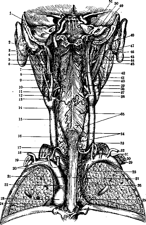
Fig. 2.
To the article Carotis arteria. middle of the artery a clearly defined narrowing of the lumen is observed—isthmus. The caliber of a. carot. com. may vary in that a bulb-like dilatation (bulbus; fig. 2) is often found either at its beginning or at the site of its division, and then it is uniformly distributed to both of its main branches. The right and left common carotid arteries, differently in origin, also have different lengths, initial direction, and relationship with surrounding organs and tissues. The left a. carot. com. is longer than the right by the height of the innominate artery, i.e., by 20-25 mm; it initially has an oblique direction, and only after emerging on the neck, it runs identically to the course of the right one; beginning in the thoracic cavity, it consequently possesses its own, inherent topographical relationships (fig. 1). In front and somewhat medially from its origin lies the beginning of the innominate artery, behind and laterally—the left subclavian artery, directly in front of its trunk crosses the left innominate vein, and between them (the artery and innominate vein) descend downward the superior cardiac branches of the n. vagi. Medially from the left a. carotis com. at some distance goes the trachea, and 172 cm medially and somewhat backward is located the esophagus. Laterally from the artery and somewhat in front goes the n. phrenicus, and behind—the n. vagus. The artery itself with its outer wall is adjacent to the mediastinal pleura, and behind it touches the prevertebral layers, being initially separated from them by the subclavian artery and the thoracic duct. The right a. carot. com. (see color plate) lies behind the right sternoclavicular joint in the intermuscular space of the sternoclavicular-mastoid muscle and on the trachea. On the neck both a. carot. com. are in similar relationships with neighboring organs, run along the trachea and esophagus, surrounded by loose connective tissue. This perivascular connective tissue space in cross-section has the form of a triangle (fig. 4). Its anterolateral wall includes the sternoclavicular-mastoid muscle and the transversely crossing the vascular-nervous bundle the omohyoid muscle; the medial wall—is formed by the trachea and larynx in front and the esophagus with pharynx behind. In the thickness of this wall is contained the recurrent nerve lying in the groove between the esophagus and trachea. Finally the posterior wall of the triangle—bony-muscular—is formed by the transverse processes of the cervical vertebrae and the muscles lying on them. From the transverse
6 7 8 9
Fig. 3. Arteries of the neck: 1-arteria vertebralis; 2-a. cervic. ascendens; 3-a. cervic. superlic; 4-a. transversa colli; 5-a. transversa scapulae; 6-a. intercost. suprema; 7-a. mammaria interne; 8-a. subclavia; 9-a. carotis communis; 10-a. cervicalis prof.; 11-a. thyreoid. inf.; 12-a. laryngea super.; 13-a. thyreoid. super.; 14-a. lingualis; 15-a. submentalis; 16-a. maxillaris ext.; 17-a. occipitalis. (After Merkel). direction, and only after emerging on the neck, it runs identically to the course of the right one; beginning in the thoracic cavity, it consequently possesses its own, inherent topographical relationships (fig. 1). In front and somewhat medially from its origin lies the beginning of the innominate artery, behind and laterally—the left subclavian artery, directly in front of its trunk crosses the left innominate vein, and between them (the artery and innominate vein) descend downward the superior cardiac branches of the n. vagi. Medially from the left a. carotis com. at some distance goes the trachea, and 172 cm medially and somewhat backward is located the esophagus. Laterally from the artery and somewhat in front goes the n. phrenicus, and behind—the n. vagus. The artery itself with its outer wall is adjacent to the mediastinal pleura, and behind it touches the prevertebral layers, being initially separated from them by the subclavian artery and the thoracic duct. The right a. carot. com. (see color plate) lies behind the right sternoclavicular joint in the intermuscular space of the sternoclavicular-mastoid muscle and on the trachea. On the neck both a. carot. com. are in similar relationships with neighboring organs, run along the trachea and esophagus, surrounded by loose connective tissue. This perivascular connective tissue space in cross-section has the form of a triangle (fig. 4). Its anterolateral wall includes the sternoclavicular-mastoid muscle and the transversely crossing the vascular-nervous bundle the omohyoid muscle; the medial wall—is formed by the trachea and larynx in front and the esophagus with pharynx behind. In the thickness of this wall is contained the recurrent nerve lying in the groove between the esophagus and trachea. Finally the posterior wall of the triangle—bony-muscular—is formed by the transverse processes of the cervical vertebrae and the muscles lying on them. From the transverse
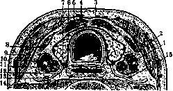
Fig. 4. Horizontal schematic section of the neck at the level of the VII cervical vertebra: 1-a. carotis com.; 2-v. jugularis int.; 3- aponeurosis superflc. cervic. (first); 4-m. sterno-hyoideus; 5-m. sterno-thyreoideus; 6- gl. thyreoidea; 7-n. recurrens sin.; 8-m. omohyoideus; 9-m. st.-cl.-mastoideus; 10-ganglion lymphatic, tractus jugularis; 11-n. vagus; 12- a. thyreoidea inf.; 13 and 15-n. sympathicus cervic; 14-deep aponeurosis of the neck. (From Testut).
Among the processes of the cervical vertebrae, the process of the sixth vertebra—Chassaignac's tuberculum, the carotid tuberculum (tuberculum caroticum)—acquires special importance in the surgery of the common carotid artery. To it the common carotid artery is pressed when it is necessary to stop the circulation in half of the neck and head. Its level corresponds to the lower edge of the cricoid cartilage. In the prevertebral aponeurosis, behind and slightly lateral to the artery (Testut), passes the cervical trunk of the sympathetic nerve (fig. 4-6). Such a deviation of the nerve to the lateral side from the course of the artery is not constant at different levels of the neck; below, the nerve runs medial to the artery, remaining behind it. One finger's breadth below Chassaignac's tuberculum, the thyroid artery crosses the common carotid artery from behind. The relationships of the common carotid artery and the sternocleidomastoid muscle, the companion of the artery on its entire course, change along the neck (fig. 3). The direction of the artery approaches the vertical, while the muscle runs more obliquely backward and upward and crosses the artery at an acute angle. Being immediately above the sternoclavicular joint between the two limbs of the said muscle and ascending upward, the artery emerges from under the anterior edge of this muscle, not being covered by it at its bifurcation.
The second muscle crossing the common carotid artery on its course—the omohyoid; it crosses the artery in its middle third and divides it into two parts: portio infraomohyoidea et portio supraomohyoidea (fig. 7). The first part (portion dangereuse L. Testut)—the 'dangerous' one due to the close proximity to the large vessels of the base of the neck; the second part—the place where ligation of the common carotid artery is usually performed. The common carotid artery, like other arteries of the neck, does not stretch as much as the median structures of the neck, which follow the bending spine; therefore, it describes a flatter arc than the latter, shifting backward at the middle of the height of the neck. When bending the head forward, the common carotid artery forms an arc convex laterally. When bending the head to the side, the vascular bundle shifts to the side toward which the head is bent, and on the inclined side, it shifts backward and outward. With rotation of the head, the common carotid artery on the side from which the head turns moves forward and becomes wider and more spacious; favorable conditions are created for a more voluminous flow of blood, while on the other side the artery shifts backward until it completely disappears behind
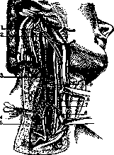
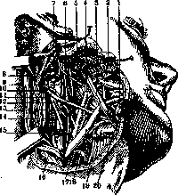
Figure 5.
Figure 6. Figure 5. Cervical sympathetic nerve. 1-a. occipitalis; 2-n. hypoglossus; 3-n. sympathicus; 4-a. thyreoidea int.; 5-window in the deep cervical aponeurosis; 6-v. jugularis int.; 7-n. vagus; 8-a. carotis communis; 9-oesophagus. (From Testut-Jacob.) Figure 6. Ganglion cervical, sup. n. sympathici. 1 and 2-cut m. digastricus; 2-gl. parotis turned upward and forward; 3-m. stylo-hyoideus; 4-a. carotis ext.; 5-gl. glosso-pharyngeus; 6-a. carotis int.; 7-cut m. digastricus; 8-outer branch of n. accessorii Willisi; 9-upper sympathetic ganglion; 10-ganglion n. vagi; 11-a. occipitalis; 12-n. hypoglossus; 13-n. laryngeus sup.; 14-n. sympathicus; 15-v. jugularis int.; 16-m. sterno-cleido-mast.; 17-n. hypoglossus, ramus descendens; 18-v. facialis com.; 19-os hyoideum. (After Testut.)
the common carotid artery. At this level, in front, the artery is covered by the lateral lobes of the thyroid gland (fig. 1). In the vascular bundle, the common carotid artery occupies the most median position; laterally to it lies the internal jugular vein, and behind them in a groove passes the n. vagus; rarely (and more on the right) the nerve is located in front of the vessels; along the anterior surface of the artery descends the descending branch of the hypoglossal nerve (ramus descendens n. hypoglossi) (fig. 7). The artery, vein, and nerve are covered by a common sheath—the vascular sheath; at the same time, all these elements of the vascular-nerve bundle are isolated from each other by a secondary partition, an individual fascicular sheath. Here, closely adjacent to the internal jugular vein, stretches a chain of lymph glands, the so-called tractus jugularis; as everywhere, these glands lie mainly around the vein; but they are not pressed directly against it, but also lie in front of the artery, behind it, and beside it (fig. 4 and 8). Changes in the position of the artery with movements of the neck. The stated relationships of the common carotid artery with the surrounding organs change considerably with movements of the neck. When throwing the head back, the soft tissues of the neck shorten behind, gathering in transverse folds; the anterior ones are stretched. The lateral bundle of vessels and nerves, due to its position, does not have the
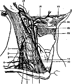
Figure 7. Mutual arrangement of nerves and vessels of the neck (schematically): 1 and 5-n. vagus; 2-n. accessorius Willisi; 3-ganglion sympathic. sup.; 4-a. carotis int.; 6-n. sympathicus; 7-a. carotis communis; 8-middle ganglion of n. sympathici; 9-lower ganglion of the same nerve; 10-v. jugularis int; 11-m. omohyoideus; 12-ramus descendens n. hypoglossi; 13-n. laryngeus sup.; 14-n. hypoglossus; 15-n. glosso-pharyngeus; 16-a. max. interna. (From Testut-Jacob.)
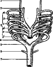
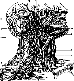
Figure 8. Lymphatic glands of the neck: 1-lymphog. parotideae; 2-lgl. auriculares anteriores; 3-lgl. auric, post.; 4-lgl. occipital.; 5-lgl. cervicales profund. superior.; 6-lgl. supraclaviculares; 7-lgl. omohyoidea; 8-lgl. cervical. prof, int.; 9-lgl. submentales; 10-lgl. submaxillares. (From Bier-Braun-Kummell.)
necessary tension; on the side toward which the head is turned, the vascular bundle lies for a great length uncovered by the sternocleidomastoid muscle, on the second side it is completely hidden under the same muscle (Delitsin). On cross-sections of corpses frozen with the head turned in such a way, it is easy to verify the complete compression of the common carotid artery and jugular vein; in a living person, of course, this phenomenon is not expressed as sharply as in a corpse (Melnikov). On the first side, the vascular
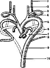
Figure 9. Figure 9. Aortic arches of humans: 1-a. carot. int.; 2-a. carot. ext.; 3, b, c, d, e, f-aortic arches; 4-a. pulm. sin.; 5-a. dors.; 6-a. subclav.; 7-aorta communis. Figure 10. 1-a. carot. int.; 2-a. carot. ext.; 3-arch of a. carotis; 4-a. carot. com.; 5-arch of aorta; 6-arch of a. pulmonal.; 7-a. pulm. dextr.; 8-a. subclavia; 9-aorta dorsalis; 10-aorta thoracica. (Schemes of Kollmann according to Aschoff.) Embryology (figs. 9-12). In the human embryo, rudiments of six pairs of primary arterial arches can be found, which however soon undergo retrograde development and therefore cannot be detected in the adult. In the region of the respiratory tract, when both common carotid arteries depart from the aorta by a single trunk. Krause reports a case where the right a. carot. com. went obliquely from left to right between the esophagus and the spine, originating from the arch of the aorta to the left of the latter. Sometimes a. carot. com., occupying its normal position, is extremely tortuous. The length of the common carotid artery is also not a constant value; it is in direct dependence on the level of the arch of the aorta, which varies extremely: from the lower edge of DIV to the lower edge of DV (Kupriyanov). Correspondingly to this, there is a whole gradation of the extension of the common carotid artery: from shorter with a high position of the aortic arch to
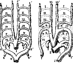
Figure 11.
Figure 12.
Fig. 11. Aortic arches (primary position): I-VI-aortic arches; 1-arterial trunk; 2, 2'-aortae ascendentes (right, left); 3, 3'-aortae descendent. (right, left); 4, 4'-a. car. Interna (right, left); 5, 5'-a. car. externa (right, left); 6, 6'-a. subclav. (right, left); 7, 7'-a. pulm. (right, left); 8-trunk resulting from the fusion of two descending aortas. Figure 12. Aortic arches (final position): I-VI-aortic arches; 1-aorta; 2-a. pulm. before division; 3, 3'-a. pulm. (right, left); 4-ductus arteriosus (Botalli); 5-arch of aorta; 6-a. anonyma; 7, 7'-a. subclav. (right, left); 8, 8'-a. carotis com. (right, left); 9, 9'-a. car. int. (right, left); 10, 10'-a. carot. ext. (right, left); 11-aorta descendens. (According to Testut.) longest with a low position of the arch; in this case elongation is observed more often than shortening; thus, Klein (according to Tikhomirov) in 295 corpses found elongation in 10, and shortening in 5. With age, arteria carotis communis elongates and becomes somewhat tortuous. The division of a. carot. com. into external and internal carotid arteries can be either higher than usual - at the level of the hyoid bone (and even higher) or lower-at the height of the middle of the larynx, the cricoid cartilage (and even lower); in the case of Morgagni, the common carotid artery had a length of only 41 mm and divided into its terminal branches in the lower part of the neck.-In the region of branching of a. carot. com. There are known cases of the independent origin of a. carotis ext. et int. either from the innominate artery or from the aorta. It happens that a. carot. com. does not divide, but goes as a single trunk, thus entering the canalis caroticus, giving off along the way branches that usually originate from the external carotid artery. Testut describes a case of the internal carotid artery originating from the external carotid artery above the origin of the latter's external maxillary artery. In exceptional cases, from the common carotid artery arise aa. thyreoid. sup., pharyng. ascend., vertebralis, thyreoid. inf. and laryng. sup. (Testut). The external carotid artery (figure 13), separating from the common carotid artery at the level of the upper edge of the thyroid cartilage, reaches the neck of the articular process of the lower jaw and in turn divides into two terminal branches: the internal maxillary and the superficial temporal. The external carotid artery supplies blood to the upper-anterior part of the neck, the face, and the walls of the skull.-The direction of a. carot. ext. Initially it lies in front of and inside a. carot. interna (fig. 15); from here it goes upward, deviating slightly outward; at the angle of the lower jaw it passes into a vertical direction, covered by the posterior belly of m. digastricus and m. stylo-hyoideus. Above a. carot. ext. it penetrates into the parotid salivary gland. The caliber of a. carot. externae is usually equal to the caliber of a. carot. internae; in childhood the latter is wider than the former. M. digastricus divides a. carot. ext. into 2 segments: the lower-from the thyroid cartilage to the crossing with the muscle and the upper-from the said crossing to its final branching. The first (lower) part of a. carot. ext., when examined in cross-section at the level of CIV, is seen lying in a triangular space (trigonum caroticum), the posterior wall of which is formed by the transverse processes of the cervical vertebrae, prevertebral muscles and m. scalenus ant.; the inner wall is the pharynx (its middle constrictor); on the front-lateral side a. carot. ext., listing layers from depth to surface of the neck, is covered by cellular tissue and lymph glands, the posterior belly of m. digastricus, the anterior edge of the sternocleidomastoid muscle with the first cervical aponeurosis covering it, the superficial muscle (platysma), subcutaneous tissue (with superficial veins and nerves) and skin. The distinguishing feature of a. carotis externae from a. carotis internae, lying closely adjacent to each other, is as follows: the first gives branches, while the second does not give branches on the neck. Laterally from a. carot. ext. go the internal jugular vein (vena jugularis int.) and the facial vein (v. facialis communis) flowing into it; these veins merge with each other at an acute angle, open upward; the third side of the resulting triangle, called the triangle of Farabeuf, is the n. hypoglossus, passing obliquely, from behind above, downward forward, between a. carot. ext. and the internal jugular vein (fig. 16). The second (upper) part of a. carot. ext. lies deeper; above the crossing with the posterior belly of m. digastricus it passes in the bifurcation of the styloid process muscles: between m. stylo-hyoideus laterally from the artery and m. stylo-glossus and m. stylo-pharyngeus on the medial side of the artery (fig. 17 and 18). Being in close proximity to the wall of the pharynx, a. carot. ext. passes near the lateral surface of the tonsils, between them and the angle of the lower jaw. Figure 13 and 14. Arteries of the neck. FIG. 13. 1-aa. palpebrales laterales; 2-glandula lacrimalis superior; 3 and 4-a. temporalis profunda anterior et posterior; 5-m. temporalis; 6-fatty mass above the cut and separated superficial lamina of fascia temporalis; 7-a. masseterica; 8-m. pterygoideus externus; 9-a. meningea media; 10-a. temporalis media; 11-a. temporalis superficialis; 12-a. maxillaris interna; 13-a. alveolaris inferior; 14-lig. spheno-mandibulare; 15-n. alveolaris inferior; 16-a. stylo-mastoidea; 17-ramus mylo-hyoideus; 18-a. auricularis posterior; 19-m. digastricus (posterior belly); 20-a. lingualis; 21-a. buccinatoria; 22-a. occipitalis; 23-a. carotis externa; 24-a. maxillaris externa; 25-a. sterno-cleido-mastoidea; 26-a. lingualis; 27-membrana hyo-thyreoidea; 28-a. thyreoidea superior; 29-ramus posterior; 30-a. carotis interna; 31-ramus anterior; 32-a. carotis communis; 33-m. thyreo-hyoideus; 34-lig. crico-thyreoideum (medium); 35-ramus crico-thyreoideus; 36-a. laryngea superior; 37-m. hyo-glossus; 38-m. thyreo-hyoideus; 39-ramus hyoideus a. lingualis; 40-a. submentalis; 41-m. mylo-hyoideus; 42-a. maxillaris externa; 43-a. mentalis; 44-a. labialis inferior; 45-a. labialis superior; 46-m. pterygoideus internus; 47-a. alveolaris superior posterior; 48-a. infraorbitalis; 49-a. alveolaris superior anterior; 50-a. dorsalis nasi; 51-a. frontalis; 52-a. supraorbitalis. Figure 14. 1-a. ethmoidalis anterior; 2-a. ethmoidalis posterior; 3-aa. nasales posteriores septi (from a. spheno-palatina); 4-a. canalis pterygoidei (arteria Vidiana); 5-fascia pharyngo-basilaris; 6-tuba auditiva (pars cartilaginea); 7-a. carotis interna in canalis caroticus; 8-ramus carotico-tympanicus; 9-paries labyrinthicus of the tympanic cavity; 10-m. levator veli palatini; 11-m. constrictor pharyngis superior; 12-m. pharyngo-palatinus; 13-a. pharyngea ascendens; 14-ramus tonsillaris; 15-tonsilla palatina; 16-a. palatina ascendens; 17-a. maxillaris externa; 18-m. stylo-glossus; 19-a. lingualis; 20-a. carotis externa; 21-a. carotis communis; 22-ramus hyoideus a. lingualis; 23-m. genio-hyoideus; 24-a. sublingualis; 25-a. profunda linguae; 26-m. genio-glossus; 27-m. glosso-palatinus; 28-glandula lingualis anterior; 29-rami dorsales linguae; 30-a. naso-palatina; 31-septum nasi; 32-aa. nasales anteriores septi (from aa. ethmoidales); 33-a. meningea anterior. (According to Toldt.) (To the illustrations of the article Carotis arteria.)
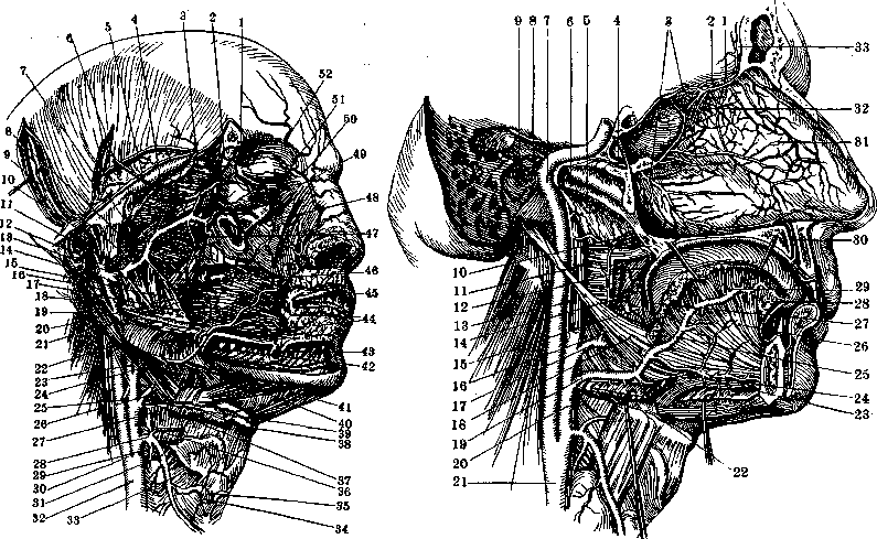
Figure 14. To the article Carotid Artery, Above the point of passage through the bundle of styloid muscles a. carotis externa penetrates into the parotid gland and goes vertically through the deep layer of the gland. More superficially and laterally from a. carot. ext. in the thickness of the gland passes the external jugular vein, appearing on the neck at the lower pole of the gland. The facial nerve has no close relation to a. carot. ext. In the thickness of the parotid gland along a. carot. ext. lie deep lymph glands (figs. 8 and 19).-Variations of origin. Figure 15. Mutual position of the origins of the external and internal carotid arteries; horizontal transverse section of the neck along the upper edge of the hyoid bone LOWER segment of the section: 1-n.vagus; 2-a. carot. int.; 3-v. jugularis int.; 4-a. carotis externa; 6-lymphatic gland; 7-branch of v. hypoglossus; 9-a. lin
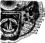
8-r. facialis communis; 10-m. hyo-glossus. (After Testut) A. carot. ext. sometimes originates from the aortic arch; variations in the height of origin of a. carot. ext. are reduced to variations in the length of the common carotid artery. Sometimes a. carot. ext. is absent and is replaced by branches of the artery of the same name from the other side or branches originating from the common carotid artery.-Variations of course. A. carot. ext. lies more superficial than usual, passing along the lateral surface of m. digastrici et stylo-hyoidei or between these muscles (Gruber; cited by Tikhomirov).-Variations of branching. The number of branches either decreases or increases. A. carot. ext. has 8 branches, of which 6 are lateral and 2 are terminal; 3 branches go forward: Figure 16. Syntopia of a. carotis ext. at its origin (Farabeuf's triangle): 1-m. st,-cl.-mastoid., covered by the first cervical aponeurosis; 2-v. jugularis int.; 3-a. occipitalis; 4-a. carot. int.; 5-a. carot. ext.; 6-v. facialis commun.; 7-a. thyreoid. sup.; 8- aponeurosis cervicalis superfic. (first); 9-n. laryngeus super.; 10-os hyoideum; 11-m. hyoglossus; 12-n. hypoglossus; 13-a. maxillaris externa; 14-m. digastricus. (After Testut-Jacob.) superior thyroid (a. thyreoidea sup.), lingual (a. lingualis) and maxillary (a. maxillaris ext.) (fig. 13); two go backward: occipital (a. occipitalis) and posterior auricular (a. auricularis post.); one branch goes inward-ascending pharyngeal (inferior-of French authors) (a. pharyngea ascend.); two terminal branches-superficial temporal (a. temporalis superficialis) and internal maxillary (a. maxillaris interna).-A. thy
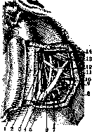
reoidea sup. originates from the anterior semicircle of a. carot. ext. somewhat higher, and sometimes at the level of the bifurcation of a. carot. com., it goes horizontally forward 1__tl „ ,_
and inward, then bends-
downward, descending z^x14>111 / F go k sootvUcheshecheye do- ^ёshkSh / ' le Щитовидной желе
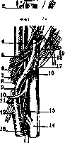
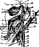
Figure 17.
Figure 18. Figure 17. Fork of styloid muscles and carotid arteries (semi-schematic): 1-n. vagus; 2 and 6-n. glosso-pharyng.; 3 and 10-n. hypogl.; 4, 9 and 11-a. carot. ext.; 5-m. stylo-glossus; 7-m. stylo-pharyng.; 8-a. maxill. ext.; 12-a. lingual.; 13-a. thyreoidea sup.; 14-a. carot. com.; 15-v. jugul. int.; 16-ram. descend, n. hypogl.; 17-horn of hyoid bone (cut); 18-m.stylo-hyoideus; 19-a. occi-pit.; 20-a. auricularis; 21-ramus n. spinalis. (After Testut) Figure 18. Fork of carotid arteries and styloid process muscles. Branches of a. car. ext. 1-m. pterygoideus ext.; 2-a. maxillaris int.; 3-m. pterygoideus int.; 4-m. stylo-glossus; 5 and 23-n. glosso-pharyngeus; 6-m. stylo-pha-ringeus; 7-section of lower jaw; 8 and 11-m. stylo-hyoideus; 9-a. maxillaris ext.; 10-m. hyo-glossus; 12-os hyoideum; 13-a. carotis com.; 14-a. thyreoid. sup.; 15-n. laryngeus sup.; 16-a. carotis ext.; 17-a. carotis int; 18-n. hypoglossus; 19 and 20-a. occipitalis; 21-m. digastricus (posterior belly); 22-a. auricularis post.; 24-pr. mastoideus; 25-a. carotis int.; 26-pr. condyloideus mandibulae; 27-a. temporalis sup.; 28-a. lingualis. (After Testut) zy. - Variations of origin. A. thyr. sup. may originate from a. carot. com.; often it originates with a common trunk with the lingual artery or with the maxillary; Figure 19. Horizontal section of the neck through the middle of gl. parotidis: 1-proces-sus styloideus with muscles attached to it; 2-aponeurotic plate between the pharyngeal wall and m. stylo-pharyn-geus, the so-called "wings of the pharynx"; 3-aponeurotic plate sagittally located between the pharyngeal wall and the deep cervical aponeurosis; 4-ganglion sup. n. sympathici; 5-n. vagus; 6-n. accessorius Willisi; 7-n. glosso-pharyngeus; 8-v. jugularis int.; 9-aponeurosis gl. parotidis; 10-gl. parotidea profund.; 11-a. carotis ext. (After Testut) a. thyr. sup. may be completely absent, being replaced by a branch of a. thyr. inferior of the same or opposite side, or a. thyr. sup. is duplicated.-A. 1 i n-gualis originates from the anterior semicircle of a. carot. ext. somewhat higher than a. thyr. sup. near the greater horn of the hyoid bone.-Variations. A. lingualis sometimes originates from a. carot. ext. with a common trunk with a. maxil. ext. or with a. thyr. sup. (less often); sometimes it emerges from the trunk of a. carot. ext. higher than a. maxil. ext. (Teile; cited by Tikhomirov). A. lingualis may not be present; then it is replaced by branches of a. maxil. ext. or a. lingualis of the opposite side (Testut).-A. maxil-laris ext. (facial, l'art ere faeiale- French authors) (fig. 13) originates from the anterior semicircle of a. carot. ext. somewhat higher than a. lingualis. Variations: a. maxil. ext. often (25%) originates with a common trunk with a. lingualis. The following 2 arteries go backward from a. carot. ext.: a. occipitalis begins from the posterior semicircle of a. carot. ext. (fig. 18), approximately at the level of a. lingualis and a. maxill. ext. Variations of origin. A. occipit. sometimes originates lower than a. maxil. ext.; it originates together with a. lingualis from a common trunk for them; it may originate from the trunk of the subclavian artery via the deep cervical artery or the thyrocervical trunk (tr. thyreo-cervicalis) (Rauber); in very rare cases a. occipit. originates from the internal carotid artery or it is duplicated.-A. auricul. post, begins from the same posterior semicircle of a. carot. ext. somewhat higher than the preceding artery. Variations of origin. A. auric, post, sometimes is only a branch of a. occipit.-A. pharyngea ascend, (figs. 2 and 14) (art. pharyngienne inf. French authors, pha-ryngo-meningea Theile)-the smallest of the branches of a. carot. ext.-originates from the inner side at the level of the origin of a. lingualis. Variations of origin. A. pharyngea asc. may begin from a. occipit., from a. carot.int., a. maxill. ext., a. thyr.sup.; originating from a. carot. ext., it may begin lower than a. thyr. sup. (Tiedemann; cited by Tikhomirov) or even from the bifurcation of a. carot. com. (Theile) (fig. 2).-A. tempo r. super f.- one of the terminal branches of a. carot. ext., begins at the level of the neck of the articular process of the lower jaw (see Temporal region).--The second terminal branch of a. carot. ext. is a. maxill. int. (fig. 13), originating at the medial side of the neck of the articular process of the lower jaw in the parotid region; it extends to the pterygo-palatine fossa (fossa pterygo-palatina), where it ends with a terminal branch, the sphenopalatine artery (a. spheno-palatina). Variations of origin. The internal maxillary artery separates from the external carotid above the zygomatic arch, being as it were a branch of the superficial temporal artery (Münz); in rare cases the internal maxillary artery is a branch of a. carot. int., occipital" (Rauber, Hoffmann) or maxillary (Klein). The internal carotid artery is the second branch of the bifurcation of a. carot. com.; the internal carotid artery supplies blood to the anterior and upper parts of the brain and the orbit with its contents. This artery has a lumen size directly proportional to the degree of development of the brain: therefore it is relatively larger in humans than in other animal species, and relatively larger in children than in adults.-Course of the artery. At first the artery lies
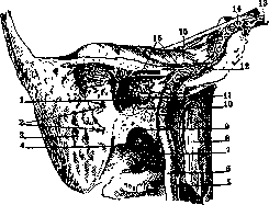
Figure 20. Carotid canal: 1-a. tympanica post.; 2-rami mastoidei; 3-cellulae mastoi-deae; 4-a. stylo-mastoidea; 5-a. pharyng. ascend.; 6-a. carot. int.; 7-a. tympanica int.; 8-m. longus capitis; 9-fossa jugularis; 10-a. tympanica inf.; 11-ramus carotico-tympani-cus; 12-plexus venosus caroticus internus; 13-v. ophthalmica sup.; 14-sinus cavernosus, 15-ramus petrosus superflc. le-a. tympanica sup. (After Spalteholz).
somewhat behind and externally from the external carotid artery (fig. 15); then it changes direction, bending around the external carotid artery at an acute angle from the outside inward toward the pharyngeal wall (fig. 14). Approaching the pharynx, the artery goes vertically upward and enters the carotid canal (fig. 20), through which it passes for its entire length. From the carotid canal, the internal carotid artery enters the cranial cavity, lying on the fibrocartilaginous plate (fibrocartilago basalis) that fills the anterior lacerated foramen (foramen lacerum anterius); then the artery enters the groove of the cavernous sinus (sinus cavernosus) (fig. 21) and makes an S-shaped bend in it. Upon exiting the sinus groove, the artery sharply deviates upward and backward, and posterior to the optic foramen it pierces the dura mater, where it gives off its lateral branch—the ophthalmic artery (a. ophthalmica)—and divides into four terminal branches: 1) a. cerebri anterior, 2) a. cerebri media, 3) a. chorioidea, and 4) a. communicans post. Topography of the internal carotid artery. Ascending from the bifurcation, the internal carotid artery enters the suprascapular region (regio carotidea sup.) (first part of the artery), then the retrostyloid space (spatium retrostyloideum) (second part); here it enters the carotid canal (third part) and forms two bends. Upon emerging into the anterior lacerated foramen (for. lacerum ant.), the artery is already intracranial and here, located in the cavernous sinus (fourth part), it makes two new bends. In the first part, the internal carotid artery lies externally and somewhat posteriorly to it (fig. 15); along the vein runs a chain of deep lymph nodes; a little above its origin, the internal carotid artery is crossed by the common facial vein (v. facialis communis); higher and anteriorly, the artery's path is crossed by the posterior belly of the digastric muscle; on the lateral side of the artery, between it and the internal jugular vein, passes the hypoglossal nerve; here its descending branch originates. Posteriorly, between the artery and the internal jugular vein, lies the vagus nerve; posteriorly and externally from it goes the hypoglossal nerve, and somewhat lower, on the medial side, the superior laryngeal nerve (n. laryngeus super.) curves around the artery (fig. 6 and 16). The external and internal carotid arteries, as they ascend, move away from each other; in this, the external carotid artery changes direction more than the internal one, deviating forward; the latter continues its ascending vertical direction, entering the space between the pharynx and the muscles of the styloid process, covered on the lateral side by the deep lamina of the fascia of the parotid gland. Medially from the artery in the sagittal plane is located a fascial lamina extending from the pharyngeal aponeurosis to the deep cervical fascia. Thus the artery is separated from the retropharyngeal cellular tissue (fig. 19). The posterior wall of the bed of this (second) part of the internal carotid artery is formed by the deep cervical aponeurosis with the underlying muscles and the transverse processes of the vertebrae. The anterior wall of the bed is formed by the stylopharyngeal aponeurosis (aponeur. stylo-pharyngeus) [so-called wings of the pharynx (Jonnesco), aileron du pharynx], stretched in the frontal plane—from the lateral surface of the pharynx to the styloid process and its muscles (fig. 19), clothed with this aponeurosis. The aponeurosis extends externally, reaches and envelops the digastric muscle and weaves into the deep lamina of the aponeurotic sheath of the sternocleidomastoid muscle. Throughout the height behind the styloid space, the elements of the vascular-nervous bundle (v. jugul. int., n. vagus, a. carotis int., and n. sympathicus) do not change their relationships and, only approaching the site of immersion into the skull, they move away from each other and change their mutual relationships: the vagus nerve approaches the internal jugular vein, which in turn moves posteriorly away from the internal carotid artery. At the base of the skull, posterior and close to the vascular-nervous bundle is located the superior sympathetic ganglion (fig. 6); below it remains somewhat distant from the bundle, separated from it by the deep cervical aponeurosis. Along the internal jugular vein follows a chain of lymph nodes. The third nerve of this area, spirally winding around the lateral side of the internal carotid artery, is the glossopharyngeal nerve; it passes, like the hypoglossal nerve, between the artery and the vein posteriorly superiorly, forward and downward. Third part of the internal carotid artery. The artery enters the external opening of the bony canal, located on the inferior surface of the pyramid of the temporal bone, anterior to the styloid process and the jugular foramen (foramen jugulare) and posterior to the junction of the cartilaginous part of the Eustachian tube with the bony part. The artery ascends vertically for 7-10 mm; here it has in front of it the bony canal of the Eustachian tube and the semicanal of the tensor tympani muscle. Posterior to the artery is the canaliculus tympani, above the artery
Figure 21. A. carotisint.
1 - hypophysis; 2
3 - sphenoid.; 4-sinus
5 - sinus cavernosus. (After Spalteholz.)
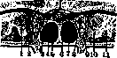
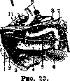
Figure 22.
Figure 22. Frontal section of the skull through sinus cavernosus et sphenoidalis: 1 and 10-oculomotorius nerve; 2-trochlearis nerve; 5-abducens nerve; i-sinus cavernosus; 5-sinus sphenoidalis; 6-hypophysis; 7-opticus nerve; 8-a. carotis interna; 9-maxillaris nerve; 11-brain. (After Testut.) Figure 23. A. carotis interna in sinus cavernosus: 1-ophthalmica vein; 2 and 5-abducens nerve; 3-maxillaris nerve; 4-sinus cavernosus; 6-internal opening of canalis caroticus; 7-a. carotis interna; 8-clinoideus post. opening; 5-a. carotis interna: 10-ophthalmica artery; 11-clinoid. ant. process. (After Testut.) The cochlea is located, externally-the anterior part of the tympanic cavity, internally-compact bone. On this segment the artery gives off branches (aa. carotico-tympanicae), which exit through canals of the same name. The artery, making a bend, passes into a horizontal direction, going forward and inward along the axis of the pyramid. Above the artery lies Gasser's ganglion, separated from it by a bony plate; medially and posteriorly is the inferior petrosus sinus (in the cranial cavity); anteriorly-externally the artery approaches the cartilaginous part of the tensor tympani muscle tube and the chorda tympani. The artery does not completely fill the bony canal, leaving sufficient space for the veins and sympathetic nerves surrounding it (plexus venosus caroticus internus and plexus caroticus internus); the latter is especially powerfully developed in the cavernous sinus, where it is called the cavernous plexus (plexus cavernosus) (figs. 20, 21 and 22). The fourth-intracranial part of a. carotis interna. Upon entering the cranial cavity, the artery passes into the bifurcation of the dura mater, into the so-called sinus cavernosus, into which it enters from behind; here the artery is secured to the walls of the sinus by fibrous cords [ligamenta carotica (Trolard)] and lies in the center of the sinus (fig. 23), surrounded by venous blood (Testut); the artery touches the walls of the sinus only during pulsation. In the sinus the artery first goes vertically, then horizontally in the sagittal plane; upon approaching the anterior clinoid process, the artery turns upward and pierces the upper wall 41» of the sinus. On the neck the internal carotid artery does not give off branches; its first branches depart in the caroticanal; a. carotico-tympanica-for the tympanic cavity and periosteal branches-for the walls of the canal itself. In the carotid canal small branches depart to the surrounding parts (to the walls of the sinus and nerves within it, to Gasser's ganglion and its three branches) and finally an anastomosis-to a. meningea media and the dura mater in the occipital region. Upon exiting the cavernous sinus the internal carotid artery gives off the a. ophthalmica, very rich in branches; the terminal branches of a. carotis interna are listed above. Variants of a. carotis interna. Sometimes a. carotis interna originates from the aortic arch, sometimes it is completely absent, being replaced by branches of a. carotis interna of the other side or branches of a. maxillaris interna. Blood supply to the brain in such cases is possible only thanks to constant anastomoses of homonymous systems of one side or the other, or heteronymous systems of the same side, for example, the system of a. carotis interna of the right and left sides or the system of a. carotis interna with the system of a. carotis externa of the same side (Tikhomirov).-Variants of course and caliber. A. carotis interna on the neck is generally straight, but may be more or less curved, with the curves varying in number, direction, and extent, and some of the curves run along the lateral surface of the tonsils, the removal of which in such cases may be dangerous. The calibers of the artery also vary considerably, for example, a. carotis interna is sometimes one-third the caliber of a. vertebralis (Hyrtl).-Variants of branching. A. carotis interna sometimes gives off branches: laryngeal, occipital, lingual, ascending pharyngeal, facial transverse, and middle meningeal (a. meningea media). Pathological anatomy. Although pathological changes in a. carotis communis and its branches do not differ in any particular way from changes observed generally in the arterial system, it is still necessary to note that the frequency of certain processes observed in the system a. carotis communis and its branches, besides everything else, depends on the histological structure of the wall (type of vessel-muscular, mixed, or elastic); secondly on the anatomical position of the vessel and thirdly on the topographical relations with surrounding organs. Here are mainly meant acute inflammatory, as well as neoplastic processes, developing somewhere near a. carotis communis or its branches and in their further development passing to the aforementioned vessels. Thus, in scarlet fever, purulent processes in the glands and cellular tissue of the neck are often observed, which subsequently pass either to a. carotis communis or (more often) to its branches, resulting in ulceration of the vessel with subsequent sometimes fatal bleeding. Malignant tumors (cancers and sarcomas), developing on the neck and originating either from connective tissue or from organs located here (lymph glands, thyroid gland), as well as malignant tumors of the mediastinum (sarcomas and cancers of the thyroid gland and others), in their development not only deform the vessels, compressing them until complete obliteration of the lumen, but also grow into them and thus on the one hand give rise to generalization of the tumor (metastases), and on the other hand, by destroying the vascular wall, can cause bleeding, sometimes fatal. Clinic. Surgical diseases of a. carotis communis and its branches. Such diseases include various injuries, for example, trauma or destruction of the artery by some pathological process that has spread from surrounding tissues and organs, and aneurysms. The most common injury to a. carotis communis is trauma, and among this type of arterial injury, gunshot wounds are in first place; the common carotid artery accounts for 51/г?'o вall gunshot wounds to vessels (Punin). In general, the frequency of gunshot wounds to vessels is 7a% of the total number of wounds. Neck vessels are sometimes wounded from within: a swallowed bone that has stopped in the esophagus, when forcibly pushed through, tears the esophagus and the common carotid artery; such a case was described by Dittmann. A. carotis interna is sometimes injured during tonsillectomy, when the tonsils are pulled too strongly into the oropharyngeal opening. As for other injuries to a. carotis communis (for example, destruction of the vessel wall by existing purulence near the artery, malignant tumor or other disease), they are not common. Thus, according to Pilz's data, bleeding from a. carotis communis was caused by purulence 17 times out of 220 cases of bleeding he collected for other reasons. The consequence of vessel injury is bleeding, external or into a simultaneously opened and nearby hollow organ (larynx, pharynx, pleura) or into the cellular tissue surrounding the vessel. Injuries to a. carotis communis are among the most severe diseases, leading to death in 60% of cases (Fischer, according to Tikhow): out of 361 cases, death occurred 201 times. The outcome sharply worsens with simultaneous injury to a. carotis communis and the internal jugular vein: death usually occurs immediately. Treatment. The best and surest way to stop bleeding is ligation or suture of the artery. In case the situation does not allow proceeding to the operation of ligation of the vessel, it is necessary to stop the bleeding either by pressing the wound with a finger (digital pressure) or by pressing the vessel at a neutral point from the injury. The common carotid artery is pressed against the transverse processes of the cervical vertebrae with the thumb of the hand, the other fingers encircling the neck from behind. Bertrand achieved recovery in one severe case of a puncture wound to a. carotis communis by digital pressure, which lasted for three days. Ligation of the injured vessel is done double and is a sure means against the immediate danger of bleeding, but it by no means can be considered an innocuous procedure (Dyakonov and Lysenkov). According to Pilz, 914 cases of ligation of a. carotis communis gave 40% mortality, according to Le Fort, among French surgeons on 435 cases-43% mortality. In addition, ligation of a. carotis communis gave 32% cerebral phenomena, half of which with fatal outcome (Pilz, according to Dyakonov and Lysenkov). In the immediate hours after ligation, anemia occurs in the corresponding half of the neck, face and head, strongest noise in the healthy ear, dilation of the pupil of the same side; nausea, sometimes vomiting and dizziness appear; with favorable course, after 10-14 hours, only general weakness and slight headache remain from all these phenomena; the noise in the ear of the healthy side lasts the longest. With unfavorable course, paralysis of the limbs, hearing impairment appear, then certain defects in the psyche appear, indicating deep disorders of brain nutrition: disintegration in the brain, red or softening depending on whether the reverse venous flow plays a role or not. The rarity of such disorder in ligation of a. carotis communis is explained by the fact that very soon a blood flow forms through the collaterals of the a. carotis externa system into the internal carotid artery. How soon does collateral circulation arise in ligation of vessels?
This question is resolved on the basis of Pirogov's research as follows: after ligation of the aorta in animals, blood begins to flow from the severed femoral arteries just one second after applying the ligature, first in drops and then in a stream. The direction of blood flow in ligation of a. carot. com. is recognized, based on Guinard's research, to be from a. carot. ext. to a. carot. int., which is disputed by the work of Marquis and Lefeuvre; these authors assert that blood flows from a. carot. int. to a. carot. ext.; therefore, they speak against ligation of a. carot. com. Their work is based on experiments on rabbits and dogs; apparently the conditions of cerebral circulation in humans and experimental animals are not the same; the following fact is interesting: these animals tolerate simultaneous bilateral ligation of a. car. com. and vertebral arteries well. Ligation of both common carotid arteries results in mortality only in 22%, and still such ligation should be resorted to only in extreme cases and with a significant time interval between the ligation of the common carotid artery on one side and the other, which allows collateral circulation to develop. According to Ceci, complications from the brain can be weakened if the internal jugular vein is ligated along with the artery, as a result of which cerebral anemia does not develop. Circulation after ligation of a. carot. com. is restored 1) through the branches of a. carot. com., connecting with the branches of a. carot. com. of the other side and the branches of the subclavian artery of the other side (deep cervical with occipital, superior thyroid with inferior, vertebral with occipital), 2) through the branches of the ophthalmic artery (branch of a. carot. int.) with the branches of a. carot. ext. - a. maxill. ext. and a. tempor. superf. of the same side and 3) through the arterial circle of Willis. This requires proper development and good condition of the arteries of the said circle, which is not always the case due to its frequent anomalies, for example when the posterior communicating arteries (art. communicantes posteriores) are completely absent. The anatomical condition of this circle according to Walker's data is as follows: in brachycephalics, additional aa. communicantes post. are often observed; in mesocephalics, the left a. communicans post. is thicker than the right in 70% of cases; in dolichocephalics, the circle is often open, moreover more often on the right; therefore ligation of a. carot. com. is safer to perform in brachycephalics, very dangerous in dolichocephalics, and in mesocephalics safer on the left side (quoted from Moskalenko). Operations. The first (reliable) ligation of a. carot. com. was performed in 1792 by Lynn (the patient died after 14 days). With a favorable outcome, Cooper first ligated this artery in Guy's Hospital in 1805. Since then, the number of ligations of a. carot. com. amounts to thousands. The vessel is ligated by applying ligatures to both ends of the vessel at the site of its incision or by applying ligatures at a distance of 1/2-1 cm from each other along the vessel with mandatory incision between them.
Ligation of a. carot. com. is performed in two areas of the artery: 1) in loco electionis - the ligature is placed 1-2 cm below the bifurcation; 2) " " " in incisione praeparati - Fig 24 - Incisions for exposing the carotid artery during operations on the neck: the artery is ligated at a) the incision for ligating the lower part of the trunk, "and a, carot. ext. et int.; this point is located" "b - normal incision but not lower than ~ cm from its" "c - incision for ligating the beginning. For the right" "a. carot. com.; c - incision for ligating the a. carot. on 2 cm above the edge of the clavicle, and for the left - at the level of the upper edge of the sternum. According to Pirogov, a. carot. com. can be exposed in 4 different places: 1) in regione supra-hyoidea (trigonum caroticum); 2) in fossa sterno-mastoideo-laryngea; 3) in fossa sterno-mastoideo-tracheali; 4) in spatio intercrureo.(Incisions for ligating the common carotid artery, external and internal carotid arteries - fig. 24 and 25.) Ligation of a. carot. com. in trigono carotico, according to Cooper, in loco electionis. The patient lies with the head thrown back and somewhat deviated to the healthy side, with a roll under the neck; the most convenient place for ligation of a. carot. com. is at the level of the cricoid cartilage where the anterior edge of the sternocleidomastoid muscle intersects with the omohyoid muscle (fig. 24 and 25). The skin incision is made along the anterior edge of the sternocleidomastoid muscle for a length of 5-6 cm, so that it ends somewhat below the level of the cricoid cartilage, or else with the aim of obtaining a less noticeable scar, the incision is made horizontally
The platysma is divided in the direction of the fibers, the edges of the wound are pulled apart with hooks, and the anterior edge of the sternocleidomastoid muscle is exposed. The latter is pulled back and up with a hook, and the carotid sheath is opened. The common carotid artery is found in the thickness of the sheath, surrounded by a plexus of nerves and the internal jugular vein. The vein is separated from the artery and pulled back. The artery is isolated for a length of 1-2 cm, and two ligatures are applied to it: one below the site of incision, the other above. The artery is incised between the ligatures, and the proximal end is tied with a catgut ligature. The distal end is left open or tied with silk. The wound is sutured in layers. Ligation of a. carot. com. in fossa sterno-mastoideo-laryngea. The incision is made along the anterior edge of the sternocleidomastoid muscle, 2-3 cm above the clavicle for the right artery and at the level of the upper edge of the sternum for the left. The subsequent technique is the same as in the previous method. Ligation of a. carot. com. in fossa sterno-mastoideo-tracheali. The incision is made along the anterior edge of the sternocleidomastoid muscle, 3-4 cm above the clavicle for the right artery and 1-2 cm above the upper edge of the sternum for the left. The subsequent technique is the same as in the previous methods. Ligation of a. carot. com. in spatio intercrureo. The incision is made along the anterior edge of the sternocleidomastoid muscle, 4-5 cm above the clavicle for the right artery and 2-3 cm above the upper edge of the sternum for the left. The subsequent technique is the same as in the previous methods.
Ligation of the external carotid artery. The incision is made along the anterior edge of the sternocleidomastoid muscle, 2-3 cm above the upper edge of the sternum. The subsequent technique is the same as in the previous methods. Ligation of the internal carotid artery. The incision is made along the anterior edge of the sternocleidomastoid muscle, 1-2 cm above the upper edge of the sternum. The subsequent technique is the same as in the previous methods. Complications of ligation of the common carotid artery. 1) Shock. 2) Hemorrhage from the proximal end of the artery. 3) Thrombosis of the internal carotid artery with subsequent embolism of the cerebral vessels. 4) Compression of the vagus nerve with subsequent paralysis of the vocal cords. 5) Compression of the sympathetic nerve trunk with subsequent Horner's syndrome. 6) Infection of the wound. 7) Laryngeal edema. 8) Paralysis of the hypoglossal nerve. 9) Paralysis of the spinal accessory nerve. 10) Paralysis of the phrenic nerve. 11) Paralysis of the recurrent laryngeal nerve. 12) Paralysis of the superior laryngeal nerve. 13) Paralysis of the cervical sympathetic trunk. 14) Paralysis of the brachial plexus. 15) Paralysis of the diaphragm. 16) Paralysis of the intercostal muscles. 17) Paralysis of the abdominal muscles. 18) Paralysis of the lower extremities. 19) Death. The mortality rate after ligation of the common carotid artery is 22%. The most common complications are hemorrhage from the proximal end of the artery and thrombosis of the internal carotid artery with subsequent embolism of the cerebral vessels. The most serious complication is death. The most common cause of death is cerebral embolism. The second most common cause of death is hemorrhage from the proximal end of the artery. The third most common cause of death is shock. The fourth most common cause of death is laryngeal edema. The fifth most common cause of death is infection of the wound. The sixth most common cause of death is paralysis of the phrenic nerve. The seventh most common cause of death is paralysis of the diaphragm. The eighth most common cause of death is paralysis of the intercostal muscles. The ninth most common cause of death is paralysis of the abdominal muscles. The tenth most common cause of death is paralysis of the lower extremities. The eleventh most common cause of death is paralysis of the brachial plexus. The twelfth most common cause of death is paralysis of the cervical sympathetic trunk. The thirteenth most common cause of death is paralysis of the vagus nerve. The fourteenth most common cause of death is paralysis of the hypoglossal nerve. The fifteenth most common cause of death is paralysis of the spinal accessory nerve. The sixteenth most common cause of death is paralysis of the recurrent laryngeal nerve. The seventeenth most common cause of death is paralysis of the superior laryngeal nerve. The eighteenth most common cause of death is paralysis of the sympathetic nerve trunk. The nineteenth most common cause of death is Horner's syndrome. The twentieth most common cause of death is compression of the vagus nerve. The twenty-first most common cause of death is compression of the sympathetic nerve trunk. The twenty-second most common cause of death is compression of the hypoglossal nerve. The twenty-third most common cause of death is compression of the spinal accessory nerve. The twenty-fourth most common cause of death is compression of the recurrent laryngeal nerve. The twenty-fifth most common cause of death is compression of the superior laryngeal nerve. The twenty-sixth most common cause of death is compression of the phrenic nerve. The twenty-seventh most common cause of death is compression of the brachial plexus. The twenty-eighth most common cause of death is compression of the diaphragm. The twenty-ninth most common cause of death is compression of the intercostal muscles. The thirtieth most common cause of death is compression of the abdominal muscles. The thirty-first most common cause of death is compression of the lower extremities. The thirty-second most common cause of death is compression of the cervical sympathetic trunk. The thirty-third most common cause of death is compression of the vagus nerve. The thirty-fourth most common cause of death is compression of the sympathetic nerve trunk. The thirty-fifth most common cause of death is compression of the hypoglossal nerve. The thirty-sixth most common cause of death is compression of the spinal accessory nerve. The thirty-seventh most common cause of death is compression of the recurrent laryngeal nerve. The thirty-eighth most common cause of death is compression of the superior laryngeal nerve. The thirty-ninth most common cause of death is compression of the phrenic nerve. The fortieth most common cause of death is compression of the brachial plexus. The forty-first most common cause of death is compression of the diaphragm. The forty-second most common cause of death is compression of the intercostal muscles. The forty-third most common cause of death is compression of the abdominal muscles. The forty-fourth most common cause of death is compression of the lower extremities. The forty-fifth most common cause of death is compression of the cervical sympathetic trunk. The forty-sixth most common cause of death is compression of the vagus nerve. The forty-seventh most common cause of death is compression of the sympathetic nerve trunk. The forty-eighth most common cause of death is compression of the hypoglossal nerve. The forty-ninth most common cause of death is compression of the spinal accessory nerve. The fiftieth most common cause of death is compression of the recurrent laryngeal nerve. The fifty-first most common cause of death is compression of the superior laryngeal nerve. The fifty-second most common cause of death is compression of the phrenic nerve. The fifty-third most common cause of death is compression of the brachial plexus. The fifty-fourth most common cause of death is compression of the diaphragm. The fifty-fifth most common cause of death is compression of the intercostal muscles. The fifty-sixth most common cause of death is compression of the abdominal muscles. The fifty-seventh most common cause of death is compression of the lower extremities. The fifty-eighth most common cause of death is compression of the cervical sympathetic trunk. The fifty-ninth most common cause of death is compression of the vagus nerve. The sixtieth most common cause of death is compression of the sympathetic nerve trunk. The sixty-first most common cause of death is compression of the hypoglossal nerve. The sixty-second most common cause of death is compression of the spinal accessory nerve. The sixty-third most common cause of death is compression of the recurrent laryngeal nerve. The sixty-fourth most common cause of death is compression of the superior laryngeal nerve. The sixty-fifth most common cause of death is compression of the phrenic nerve. The sixty-sixth most common cause of death is compression of the brachial plexus. The sixty-seventh most common cause of death is compression of the diaphragm. The sixty-eighth most common cause of death is compression of the intercostal muscles. The sixty-ninth most common cause of death is compression of the abdominal muscles. The seventieth most common cause of death is compression of the lower extremities. The seventy-first most common cause of death is compression of the cervical sympathetic trunk. The seventy-second most common cause of death is compression of the vagus nerve. The seventy-third most common cause of death is compression of the sympathetic nerve trunk. The seventy-fourth most common cause of death is compression of the hypoglossal nerve. The seventy-fifth most common cause of death is compression of the spinal accessory nerve. The seventy-sixth most common cause of death is compression of the recurrent laryngeal nerve. The seventy-seventh most common cause of death is compression of the superior laryngeal nerve. The seventy-eighth most common cause of death is compression of the phrenic nerve. The seventy-ninth most common cause of death is compression of the brachial plexus. The eightieth most common cause of death is compression of the diaphragm. The eighty-first most common cause of death is compression of the intercostal muscles. The eighty-second most common cause of death is compression of the abdominal muscles. The eighty-third most common cause of death is compression of the lower extremities. The eighty-fourth most common cause of death is compression of the cervical sympathetic trunk. The eighty-fifth most common cause of death is compression of the vagus nerve. The eighty-sixth most common cause of death is compression of the sympathetic nerve trunk. The eighty-seventh most common cause of death is compression of the hypoglossal nerve. The eighty-eighth most common cause of death is compression of the spinal accessory nerve. The eighty-ninth most common cause of death is compression of the recurrent laryngeal nerve. The ninetieth most common cause of death is compression of the superior laryngeal nerve. The ninety-first most common cause of death is compression of the phrenic nerve. The ninety-second most common cause of death is compression of the brachial plexus. The ninety-third most common cause of death is compression of the diaphragm. The ninety-fourth most common cause of death is compression of the intercostal muscles. The ninety-fifth most common cause of death is compression of the abdominal muscles. The ninety-sixth most common cause of death is compression of the lower extremities. The ninety-seventh most common cause of death is compression of the cervical sympathetic trunk. The ninety-eighth most common cause of death is compression of the vagus nerve. The ninety-ninth most common cause of death is compression of the sympathetic nerve trunk. The one hundredth most common cause of death is compression of the hypoglossal nerve.
The one hundred and first most common cause of death is compression of the spinal accessory nerve. The one hundred and second most common cause of death is compression of the recurrent laryngeal nerve. The one hundred and third most common cause of death is compression of the superior laryngeal nerve. The one hundred and fourth most common cause of death is compression of the phrenic nerve. The one hundred and fifth most common cause of death is compression of the brachial plexus. The one hundred and sixth most common cause of death is compression of the diaphragm. The one hundred and seventh most common cause of death is compression of the intercostal muscles. The one hundred and eighth most common cause of death is compression of the abdominal muscles. The one hundred and ninth most common cause of death is compression of the lower extremities. The one hundred and tenth most common cause of death is compression of the cervical sympathetic trunk. The one hundred and eleventh most common cause of death is compression of the vagus nerve. The one hundred and twelfth most common cause of death is compression of the sympathetic nerve trunk. The one hundred and thirteenth most common cause of death is compression of the hypoglossal nerve. The one hundred and fourteenth most common cause of death is compression of the spinal accessory nerve. The one hundred and fifteenth most common cause of death is compression of the recurrent laryngeal nerve. The one hundred and sixteenth most common cause of death is compression of the superior laryngeal nerve. The one hundred and seventeenth most common cause of death is compression of the phrenic nerve. The one hundred and eighteenth most common cause of death is compression of the brachial plexus. The one hundred and nineteenth most common cause of death is compression of the diaphragm. The one hundred and twentieth most common cause of death is compression of the intercostal muscles. The one hundred and twenty-first most common cause of death is compression of the abdominal muscles. The one hundred and twenty-second most common cause of death is compression of the lower extremities. The one hundred and twenty-third most common cause of death is compression of the cervical sympathetic trunk. The one hundred and twenty-fourth most common cause of death is compression of the vagus nerve. The one hundred and twenty-fifth most common cause of death is compression of the sympathetic nerve trunk. The one hundred and twenty-sixth most common cause of death is compression of the hypoglossal nerve. The one hundred and twenty-seventh most common cause of death is compression of the spinal accessory nerve. The one hundred and twenty-eighth most common cause of death is compression of the recurrent laryngeal nerve. The one hundred and twenty-ninth most common cause of death is compression of the superior laryngeal nerve. The one hundred and thirtieth most common cause of death is compression of the phrenic nerve. The one hundred and thirty-first most common cause of death is compression of the brachial plexus. The one hundred and thirty-second most common cause of death is compression of the diaphragm. The one hundred and thirty-third most common cause of death is compression of the intercostal muscles. The one hundred and thirty-fourth most common cause of death is compression of the abdominal muscles. The one hundred and thirty-fifth most common cause of death is compression of the lower extremities. The one hundred and thirty-sixth most common cause of death is compression of the cervical sympathetic trunk. The one hundred and thirty-seventh most common cause of death is compression of the vagus nerve. The one hundred and thirty-eighth most common cause of death is compression of the sympathetic nerve trunk. The one hundred and thirty-ninth most common cause of death is compression of the hypoglossal nerve. The one hundred and fortieth most common cause of death is compression of the spinal accessory nerve. The one hundred and forty-first most common cause of death is compression of the recurrent laryngeal nerve. The one hundred and forty-second most common cause of death is compression of the superior laryngeal nerve. The one hundred and forty-third most common cause of death is compression of the phrenic nerve. The one hundred and forty-fourth most common cause of death is compression of the brachial plexus. The one hundred and forty-fifth most common cause of death is compression of the diaphragm. The one hundred and forty-sixth most common cause of death is compression of the intercostal muscles. The one hundred and forty-seventh most common cause of death is compression of the abdominal muscles. The one hundred and forty-eighth most common cause of death is compression of the lower extremities. The one hundred and forty-ninth most common cause of death is compression of the cervical sympathetic trunk. The one hundred and fiftieth most common cause of death is compression of the vagus nerve.
Related articles
Mentioned in
- Abducens Nerve
- Brain (cerebrum, a comprehensive term for the entire)
- Cardiography
- Cavernous
- Corpus Luysi
- Decapitation
- Decerebration (DECEREBRATE)
- Diabrosis
- Enophthalmus
- Friedman's Vasomotor Syndrome
- Glosso-Pharyngeal Nerve
- Hanging
- Hering Reflex
- Hypoglossus Nerve
- Laryngeus Superior Nerve
- Noises
- Ophthalmic Artery
- Parabiosis
- Pupillary Fibers
- Retropharyngeal Space
Cite this page
“Carotid Artery.” Soviet Medical Encyclopedia. English translation of Bolshaya Meditsinskaya Entsiklopediya, 1st ed. (Moscow, 1928–1936), ed. N. A. Semashko. https://sovietmedicalencyclopedia.pages.dev/article/carotid-artery/