Blood Vessels (525)
Historical document, translated for reference. It reflects medical knowledge of the 1920s–30s and is not medical advice.
Summary
This article from the 1928–1936 Soviet medical encyclopedia defines the circulatory system, distinguishing between arterial and venous systems connected by capillaries. It also details the three developmental stages of circulation in vertebrates: yolk, placental, and pulmonary.
Encyclopedia article (1928–1936)
525 I. Embryology. Blood vessels (vasa sanguinifera), a closed system of tubes of varying caliber, permeating all tissues and organs, distributing blood throughout the organism from the heart to the periphery (to the integuments, muscles, etc.) and carrying it back from the periphery to the heart. The heart and blood vessels constitute, as a whole, the system of circulatory organs, the circulatory system. Blood vessels that carry blood away from the heart and bring it to an organ are called arteries, and the entire system of vessels of this order is called the arterial system. Blood vessels that carry blood from the organs to the heart are called veins, and the entire system of these vessels is called the venous system. The vessels connecting both systems (arterial and venous) are the smallest terminal branches of the arteries and the initial branches of the veins, which bear the name capillaries, or hair-like vessels (see Capillaries). In higher vertebrates and in humans, three periods of circulation are distinguished during development. The period of yolk circulation, or primary; the placental (fetal circulation), or secondary; and the post-placental period, pulmonary, or tertiary. Each of these periods is marked by a number of structural features of the circulatory system and is characterized by the presence of special additional vessels that exist and function only for a given period of circulation and disappear or become obliterated in the subsequent period (Fig. 1 and separate table-Fig. 2). Additional blood vessels in the yolk and placental periods are located, mainly, outside the embryo
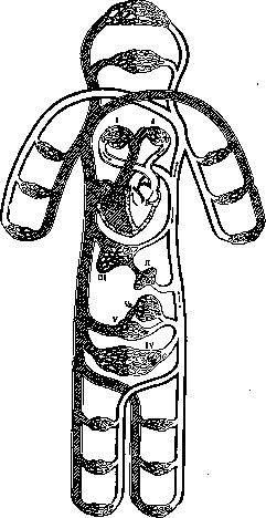
Figure 1. General scheme of circulation in higher animals: I-lungs; II-gastrointestinal tract; III-liver; IV-pelvic organs; V-kidneys; Va-renal glomeruli.
embryo, pass only for a short distance within the body of the embryo and join the blood vessels located inside its body. The yolk circulatory system in humans appears reduced and not only does not precede the placental one, but develops even somewhat later than the latter. Examining the circulatory system of a bird embryo during the yolk period, one notes well-developed paired blood vessels carrying blood (with nutrient substance) from the yolk sac to the circulatory system of the embryo and called vv. omphalomesentericae (vitellointestinal veins). They and Figure 1. Arteries of the right half of the internal female genital organs (from behind): 1-uterus; 2-oviduct; 3-ovary; 4-a. ovarica; 5-lig. rotundum uteri; 6-a. uterina; 7-a. hypogastrica; 8-vagina. (According to Hyrtl.) Figure 2. Circulatory system of the embryo: 1-v. ophthalmica; 2-a. carotis interna; 3-v. cardinalis anterior; 4-IV aortic arch; 5-aorta descendens (paired); 6-ductus Cuvieri; 7-sinus venosus; 8-v. umbilicalis sinistra; 9-v. cardinalis posterior; 10-aorta abdominalis (unpaired); 11-a. umbilicalis sinistra; 12-umbilical cord; 13-a. omphalomesenterica sinistra; 14-vv. omphalomesentericae; 15-truncus arteriosus; 16-aorta ascendens; 17-I-III aortic arches. (According to Braus.) Figure 3. Aa. comitantes nervi ischiadici (1 and 2); 3-n. ischiadicus; 4-a. perforans I; 5-a. glutaea superior; 6-a. glutaea inferior. Figure 4. 1-vasa vasorum (arteries and veins) of the aortic trunk; 2-aorta; 3-left bronchus; 4-left pulmonary vein; 5-n. vagus sin.; 6-n. phrenicus; 7-calcified lymphatic gland, to which the n. phrenicus is fused; 8-a. subclavia; 9-v. anonyma sinistra; 10-a. mammaria interna; 11-esophagus. Figure 5. Venous plexus and venous emissary: 1-v. temporalis superficialis; 2-plexus pterygoideus; 3-v. facialis profunda; 4-v. maxillaris interna; 5-v. jugularis interna; 6-v. jugularis externa; 7-v. vertebralis; 8-emissarium mastoideum; 9-v. occipitalis; 10-v. auricularis posterior. (Figures 3 and 5 according to Rauber, Fig. 4 according to the drawing and preparation of Yesipov.) (To the illustration for the article Blood Vessels.)
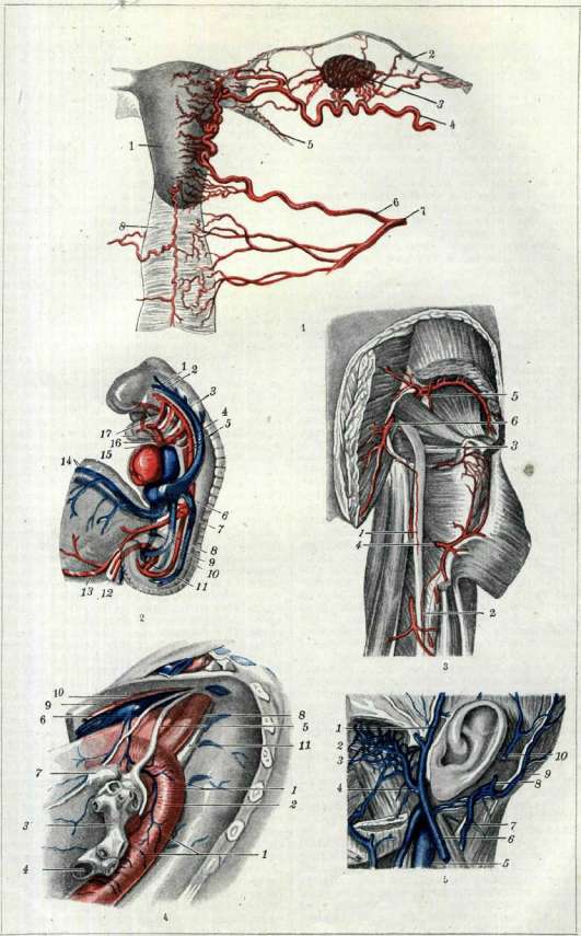
B. M. E. Vol. II. Blood Vessels, flow into the lower part of the cardiac sac, the so-called venous sinus (sinus venosus). Blood vessels carrying blood from the circulatory system from both dorsal aortas of the embryo to the yolk sac, also paired, are called aa. omphalo-mesentericae (omphalomesenteric arteries). - Vessels of the yolk sac (vesicle) in humans also appear, but since the connection of the embryo with the uterus is established very early and placental vessels develop powerfully, the aa. et vv. omphalo-mesentericae lose their significance, especially since the significance of the yolk sac in the nutrition of the human embryo is extremely small. Paired umbilical-intestinal (yolk) arteries of the human embryo are branches of the umbilical (paired) arteries (aa. umbilicales), and the yolk-intestinal (yolk) veins (two) go from the yolk sac to the heart, where they flow together with the umbilical veins into the venous sinus (sinus venosus) (see separate table, fig. 2). The period of placental circulation in humans, together with the structural features of the fetal circulatory system inherent in this period, plays a primary role in the blood supply to the developing embryo up to the birth of the fetus (see Fetus, circulation). The connection of the placenta (see Placenta) with the fetal circulatory system is maintained upon the final formation of placental vessels by one umbilical vein (v. umbilicalis; in the initial stage of development of placental vessels, umbilical veins appear in the number of two) and two umbilical arteries (aa. umbilicales). Vv. omphalo-mesentericae (dex. et sin.), just like the vv. umbilicales (dex. et sin.), with their segments take a substantial part in the formation of the hepatic venous network, in particular in the formation of the unpaired trunk, the so-called portal vein, hepatic veins, and the central segment of the v. cava inf. Upon the birth of the fetus, after the disruption of the connection of its circulatory system with the placenta, the umbilical vessels lose their significance, become obliterated, and in the adult, only the connective tissue cords corresponding to their course remain. The remnant of the v. umbilicalis, enclosed in the duplication of the peritoneum, forms the thickened edge of one of the peritoneal ligaments of the liver (lig. falciforme hep.), the so-called lig. teres, going from the navel to the anterior left notch of the liver, and its continuation, the so-called ductus venosus (Arantii), forms the lig. venosum. - Umbilical arteries (aa. umbilicales), paired from the moment of appearance until the moment of their obliteration, are in the embryo a direct continuation of the bifurcated end of the dorsal aorta, and in subsequent stages of development, a branch of the hypogastric arteries (aa. hypogastricae). They go to the umbilical end, and from there in the umbilical cord, spirally bypassing the v. umbilicalis, to the placenta, where they branch out in its villi. After obliteration, the aa. umbilicales leave two cords corresponding to their course, lifting the peritoneum of the anterior abdominal wall into two folds: lig. vesico-umbilicale lat. dex. et sin., having an ascending direction from the pelvic region to the umbilical region. During the periods of yolk and placental circulation, the development of arterial and venous vessels occurs within the embryo itself, the course of which is related to the development of the heart, individual organs, and body parts. In the earliest stages of the embryo, paired cardiac tubes, laid in the cervical region between the layers of the so-called ventral mesentery, represent the primordium of the heart, lying consequently between the abdominal wall and the foregut. The cardiac tubes in subsequent moments approach with their medial surfaces and fuse, forming an unpaired cardiac tube (heart), which in the further course of development undergoes complex changes. The anterior (cranial) end of the cardiac tube, narrowing, forms the so-called conus arteriosus, which continues into the main arterial trunk of the embryo - truncus arteriosus. The main changes in the further stage of development of the arterial system are undergone by its anterior (cranial) section. The arterial (aortic) arches in humans (just as in higher vertebrates) then either become empty and regress or develop powerfully, gradually forming the aortic arch with its main branches and the pulmonary artery. The continuation of the ventral aorta gives rise to the formation of the aa. car. ext. In fish, a reduction of the I and II pairs of branchial arterial arches was noted; in humans and mammals, the I and II pairs of branchial arches also regress (see Aorta, Botallo's duct); the III pair of arterial arches gives rise to the aa. carot. int.; the IV pair on the left side forms the arcus aortae, continuing into the aorta descendens, while on the right - the art. anonyma and the initial segment of the art. subclaviae dext.; the V pair of arterial arches is completely reduced; the VI pair, having divided accordingly by a septum, gives rise to the ascending aorta (aorta ascendens), the art. pulmonalis, and on the left, to Botallo's duct, which subsequently becomes empty and forms the so-called ligamentum arteriosum. The art. subclavia sin. develops from a segmental artery of the dorsal aorta. The arterial system in the course of its development sharply loses its (initially) symmetrical character and becomes asymmetrical. The aortic arch formed at the expense of the left aortic (arterial) arch goes to the dorsal side of the embryo and passes into the dorsal spinal aorta, which is subdivided upon the completion of the processes of formation of the arterial system into the descending or thoracic aorta (aorta thoracalis) and the abdominal (aorta abdominalis). The caudal end of the spinal aorta is bifurcated and in the placental period, with its two terminal branches, gives, as indicated, rise to two umbilical arteries, while after their obliteration it is preserved in the form of the iliac arteries (aa. iliacae comm.), giving off branches to the walls and organs of the pelvis, as well as to the lower extremities. The initial primordium of the entire venous system, including extraembryonic veins (placental and yolk), is represented by 4 pairs of veins: 2 anterior cardinal veins (vv. cardinales ant., s. vv. jugulares primitivae), 2 posterior cardinal veins (vv. cardinales post.), 2 umbilical veins (vv. umbilicales), and 2 yolk-intestinal veins (vv. omphalo-mesentericae) (see separate table, fig. 2). The latter two pairs were considered above. The anterior and posterior cardinal veins are the main veins of the primordium of the intraembryonic venous system. The anterior cardinal veins in their development take precedence over the posterior ones, which is connected with the faster growth of the head and the cranial section of the embryo in general. In connection with the displacement of the heart from the cervical region to the thoracic, the Cuvierian ducts change their direction and from a transverse position (in relation to the body axis of the embryo) are located more obliquely, inclined, almost longitudinally. The change in the position of the Cuvierian ducts somewhat changes the course of the upper sections of the posterior cardinal (right and left) veins. At this stage of development, the Cuvierian ducts, which are as if a continuation of the anterior cardinal veins collecting blood from the head, neck, and anterior extremities (fins in fish), are called superior venae cavae (vv. cavae sup. dex. et sin.), while the posterior cardinal veins, flowing into the latter, are called azygos veins (vv. azygos dex. et sin.); they collect blood mainly from the walls of the trunk and the Wolffian bodies, the most voluminous organs of the embryo. Consequently, the venae azygos are remnants of the posterior cardinal veins. In subsequent moments of development, in connection with the division of the venous sections of the heart into right and left halves, the main mass of blood is directed into the right half of the venous system. Simultaneously, transverse anastomoses appear between the left and right veins, allowing for a freer outflow of blood to the heart through the right veins; some parts of the left cardinal veins (anterior and posterior) begin to regress. In the upper section of the venous system, lying above the heart, and then in the lower one, individual sections gradually undergo reduction, transverse anastomoses acquire greater significance, becoming wider, new venous trunks are formed in the middle part of the embryo (abdominal cavity), in particular the inferior vena cava (with its branches) and the portal vein. The lumen of the v. cavae sup. sin. gradually decreases (it is preserved until the 4th month); from the 4th month, the v. cava sup. sin. becomes obliterated along the extent from the transverse anastomosis (between the vv. cavae sup.) to the coronary sulcus of the heart, leaving only a connective tissue cord called the lig. venae cavae. The section of the v. cavae sup. sin. lying in the coronary sulcus of the heart receives the veins of the heart itself and, gradually growing into the wall of the right atrium, opens into it with an independent opening - sinus coronarius cordis, s. sinus venarum cardiacarum. The v. cava sup. dextra and the transverse anastomosis with the jugular and subclavian veins flowing into them significantly increase their lumen and in the final result form (the upper part of the v. cava sup. d. - v. anonyma dex.) the transverse anastomosis - v. anonyma sin., and the part of the v. cavae sup. d. below the anastomosis forms the very trunk of the v. cava sup., flowing into the right atrium. - Vv. jugulares primitivae develop into vv. jugulares ext.; the developing brain and its venous network condition the development of new trunks - vv. jugulares int.
Due to the obliteration of the left superior vena cava, the upper part of the left azygos vein flowing into it also undergoes atrophy, and all blood from the remaining upper part of the left azygos vein is directed through the upper anastomosis (between the right and left azygos veins) into the right azygos vein. The lower (posterior) segment of the left azygos vein, from the anastomosis to the point where the left renal vein flows into it (where a small section of the left azygos vein is preserved), as well as slightly below this point, also obliterates, thus remaining functional only for a short distance above and below the described anastomosis. The left azygos vein, sharply altered, forms the hemiazygos vein, which flows through the preserved transverse anastomosis into the right azygos vein (called simply the azygos vein). Sometimes, the preservation of the upper part of the left azygos vein is observed, i.e., the hemiazygos vein, which connects to the left anonymous vein; this connecting vein is called the accessory hemiazygos vein. After the obliteration of the lower segment of the left azygos vein, the veins from the left half of the pelvis and the lower limb flow through the dilated lower transverse anastomosis (the left common iliac vein) into the terminal section of the right azygos vein (inferior vena cava). The right azygos vein is preserved almost throughout its entire length, transforming in the upper half into the azygos vein, and in the lower half into the inferior vena cava, obliterating only in a small section in the lower half above the point where the right renal vein flows into it. The course of development of the inferior vena cava and the portal vein system is more complex. The development of both veins is connected with the development of the venous network of the liver, and in some parts, with the gastric and umbilical veins. The lower (posterior) segment of the inferior vena cava is formed at the expense of the pre-renal section of the right azygos vein (posterior cardinal vein); the upper (anterior) section is laid down independently in the form of an unpaired trunk, connected to the lower section at the point where the right renal vein flows in. At this place, between the right and left azygos veins, a transverse (2nd middle) anastomosis was noted, which is preserved and connects the remainder of the left renal vein (which flowed into the left azygos vein) with the inferior vena cava, forming the left renal vein, which flows into the inferior vena cava. The most central segment of the inferior vena cava from the liver to the point where it flows into the right atrium is formed from the central sections of the vitelline veins, which represent in the final moments the efferent veins of the liver (hepatic veins). With the development of the inferior vena cava, the efferent veins of the liver receive the name hepatic veins. The afferent veins of the developing liver (afferent veins) are formed at the expense of the vitelline veins (omphalomesenteric veins). The formation of transverse anastomoses between the right and left vitelline veins, then the expansion of these veins in some sections (between the anastomoses) and their obliteration in others, leads to the formation of the portal vein, which, as indicated, connects by an anastomosis with the umbilical (left) vein. Subsequent changes in the entire venous system boil down to the further development of branches, the formation of plexuses, etc., and the development of the latter proceeds together with the development of the corresponding organs, limbs, and other parts of the body. The sharp asymmetry of the venous system with its predominant development on the right side is understandable from a consideration of the course of its development. The pulmonary veins are connected with the development of the heart and lungs themselves (see Heart). Flowing in the initial stages of development into the left atrium as a common trunk, they gradually, as they are overgrown by the walls of the atrium, break up into four separate trunks with separate points of entry. II. General anatomical outline. Arterial system, arteries (arteriae). All factors, both external and internal, influencing the processes of development, growth, and formation of the body, its parts (and organs), and conditioning the development of a specific human constitution, must equally exert an influence on the formation of the circulatory system. This includes the influence of heredity, endocrine glands, external conditions in which the development of the organism takes place, profession, etc. Disturbances in the course of the formation and growth processes of the body as a whole or its parts also exert their influence on the general plan of construction (dimensions, etc.) of the circulatory system, in particular the arterial one. The practical significance for a physician of establishing the connection between the structure of the circulatory system and the structure of the body, its dimensions, proportions, and external forms, is not subject to doubt and can help (with certain corrections) to some extent to predict the picture of the distribution of blood vessels (in particular arteries) of a given region. It should also be noted that there are sexual and age-related features of the structure of the circulatory system and its branches in different periods of a person's life, starting from the earliest years and ending with old age. Approaching the structure of the body from the point of view of its segmentation, it is possible to notice the presence of segments in the structure of the circulatory system (arteries) as well. This is noticeably revealed in the circulatory system of lower vertebrates and in higher ones, up to and including humans. An example of such a vascular segment can be the segment of the thoracic aorta with the intercostal arteries departing from it. In individual large trunks, as well as in their branches, a different caliber is observed. Before dividing into its branches, a specific arterial trunk normally maintains the same caliber, diameter, throughout its length, which is confirmed by the following: carotid arteries of animals with very long necks (camel, giraffe), which are devoid of branches along their length, maintain the same transverse dimension from the beginning to the place of their division (Hyrtl). The diameter of the aorta (according to Henle) is 28 mm, the pulmonary artery is 30 mm. The right pulmonary artery has a transverse dimension of 21 mm, the left of 19 mm, the subclavian artery of 10.5 mm (9-12 mm), the common iliac artery of 12.5 mm (Lesgaft). Then (according to Lesgaft) come arteries with a diameter of 8 mm (common carotid, axillary, celiac, superior mesenteric, external iliac, femoral arteries); arteries with a diameter of 6 mm (brachial, external and internal carotid, hepatic, splenic, renal, hypogastric, deep femoral arteries). The diameter of the radial, internal maxillary, ulnar, coronary arteries of the left ventricle, inferior mesenteric, gluteal, posterior tibial, and others is equal to 5 mm. Finally, some arteries have a diameter equal to 3.5 mm (coronary arteries of the heart) and 2 mm (internal spermatic artery). The table shows the circumference of large arterial vessels in mm in connection with age (according to Rauber). The thickness of arterial walls depends mainly on the degree of development of the middle layer (tunica media, or muscular layer) and is connected with the caliber of the vessel. For example, the wall thickness of the ascending aorta is approximately equal to 1.6 mm, the pulmonary artery to 1.1 mm, the anonymous artery to 0.3 mm, the common iliac artery to 0.3 mm (Rauber). The thickness of individual layers of the vascular wall is also different: the middle layer (media and intima) for the aorta in the region of the valves is 1.4 mm (men) and 1.3 mm (women) (Schiele-Wiegand); the thickness of the intima of large arteries is on average equal to 0.3 mm (Henle); the adventitia is 0.3-0.4 mm. With age, a significant thickening of the intima occurs (physiological hyperplasia). Branching of large arterial branches into smaller ones, as well as the branching of arteries of a specific organ before entering the organ and inside the organ itself, occurs according to three types: magistral, scattered (Fig. 2), and mixed (see Artery). According to the method of branching, arteries are divided into terminal and anastomosing (see Artery). In pathology, such arteries are sometimes designated in the same way as functionally terminal (see Infarct). Between these two types, a transitional one is also noted, when the arteries of an organ form a network of anastomoses on the periphery of the organ, and inside the organ they run as terminal arteries (e.g., arteries of the adrenal glands, thyroid gland, brain; according to Krasusskaya). The angles at which arterial branches depart from the main trunks are most often acute, significantly less often right, and even more rarely obtuse (the angles are open in the direction of the course of the main trunk). Acute angles are noted more often during the departure of branches that have a comparatively large extent (length), such as, for example, the internal spermatic artery; right angles during short arteries, for example, the renal artery; obtuse angles predominantly in so-called recurrent arteries (recurrent arteries) [see separate table (pp. 375-376), Fig. 1]. In some cases, recurrent arteries depart from the main arterial trunk at a right or even acute angle and only then, bending, go in a direction opposite to the course of the main artery.
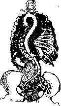
The numerical data for the angles of departure of large arterial branches (in degrees), according to Valentin, are as follows: the angle between the a. anonyma and the a. carot. dextra is 38°, between the aorta and the a. carot. sin. is 81°, between the aorta thoracalis and the a. subclavia sin. is 102°, between the aorta abdom. and the a. renalis dextra (lower angle) is 87°, and between the aorta abdom. and the a. renalis sin. (lower angle) is 88° (Rauber). At the point where a branch departs from the main artery at an acute angle, a protruding sharp ridge—the so-called spur (spur)—is observed on the side of the lumen of the main trunk, against which the blood flow seems to break. Arterial branches to individual organs, anatomical formations, regions, and parts of the body (Lesgaft, Rauber) in the first period of an organ's development follow the shortest path; when an organ is displaced during the process of its development, the origin of the vessel is often located at a considerable distance from the organ (e.g., a. spermatica int., a. ovarica) (Krasusskaya). The main arterial trunks lie on the concave side of the body or limb, between muscles, deep against the bones. On the limbs, they are located on the inner side of the joints, which excludes the possibility of their severe stretching (almost to the loss of the lumen), for example, during flexion, as could be observed if the arteries were located on the outer side. In some cases, the course of an artery is observed to have bends in connection with the curvatures of the bony base upon which it lies; for example, in the case of a curvature of the spine, the aorta makes bends, following the curvatures of the latter (Fig. 3). The arteries of some organs have a very tortuous, often spiral course. This tortuosity is normal and is observed mainly in organs with changing volume, which expand and contract [for example, in the uterus (see separate table, pp. 391-392, Fig. 1), in the urinary bladder], and then in arteries to relatively easily movable parts and formations [for example, aa. umbilicales (see separate table, pp. 391-392, Fig. 2)]; furthermore, a tortuous course can be noted in the a. spermatica int. (or in the a. ovarica), and a spiral twisting of the main trunk and branches—in the vessels of the lungs (spirals of a large radius with a wide chest and a small radius with a narrow one). With age, along with other changes in the arteries, changes in their course occur in the sense of greater tortuosity. Senile changes contribute to the appearance of sharper tortuosity (a. lienalis) or the appearance of bends in straight trunks (e.g., aorta abdominalis).
Individual organs and individual anatomical formations receive arterial branches either from a single source (e.g., kidney, spleen, etc.) or simultaneously from two or more sources (e.g., adrenal glands, brain, pancreas, thyroid, joints, etc.). For some anatomical formations, more or less constant one or several arteries are almost always observed, while for other formations (e.g., muscles, skin, etc.), arterial branches come from nearby sources, usually not fitting into the scheme of arterial distribution. The penetration of arteries into the mass of an organ is accomplished either by a common trunk (without preliminary division) or by several branches. The latter, near the organ or on its surface, in some cases give off connecting branches—anastomoses—either to the homonymous arteries of the opposite side (e.g., arteries of the thyroid gland) or to branches approaching the organ from another source of blood supply (artery of the stomach, aa. thyreoid. sup. et inf.). It is extremely characteristic for the arterial system to have more or less constant anastomoses (see) and collaterals in most regions and organs, both outside the organ and within its mass. Arterial anastomoses in some cases allow for a more uniform distribution of blood in the vessels, while in other cases they allow the mass of blood from one source of blood supply or from the blood vessels of one side to supply blood to the opposite part of the organ if, for example, the arteries of another source of blood supply or the other side are underdeveloped, and thereby compensate for the abnormal blood supply to the organ (in the absence of the a. thyreoidea inf., for example, an increase in the caliber and network of the a. thyreoidea sup. of the corresponding side is observed). Anastomosing type of arteries in this respect differs sharply from arteries of the terminal type of branching. Due to the presence of anastomotic branches, various types of groupings of arterial branches are formed in the form of circles, arcs, networks, etc., or a fusion of arterial trunks is observed, for example, from two into one. In anatomy, some of the anastomotic branches are given separate names, while others do not have them. Examples of arterial anastomoses include the a. communicans ant. (connects the aa. cerebri ant.), the a. communicans post. (connects the a. cerebri post. and the a. carot. int.), and the aa. communic. post. (connect the system of branches of the aa. vertebrales and the aa. carot. int., as well as the arteries of the brain of the right and left sides, and form at the base of the brain the so-called circulus arteriosus Willisii). Arterial circles, but of a somewhat different type (and construction), are also formed in other places, for example, around the circumference of the oral opening, having developed at the expense of the upper and lower labial arteries (aa. labiales sup. et inf.) of the right and left sides. Furthermore, typical anastomoses are the so-called a. radialis superficialis, connecting the a. radialis with the superficial arterial palmar arch, forming the a. ulnaris; a large number of connecting arterial branches in the arteries of the intestine (mesenteric arteries), where, thanks to anastomoses, typical arterial arcs (and loops) are formed, arranged in one, two, or three rows (I, II, and III order), which to a certain extent depends on the length, shape, and arrangement of the intestinal canal (in connection with the nature of the food consumed by different animals; e.g., in carnivores—1 arc, in humans—2-3 and more) (Krasusskaya). The formation of arterial arcs is one of the most frequent forms of the arterial network type; it is found on the stomach, hand, foot, and in other places and organs.
Collaterals can be reduced to certain types: on the limbs—local, short arcs around the main trunk (muscular, bony, vasa vasorum), and distant (networks of various regions), connecting individual systems (for example, a. transversa scapulae and a. subscapularis); in internal organs—intra-organ and extra-organ (distant) collaterals. Among the forms of distribution of arteries connected by numerous anastomoses, one includes the so-called networks (rete), of which the most typical are the networks of arteries nourishing various joints (rete articulare) [see separate table (pp. 375-376), Fig. 1]. Arterial networks with numerous anastomoses are observed in the subcutaneous fatty layer and in the skin itself. In some cases, it is observed that an artery, having divided into two branches, almost immediately passes into one trunk, forming a so-called "islet." Knowledge of more or less constant arterial anastomoses-collaterals is of significant interest to the physician. Of no less interest is familiarity with the potential properties of the arterial system in terms of the possibilities for the formation of bypass (collateral) pathways, new collaterals, and with those capabilities that arteries possess in terms of replacing an excluded (e.g., by a thrombus, ligation) source of arterial blood supply to a certain region in the sense of expanding and proliferating existing, but under normal conditions underdeveloped, anastomotic branches. When ligating an artery, the main role in restoring blood supply belongs to the so-called indirect anastomoses, formed at the expense of branches of neighboring arteries. Direct anastomoses (junctions) between both stumps of the ligated trunk develop significantly less often. The most essential role in the development of indirect collaterals-anastomoses is played by muscular arteries, and a lesser role by arteries nourishing nerves (aa. nutriciae nervorum), and partly by cutaneous arteries (Fig. 4). In addition to anastomoses between arterial branches, in some organs, direct anastomoses between arteries and veins (arteriovenous anastomoses) are observed, first noted by Sucquet and named
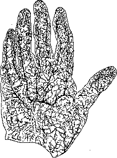
Figure 3. Course of the aorta in connection with the curvature of the spine. Figure 4. Arteries of the skin of the palmar surface of the hand.
derivative channels (canaux derivatifs, s. apparatus derivatorius). These anastomoses exist between vessels of medium and small caliber. According to Hoyer (H. Hoyer), such anastomoses in animals are found on the auricles, at the tip of the nose, on the lips, on the fingers of the forelimbs and hindlimbs, at the tip of the tail, and in the cavernous bodies of the genital organs; in humans, they are found in the terminal digital branches and the cavernous bodies. According to Testut, numerous arteriovenous anastomoses are observed in the pia mater of an adult; Geberg (A. Geberg) discovered them in the renal capsule (of a dog) (Rauber). Arteriovenous anastomoses have also been established in the kidney (between the arcuate arteries and veins of the latter), in the hoof of a calf, and between the pulmonary and bronchial arteries of a child (Krassuskaya). Krassuskaya regards these anastomoses as safety valves in the event of an obstruction appearing in the capillary network. The branching of arterial branches within individual organs (the smallest twigs, arterioles, precapillaries) presents a number of features. Arteries, surrounded by connective tissue, penetrate into an organ (e.g., into the liver), divide into branches that are distributed in the connective tissue around the circumference of the lobules, then penetrate into the lobules themselves, and there pass into capillaries. The very nature of branching within an organ can be different (with or without anastomoses)—either following the same types of branching as in extra-organ branches, or being somewhat peculiar. Branches located inside an organ are grouped according to the internal structure of the organ: for example, in the pancreas, the arterial network of the so-called islets of Langerhans has one character, while the network of the surrounding tissue has another; in the medullary and cortical substance of the adrenal gland, as well as the kidney, the arterial branches are distributed differently.
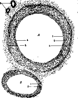
Figure 5. Vasa vasorum: A—artery, B—accompanying vein; 1—intima; 2—media; 3—adventitia; 4—internal elastic lamina; 5—external elastic lamina; 6—vasa vasorum. A completely peculiar method of branching is the so-called wonderful network (rete mirabile). A distinction is made between rete mirabile unipolare and rete mirabile bipolare. By a wonderful network, one understands in the first case (rete mirab. unipol.) a sudden disintegration of an arterial branch into the smallest ramifications in the form of a brush, which pass into capillaries; in the second (rete mirab. bipol.)—a division of arterial branches into the smallest ramifications, forming an arterial network (wonderful network), but not passing directly into capillaries, but rather constituting one vessel again, which gives off capillaries. In the latter case (r. mirab. bipol.), the vessel before the formation of the "wonderful network" is called the vas afferens, and after the "wonderful network"—as if carrying blood away from it—the vas efferens. In humans, only the Malpighian corpuscles of the kidneys are classified as formations in the form of a wonderful network, whereas in animals they are also observed in other places. Arteries to muscles usually approach them from their inner side and are distributed in the muscular tissue, mainly following the course of the muscle fibers. Arterial branches to nerves (e.g., spinal nerves), penetrating them near the place where the nerve emerges to the periphery, go with it and accompany it; hence the name of these arteries—accompanying arteries (or nutrient arteries of the nerves) [see separate table (pp. 391-392), figure 3]. The walls of the arteries are supplied with blood through the vasa vasorum [see figure 5 and separate table (pp. 391-392), fig. 4]. From the capillaries of this vascular network, veins carrying blood away from the arterial walls are formed, which (usually in the number of two) accompany each artery. In the walls of the arteries, there are segmentally located nerve plexuses rich in branches, intended for the supply of their muscular layer (smooth muscles). Large arteries, as well as their branches, are subject to a significant degree to variations (variants, anomalies), many of which are of practical interest. In many cases, their appearance is associated with a temporary disturbance in the embryonic life of the growth and development of individual organs and parts of the body, with a disturbance in the development of individual parts of the arterial system (Tikhomirov), and also reflects one or another stage of embryogenesis. With regard to individual arteries, variants of the origin of a given artery, displacement of the place of its beginning, variants of the course, caliber, and branching in the sense of an increase (resp. decrease) in the number of usually departing branches or the complete absence of one or another vessel are observed. The frequency of the appearance of variants for individual arteries and their character are different. Some variants, especially of large trunks, are extremely rare and are of more morphological interest, while others are encountered relatively frequently and, by their topo-
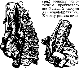
Figure 6. Irregular
Figure 7. Increase in the number
position and course of the aortic arch
of branches of the aortic arch (from a different source
(origin of the vertebral artery from it).
are, for example, the right-sided position of the aortic arch or its irregular course in relation to nearby organs and anatomical structures with a simultaneous variant of the order of origin of its branches from the aortic arch (figure 6). More frequent is, for example, a decrease or (more often) an increase in the number of branches departing from the aorta. As an example, one can take the case of the origin of the vertebral artery (a. vertebralis) from the aortic arch (figure 7) or the case of the origin of the lowest thyroid artery (a. thyreoidea ima) from it (figure 8). The latter
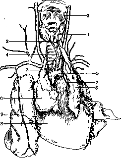
Figure 8. A. thyreoidea ima: 1—thyroid gland; 2—superior thyroid artery (left); 3—inferior thyroid artery (right); 4—lowest thyroid artery; 5—aortic arch; 6—right and left lobes of the thymus gland; 7—thymic arteries; 8—heart; 9—right lung.
can also depart from the brachiocephalic trunk, internal mammary artery, thyrocervical trunk, or common carotid artery. The lowest thyroid artery is observed in approximately 10% of cases (Braus, Rauber). Variants of the branching of arterial branches are observed relatively frequently. For example, the inferior thyroid artery can be observed doubled (usually on one side), can be poorly developed, or be completely absent. The connection of irregular origin, course, change in caliber of branches, etc., depending on the disturbance of the course of development of an organ, clearly appears, for example, in the case of the so-called horseshoe kidney (figure 9). Among the most frequently observed variants of the arteries of the limbs is the high position of the place of division of the brachial artery into its main branches (ulnar and radial arteries)—an example of the scattered type, according to Shevkunenko (Geselevich). Besides the variants appearing
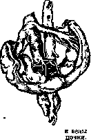
Figure 9. Arteries of a horseshoe
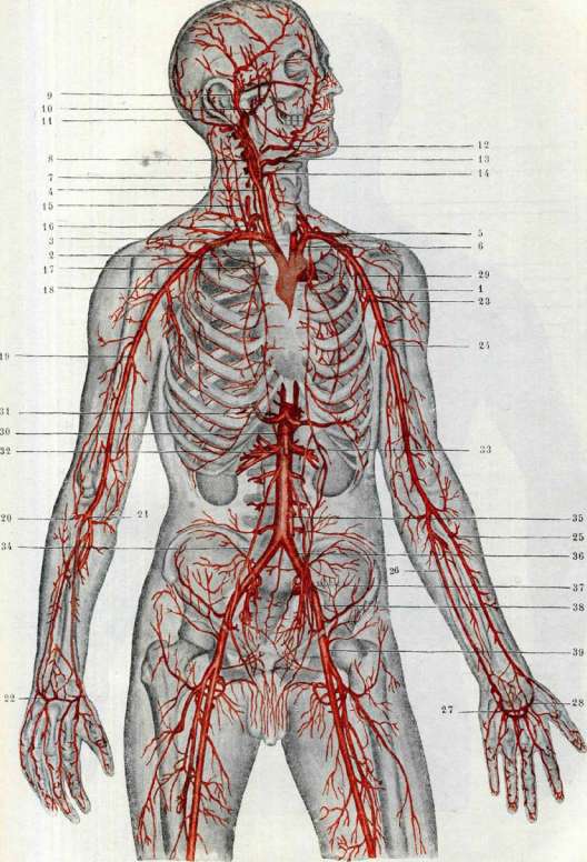
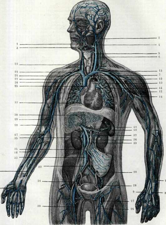
As a result of disturbances in the embryonic development of the circulatory system, variants of arteries are observed that correspond to vessels normally encountered in one or another phylogenetic group, for example, a double aortic arch, a right-sided position of the arcus aortae, the origin of branches from the latter following the pattern of such in certain animals (e.g., truncus bicaroticus), the origin of the a. anonyma and the a. carotis comm. sin. from a common trunk, etc. Venous system, veins (venae). The venous system, as a result of the complex course of the embryonic development of its individual parts, turns out to be constructed asymmetrically, with the arrangement of large venous trunks on the right, with varying lengths of right and left veins of the same name, etc. The structure of the venous system, just like the arterial system, is related to the general constitutional type of body structure, but since, in relation to the venous system and its individual sections and branches, typical structural features have not been sufficiently developed, it is more difficult to speak about the arrangement and character of veins in connection with one or another body type. There is no complete correspondence in the course, arrangement, number of branches, etc., between the arterial and venous systems. The majority of veins bear names that do not correspond to the names of the arteries of the corresponding regions, and only veins that precisely follow the course of a specific artery and lie in immediate proximity to it retain the same name as the artery (for example, a. brachialis-v. brachialis, a. mesenterica inferior-v. mesenterica inferior, etc.). The entire venous system can be divided into separate sections in connection with certain features of their structure. It is necessary to distinguish the veins of the soma (proper veins of the body walls, veins of the limbs, etc.), the veins of the viscera—visceral, of the neck and abdominal cavity (including the pelvic cavity), and the veins of the brain and its membranes (veins of the head). The structure of the vein walls—see Veins. The course of a vein is usually straight or slightly arcuate (excluding veins participating in the formation of so-called plexuses), and even where the corresponding artery has a comparatively tortuous course, the vein is more straight, and therefore shorter (e.g., v. spermatica int., v. lienalis, etc.). Veins of medium and small caliber accompany the corresponding artery in pairs (even the veins of the vasa vasorum); large arteries (e.g., a. axillaris, a. femoralis, etc.) are accompanied by one vein; it is exceptionally rare to note one vein accompanying two arteries of the same name (e.g., aa. dorsales penis dextra et sinistra are accompanied by one v. dorsalis penis). Veins, especially superficial ones, have a large number of anastomoses. In some regions and organs, they form connecting branches between themselves, which, together with the tortuous course of the latter, gives a picture of a very rich venous network; these are plexuses, plexus venosus (e.g., venous plexuses of the pelvic organs, uterus, urinary bladder, plexuses in the spatium epidurale) [see separate table (pp. 391-392), figure 5]. Human arterial system. 1-aorta ascendens et arcus aortae; 2-a. anonyma; 3-a. subclavia dextra; 4-a. carotis communis dextra; 5-a. carotis communis sinistra; 6-a. subclavia sinistra; 7-a. carotis interna dextra; 8-a. carotis externa dextra; 9-a. temporalis superficialis dextra; 10-a. maxillaris interna dextra; 11-a. occipitalis dextra; 12-a. maxillaris externa dextra; 13-a. lingualis dextra; 14-a. thyreoidea superior dextra; 15-a. vertebralis dextra; 16-truncus thyreo-cervicalis dexter; 17-a. mammaria interna dextra; 18-a. axillaris dextra; 19-a. brachialis dextra; 20-a. radialis dextra; 21-a. ulnaris dextra; 22-arcus dorsalis; 23-a. axillaris sinistra; 24-a. brachialis sinistra; 25-a. radialis sinistra; 26-a. ulnaris sinistra; 27-arcus volaris superficialis; 28-arcus volaris profundus; 29-aorta descendens; 30-aorta abdominalis; 31-a. coeliaca; 32-a. renalis dextra; 33-a. mesenterica superior; 34-a. spermatica interna dextra; 35-a. mesenterica inferior; 36-a. iliaca communis sinistra; 37-a. hypogastrica sinistra; 38-a. iliaca externa sinistra; 39-a. femoralis sinistra. Human venous system. 1-v. facialis anterior sinistra; 2-v. facialis posterior sinistra; 3-v. facialis communis sinistra; 4-v. jugularis interna sinistra; 5-v. jugularis communis (interna) sinistra; 6-v. jugularis externa sinistra; 7-v. anonyma sinistra; 8-v. salvatella sinistra; 9-v. cephalica pollicis sinistra; 10-v. basilica sinistra; 11-v. cephalica sinistra; 12-v. axillaris sinistra; 13-v. subclavia sinistra; 14-arcus venosus; 15-vv. comitantes a. ulnaris dext.; 16-vv. comitantes a. radialis dext.; 17-v. mediana cubiti dextra; 18-v. basilica dextra; 19-vv. comitantes a. brachialis dext.; 20-v. axillaris dextra; 21-v. cephalica dextra; 22-v. subclavia dextra; 23-v. jugularis interna (communis) dextra; 24-v. anonyma dextra; 25-v. cava superior; 26-v. saphena magna sinistra; 27-v. femoralis sinistra; 28-v. iliaca externa sinistra; 29-v. iliaca communis sinistra; 30-v. femoralis dextra; 31-v. iliaca externa dextra; 32-v. iliaca communis dextra; 33-v. cava inferior; 34-v. spermatica interna dextra; 35-v. renalis dextra; 36-v. spermatica interna sinistra; 37-v. renalis sinistra; 38-vv. hepaticae; 39-v. mesenterica superior; 40-v. lienalis; 41-v. portae; 42-arcus aortae; 43-a. pulmonalis; 44-vv. pulmonales sinistrae. (To the illustration of the article Blood Vessels.)
To the article Blood Vessels.
Due to the presence of anastomoses in a number of regions, networks with arches and loops are formed, e.g., mesenteric veins, subcutaneous veins, veins of the dorsum of the hand, foot, etc. However, anastomoses rarely connect superficial veins with deep ones, e.g., in the limbs. Among the peculiar anastomoses of the venous system, one should include the so-called venous emissary veins—vv. emissariae—small trunks connecting the venous sinuses of the dura mater of the brain with the superficial veins of the head, passing through openings in the cranial bones [see separate table (pp. 391-392), fig. 5] (e.g., emissarium parietale in the foramen parietale of the parietal bone, connecting the sinus sagittalis of the dura mater with the superficial veins of the head). In some regions, anastomoses are observed connecting the right and left halves of the venous system of a given region (for example, arcus venosus juguli—a venous arch located in the neck region) and, thanks to this, forming a kind of circle—circulus venosus, e.g., in the region corresponding to the position of the circulus arteriosus Willisii. The veins of the trunk, limbs, neck, and head are divided into superficial (vv. superficiales, vv. cutaneae), subcutaneous, passing independently (independently of the arteries), lying in the mass of the subcutaneous fatty layer, with numerous anastomoses, arches, and rich in valves [see separate table (pp. 375-376), fig. 2]; deep veins (profundae), usually running together with the corresponding arteries in a common connective tissue sheath, accompanying the arteries (vv. comitantes) (fig. 10), in the number of one or two, with single veins of medium caliber gradually passing into a single vein of larger caliber, which receives the veins of its region and receives a new name corresponding to the artery. Superficial veins flow into deep ones (e.g., v. saphena magna flows into the deep v. femoralis), piercing the fascia of the given region. The veins of the internal organs flow into three main trunks of the venous system, namely: into the superior vena cava, the inferior vena cava, which constitute these and the trunks themselves, not
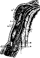
Fig. 10 Vv. comitantes: 1-a. axill.; 2-v. axill.; 3-v. cephal.; 4-a. brachial.; 5-vv. brachial. (vv. comitantes); 6-vv. comitant. a. rad.; 7-vv. comitantes a. uln.; 8-v. basil.; 9-v. mediana cubiti.
and into the portal vein. The branches of the trunks, just like the arteries, have valves; in some organs, venous plexuses (plexus venosus) are formed, while in others they strictly correspond to the course of the arteries (e.g., a. renalis-v. renalis). The portal vein system (see) differs sharply from other visceral veins in that it does not carry purely venous blood; the branches that compose it correspond to several arterial branches of the abdominal aorta, and finally, it does not flow into any larger venous trunk, but branches out in the parenchyma of the liver. The veins of the brain are characterized by the fact that they do not accompany its individual arteries, do not correspond to them in their course, and do not pour blood into larger venous trunks that carry it away from individual parts of the head, but first divert it into slit-like cavities (a peculiar accessory system to the general venous one), the so-called sinuses of the dura mater of the brain, from where the blood is removed through a large vein (v. jugularis int.), and partly into the superficial veins through venous emissaries [see separate table (cols. 391–392), figure 5]. Finally, they do not have valves. The causes for the appearance of venous variants should also be sought in disturbances of the developmental course and a number of other factors of the embryonic life period. An incorrect course of development and the final formation of an organ with a shape, size, etc., deviating from the relative norm, produces corresponding deviations in its veins as well. In other cases, the appearance of a variant is connected, as it were, exclusively with the developmental course of a specific section of the venous system, and not the organ. One distinguishes variants of the vein's course, its topographical position, absence or increase in number, the appearance of additional trunks, etc. A peculiar picture is observed in the so-called horseshoe kidney, which confirms the position regarding the connection of organ development with the development of its blood vessels (Fig. 9). The appearance of an additional venous trunk is observed, for example, between v. renalis sin. and v. iliaca comm. sin. (corresponding to v. cardinal. post. sin.) (Fig. 11), between v. portae and v. cava inf. (v. porto-renalis sin., v. porto-spermatica dextra) (Torka
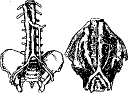
Fig. 11. Connecting venous trunk between v. renal. and v. iliaca comm. (remnant of v. cardin. post. sin.).
Figure 12. Incorrect course and low entry of v. renal. sin. (cheva). An incorrect topographical position and course of the vein is illustrated, for example, by a case in which the direction of v. renalis sin. is observed behind the abdominal aorta and its entry into v. cava inf. at the site of the fusion of vv. iliacae communes (Fig. 12), moreover at a significant distance from the site of entry of v. renalis dextrae. For the structure of B. v., as well as the pathological anatomy of blood vessels, see Artery, Veins. 411
BLOOD VESSELS
412 Table of arteries. Name of artery Point of origin Organs or region supplied Abdominalis externa, s. circumflexa ilium profunda, s. abdominalis sub- cutanea, s. epigastrica superficialis (external abdominal art.) See a. epigastrica superficialis Acetabuli (ramus) [art. of the acetabulum (branch)] A. circumflexa femoris medialis, a. circumflexa fem. lateralis Hip joint Acetabuli (BNA), ramus acetabuli [art. of the acetabulum (branch)] A. obturatoria Hip joint, lig. teres femoris, caput femoris Acromialis ramus (acromial branch) A. thoraco-acromialis Region of the shoulder girdle, acromial process and upper part of the deltoid muscle Adiposa cordis A. coronaria cordis dextra, a. coronaria dextra (BNA) A. pulmonalis Adiposa(ae) a. renalis (art. of the adipose capsule of the kidney) A. suprarenalis media et inferior, a. phrenica infer., a. renalis, aa. colicae dextra, media et sinistra, a. spermatica interna, aa. lumbales prima, secunda et tertia, aorta abdominalis Capsula adiposa renis Ad pontem [art. to the pons (Varolii)] A. basilaris Pons Varolii Alaris Halleri, s. alaris glandulosa axillaris, s. alaris thoracica [alar art. (Haller)] A. axillaris Skin, fasciae and lymph glands of the axillary fossa Alveolaris(es) (alveolar art.) A. alveolaris inferior Alveoli of the lower jaw Alveolaris(es) (alveolar art.) A. infraorbitalis Walls of the alveoli of the maxillary bone Alveolaris inferior, s. mandibularis, s. maxillaris (dentalis) inf. (inferior alveolar art.) A. maxillaris interna Soft tissues of the chin, lower lip, mm. mylo-hyoideus, buccinator, lower jaw, alveoli of the lower jaw, mucous membrane of the oral cavity, gums, teeth of the lower jaw Alveolares sup. anteriores (BNA), s. supramaxill., s. dentales superiores (anterior superior alveolar art.) A. infraorbitalis Periosteum of the maxilla, mucous membrane, sinus maxillaris, alveoli of the upper jaw, gums, teeth (canines and incisors) Alveolaris sup. post. (posterior superior alveolar art.) A. maxillaris interna Periosteum and bone substance of the maxilla, mucous membrane of the cheeks, mucous membrane of the sinus maxillaris, gums and teeth of the upper jaw (molars and premolars), mm. buccinator et masseter Anastomotica brachii (Quain), s. collateralis ulnaris inferior (anastomotic art. of the arm) See a. collateralis ulnaris inferior Anastomotica magna, s. genu suprema (great anastomotic artery) See a. genu suprema Angularis (BNA), s. nasalis post. lateralis (angular art.) A. maxillaris externa Wings and dorsum of the nose, mm. nasalis, frontalis, corrugator supercilii, procerus, orbicularis oris Anonyma, s. innominata, truncus anonymus, s. innominatus (innominate art.) [unpaired] Arcus aortae Right half of the neck, head, chest, shoulder girdle and right upper limb Anonyma iliaca, s. iliaca comm. (innominate iliac art.) See a. iliaca communis Aorta abdominalis (abdominal aorta) Continuation of aorta thoracica Organs of the abdominal cavity, reproductive organs, diaphragm, muscles of the back and contents of the vertebral canal
I Name of artery Point of origin Organs or region supplied Aorta ascendens (ascending aorta) Conus arteriosus of the left ventricle Heart Aorta descendens (descending aorta) See Thoracalis aorta Appendicularis (BNA), s. appendicalis, s. vermicularis (art. of the vermiform appendix) Ramus colicus a. ileo-colicae or directly from a. ileo-colica or ramus ilealis descend. a. mesenter. superioris Processus vermicularis (appendix) Arciformis(es) renis(um), s. arcus vasculosus minor (arcuate art. of the kidney) A. interlobaris(es) Parenchyma of the kidney Arcuata (BNA), s. metatarsea, s. tarsea lateralis ant. (arcuate art.) A. dorsalis pedis Interosseous spaces (II, III and IV) and toes of the foot (forms rete dorsale pedis) Arcus aortae (aortic arch) Continuation of aorta ascendens Neck, head, chest, upper limbs Articularis capituli fibulae propria, s. ramus fibularis (BNA) (proper articular art. of the head of the fibula) See ram. fibularis Articulares genu, s. aa. genu (articular art. of the knee) See aa. genu Ascendens ramus (ascending branch) A. transversa colli Muscles of the occipital region (partially) Atrabiliaris, s. suprarenalis media See a. suprarenalis media Auditiva externa (Weber), s. tympanica ant. (BNA) (external auditory art.) See a. tympanica anterior Auditiva interna (internal auditory artery) A. basilaris Membranous semicircular canals, sacs (spherical and elliptical) of the vestibule and cochlea Auricularis ramus (BNA) (auricular branch) A. auricularis posterior Auricle, muscles of the external ear Auricularis(es) ant. ramus(i) (anterior auricular branch) A. temporalis superficialis Auricle, muscles of the auricle, external auditory meatus Auricularis cordis dext. ramus (branch to the right auricle of the heart) A. coronaria cordis dextra Right atrium (auricle) Auricularis cordis sin. ramus (branch to the left auricle of the heart) A. coronaria cordis sinistra Left atrium (auricle) Auricularis posterior (posterior auricular artery) A. carotis externa Mm. digastricus mandibulae (venter post.), stylo-hyoideus, stylo-glossus, sterno-cleido-mastoideus, masseter, pterygoideus int., occipitalis, auriculares, stapedius, gl. parotis, tympanic cavity, mucous membrane of the cellulae mastoid. and middle ear, dura mater Auricularis profunda (deep auricular art.) A. maxillaris int. Temporomandibular joint, skin of the external auditory meatus, tympanic membrane Axillaris (axillary art.) Continuation of a. subclavia Fasciae and lymph glands of the axillary fossa, muscles of the chest, lateral thoracic wall, mammary gland (partially), shoulder joint, muscles of the shoulder girdle Azygos, s. genu media (unpaired artery) See a. genu media Basilaris, s. truncus basilaris (basilar art.) [unpaired] Formed by the fusion of aa. vertebrales Pons Varolii, cerebellum, thalamus opticus, corpora quadrigemina and the adjacent part of the brainstem, posterior section of the cerebral hemispheres (temporal and occipital lobes), internal ear 415 BLOOD VESSELS
| Name of Artery | Origin | Supplied Organs or Region | | :--- | :--- | :--- | | Bicipitalis (Gegenbaur) (biceps artery) | A. brachialis | Biceps brachii muscle | | Brachialis (BNA), s. humeralis (humeral artery) | Continuation of a. axillaris | Upper limb: mm. biceps brachii, coraco-brachialis, pronator teres, triceps brachii, flexor carpi ulnaris, brachialis (internus), humerus | | Bronchialis ant. ramus (anterior bronchial branch) | Aa. mediastinales anteriores | Bronchi | | Bronchialis post. (posterior bronchial artery) | Aorta thoracalis | Lung tissue, trachea, esophagus, and lymphoglandulae bronchiales | | Bronchialis sup. (superior bronchial artery) | Arcus aortae | Bronchi | | Buccinatoria, s. buccalis (buccal artery) | A. maxillaris int. | Mm. buccinator, masseter, pterygoideus int., caninus and adjacent facial muscles, oral mucosa, gums (of the upper jaw), upper molars | | Bulbi urethrae (BNA), s. bulbo-urethralis, s. bulbo-cavernosa, s. bulbina, s. corporis cavernosi urethrae, s. bulbosa (artery of the bulb of the corpus cavernosum of the urethra) | A. penis, a. pudenda interna | Bulbus urethrae, soft tissues of the trigonum urogenitale, pars membranacea urethrae, glandulae bulbo-urethrales, corpus cavernosum urethrae, bulbus vestibuli of women | | Bulbi vestibuli (vaginae) (artery of the bulb of the vestibule of the vagina) | A. clitoridis | Bulbus vestibuli | | Calcanei laterales rami (lateral calcaneal branches) | A. peronaea | Region of the lateral malleolus and lateral side of the calcaneus | | Calcanei mediales rami (medial calcaneal branches) | A. tibialis post. | Region of the medial malleolus and medial side of the calcaneus | | Canalis pterygoidei (Vidii), s. Vidiani [artery of the pterygoid (Vidian) canal] | A. maxillaris int. or a. palatina descendens | Upper wall of the pharynx, tuba auditiva, tympanic cavity | | Capitis femoris ant. (Anserov) (anterior artery of the head of the femur) | A. circumflexa femoris lateralis | Head and neck of the femur | | Capitis femoris medialis (Anserov) (medial artery of the head of the femur) | A. circumfl. femoris med. | Head and neck of the femur | | Capitis femoris posterior lateralis (Anserov) (posterior lateral artery of the head of the femur) | A. perforans I | Head and neck of the femur | | Capsularis, s. a. hyaloidea (capsular artery) | See a. hyaloidea | - | | Carotico-tympanicus ramus (carotico-tympanic branch) | A. carotis interna | Part of the tympanic cavity, mucous membrane of the middle ear | | Carotis communis dextra, s. primitiva (right common carotid artery) | A. anonyma | Right half of the neck and head, also organs of the corresponding regions | | Carotis communis sin., s. primitiva (left common carotid artery) | Arcus aortae | Left half of the neck and head, also organs of the corresponding regions | | Carotis externa, s. facialis (external carotid artery) | A. carotis communis | Region of the neck, head, face, and corresponding organs | | Carotis interna, s. cerebralis (internal carotid artery) | A. carotis communis | Brain (partially), crus cerebri, tuber cinereum, pituitary gland, eyeball and contents of the orbit, eyelids, soft tissues of the forehead region (partially), saccus lacrimalis, skin of the root of the nose | | Carpeus dorsalis ramus (dorsal carpal branch) | A. radialis and ulnaris | Branches to the rete carpi dorsale | | Carpeus volaris ramus (palmar carpal branch) | A. radialis and ulnaris | Ligamentous apparatus of the wrist joint region and the joint | | Caudalis, s. sacralis media (caudal artery) | See a. sacralis media | - | | Cavernosa penis, s. profunda penis (cavernous artery of the penis) | See a. profunda penis | - | | Cavi tympani, s. tympanica inf. (artery of the tympanic cavity) | See a. tympanica inf. | - | | Centralis retinae, s. Zinni (central artery of the retina) | A. ophthalmica | Inner layers of the retina of the eye | | Cerebelli inf. ant., s. media [anterior, inferior (or middle) cerebellar artery] | A. basilaris | Anterior surface of the cerebellar hemispheres (lobulus biventer and flocculus) | | Cerebelli inf., s. inf. post. [inferior (or posterior inferior) cerebellar artery] | A. vertebralis | Inferior surface of the cerebellum (lobulus semilunaris inf., tonsilla cerebelli, vermis inferior) | | Cerebelli superior (superior cerebellar artery) | A. basilaris | Superior surface of the cerebellum (lobulus quadrangularis et semilunaris sup., vermis superior) | | Cerebri anterior, s. cerebralis ant., s. corporis callosi (anterior cerebral artery) | A. carotis interna | Head of the caudate nucleus, anterior part of the internal capsule, ependyma of the anterior horn of the lateral ventricle, medial surface of the frontal and parietal lobes, corpus callosum (except its posterior end), part of the outer surface (superior and middle frontal gyri), upper part of the anterior central gyrus, part of the inferior surface (gyrus rectus), tractus olfactorius, bulbus olfactorius, lamina cinerea | | Cerebri media, s. cerebralis media, s. fossae cerebri med., s. fossae Sylvii, s. insularis (middle cerebral artery) | A. carotis interna | Globus pallidus, thalamus opticus, head of the caudate nucleus, uncus, insula (Reili), middle and inferior frontal gyri, central parietal and temporal gyri | | Cerebri posterior, s. profunda cerebri (posterior cerebral artery) | A. basilaris | Base and tegmentum of the midbrain, region of the corpora quadrigemina, cerebral peduncles, thalamus, subthalamic region, walls of the lateral and III ventricles, nuclei of the III and IV cranial nerves, their roots, substantia nigra (Soemmeringi), temporal and occipital gyri, gyrus uncinatus, gyrus lingualis, cuneus, corpora quadrigemina and posterior part of the thalamus opticus | | Cervicalis ascendens, s. ramus cervicalis trunci thyreo-cervicalis (ascending cervical artery) | Truncus thyreo-cervicalis and a. thyreoidea inferior | Mm. scaleni, m. platysma, deep muscles of the neck (longus capitis et colli, etc.), levator scapulae | | Cervicalis descendens profunda, s. ramus occipitalis, s. ramus descendens (BNA) (descending deep cervical artery) | A. occipitalis | Soft tissues of the occipital region | | Cervicalis profunda, s. princeps cervicis (Macalister) (deep cervical artery) | Truncus costo-cervicalis | Mm. scaleni, deep muscles of the neck (longus capitis et colli, etc.) and occipital region | | Cervicalis superficialis, s. transversa, s. transversalis cervicis (superficial cervical artery) | A. thyreo-cervicalis | Posterior belly of m. omo-hyoidei, lymph nodes and skin of the lateral regions of the neck, mm. sterno-cleido-mastoideus, platysma, trapezius, levator anguli scapulae, splenius, rhomboidei | | Chorioidea, s. chorioidea ant., s. inferior [anterior (or inferior) artery of the choroid plexus] | A. carotis interna | Plexus chorioideus, ventric. lateral. et ventric. tertii, region of the lateral geniculate body, optic tract, tail of the nuclei caudati, part of the nuclei lenticularis, retrolenticular part of the capsulae internae and Ammon's horn | | Chorioidea posterior (media) [posterior (middle) artery of the choroid plexus] | A. cerebri posterior | Plexus chorioideus and walls of ventriculi tertii | | Chorioidea(ae) superior(es) (superior artery of the choroid plexus) | A. ophthalmica | - | | Ciliaris(es) anterior(es) (anterior ciliary artery) | A. ophthalmica | - | | Ciliaris(es) post. brevis(es) (short posterior ciliary artery) | A. ophthalmica | - | | Ciliaris(es) post. longa(ae), s. media(ae) Cruveilhier (long posterior ciliary artery) | A. coronaria cordis sin. | - |
A. profunda femoris, A. profunda femoris, Sclera, conjunctiva bulbi, m. ciliaris, iris (forms with its branches the circulus arteriosus iridis major and circulus arteriosus iridis minor), Sclera (posterior half), chorioidea (tunica vasculosa), Corpus ciliare, iris, m. ciliaris, Lateral and posterior parts of the left ventricle, left atrium, Mm. tensor fasciae latae, glutaeus medius et minimus, quadriceps femoris, sartorius, hip joint, Mm. psoas major, adductor brevis, adductor magnus, pectineus, vastus lateralis, hip joint, See a. genu, A. axillaris, A. axillaris, Caput humeri, shoulder joint, mm. coraco-brachialis, biceps brachii, Shoulder joint, mm. deltoideus, coraco-brachialis, triceps brachii (partially), A. iliaca externa, A. femoralis, Anterior abdominal wall (partially), mm. transversus abdominis, obliquus abdom. ext. et int., iliacus, tensor fasciae latae, sartorius, Skin and fascia of the region of the spina iliaca ant. superior and above the lig. inguinale, lymphoglandulae inguinales, m. sartorius, See a. interossea recurrens, A. subscapularis or a. axillaris, Clitoridis, s. clitoridea (artery of the clitoris), Coccygea (Rauber's) (coccygeal artery), Cochleae, s. ramus cochleae (artery of the cochlea), Coeliaca, s. truncus coeliacus (celiac artery) [unpaired], Colicus ramus (branch to the colon), A. pudenda interna (its continuation), A. glutaea inferior, A. auditiva interna, Aorta abdominalis, A. ileo-colica, Mm. teres major et minor, latissimus dorsi, deltoideus, infraspinatus et supraspinatus, Clitoris, Coccyx and apex of the sacrum, Cochlea (spiral ganglion of the cochlea, osseous spiral lamina, wall of the scala tympani up to the spiral ligament), Greater part of the stomach, lower section of the esophagus, liver, gallbladder, spleen, duodenum, pancreas, greater omentum, Terminal segment of the intestin. ileum, caecum, appendix, lower third of the colon ascendens
BLOOD VESSELS
Name of artery, Place of origin, Colica dextra (right artery of the colon), Colica media, s. sup. accessoria (middle artery of the colon), Colica media accessoria (Waldeyer) (middle accessory artery of the colon), A. ileo-colica or a. colica media (in 75%); more often from the first or from the a. mesenterica superior (in 25%) 4, A. mesenterica superior, Colica sinistra (left artery of the colon), Collateralis media (middle collateral artery), Collateralis radialis (radial collateral artery), A. colica media (more often) or a. mesenterica sup., A. mesenterica inferior, A. profunda brachii, A. profunda brachii, Collateralis ulnaris inferior (lower ulnar collateral artery), Collateralis ulnaris sup. (upper ulnar collateral artery), Comitans, s. comes n. ischiadici, s. ramus (comes) ischiadiaeus a. ischiadicae (artery accompanying the sciatic nerve), Comitans n. phrenici, s. pericardiaco-phrenica (artery accompanying the phrenic nerve), Communicans peronaeae ramus (communicating branch), Communicans tibialis ramus (communicating branch), Communicans anterior (anterior communicating artery) [unpaired], Communicans posterior (posterior communicating artery), Conjunctivalis(es) anterior(es) (anterior artery of the conjunctiva of the eye), A. brachialis, A. brachialis, Organs or area supplied, Colon ascendens, Proximal section of the colon transversum (2/3), Colon transversum (middle part), Distal section of the colon transversum (1/3), colon descendens, Elbow joint, olecranon, Elbow joint, mm. extensor carpi radialis brevis, extensor carpi radialis longus, brachio-radialis, A. glutaea inferior, Elbow joint (partially), mm. brachialis int., biceps brachii, flexor carpi ulnaris, Elbow joint, m. brachialis internus, caput mediale m. tricipitis, mm. biceps brachii, flexor carpi ulnaris, A. mammaria interna, Nervus ischiadicus, Pericardium, diaphragm, pleura, A. peronaea, A. tibialis post., A. cerebri ant. dextra et sinistra, A. carotis interna, A. ciliaris ant., Conjunctivalis(es) posterior(es) (artery of the conjunctiva of the eye)
posterior) i Cornu Ammonis (artery of the Ammon's horn) Coronaria cordis dextra (right coronary artery of the heart) Coronaria cordis sinistra (left coronary artery of the heart) Coronaria labii inf., s. labialis inf. (coronary artery of the lower lip) Aa. palpebrales mediales et laterales A. cerebri post. Aorta ascendens (sinus aortae dexter) Anastomotic branch of a. peronaea with a. tibialis post. Anastomotic branch of a. tibialis post. with a. peronaea Optic chiasm, septum pellucidum and anterior commissure Anterior 1/3 of the cerebral peduncle, tuber cinereum, chiasma nn. opt., corpus Luysi, anterior 1/3 of the posterior part of the internal capsule and part of the lateral nucleus of the thalami Conjunctiva of the eye, m. ciliaris, iris Conjunctiva of the eye Hippocampus Aorta ascendens (sinus aortae sinister) Conus arteriosus, right atrium, walls of the right auricle, aorta and pulmonary artery, adipose tissue surrounding the vessels, right and left ventricles, atrioventricular bundle of His-Tawara Left atrium, walls of the aorta and pulmonary artery, right and left ventricles, septum (of the ventricles) See a. labialis inf. Name of artery Place of origin Supplied organs or region Coronaria labii sup., s. labialis sup. (coronary artery of the upper lip) See a. labialis sup. Coronaria malleolaris post. lateral., s. malleolaris post. lat. (posterior lateral malleolar coronary artery) See a. malleolaris post. lateralis Coronaria ventriculi dextra, s. gastrica dextra (right coronary artery of the stomach) See a. gastrica dextra Coronaria ventriculi inf. dextra, s. gastro-epiploica dextra (right inferior coronary artery of the stomach) See a. gastro-epiploica dextra Coronaria ventriculi inf. sin., s. gastro-epiploica sinistra (left inferior coronary artery of the stomach) See a. gastro-epiploica sinistra Corporis callosi, s. cerebri ant. (artery of the corpus callosum) See a. cerebri ant. Corporis cavernosi urethrae, s. bulbi urethrae (artery of the corpus cavernosum of the urethra) See a. bulbi urethrae Costo-cervicalis truncus (costocervical trunk) A. subclavia Deep muscles of the neck, I and II intercostal spaces Cremasterica (Cooper's), s. spermatica externa (cremasteric artery) See a. spermatica externa Crico-thyreoideus ramus (cricothyroid branch) A. thyreoidea superior M. crico-thyreoideus Cruralis, s. femoralis (femoral artery) See a. femoralis Cruralis iliaca, s. iliaca externa (femoral iliac artery) See a. iliaca externa Cubitalis, s. ulnaris (ulnar artery) See a. ulnaris Cystica (artery of the gallbladder) [unpaired] Ramus dexter or main trunk of a. hepaticae propr. Gallbladder Deferentialis, s. spermatica deferentialis (artery of the ductus deferens) A. hypogastrica or vesicalis inf. Ductus deferens, epididymis Deltoideus ramus (deltoid branch) A. profunda brachii M. deltoideus Deltoideus ramus (deltoid branch) A. thoraco-acromialis Mm. deltoideus and pectoralis major Lower teeth Dentalis(es) ramus(i) (dental branch) A. alveolaris inf. Dentalis(es) ramus(i) (dental branch) A. infraorbitalis Upper teeth Descendens ramus (descending branch) A. transversa colli Mm. rhomboideus major, rhomboideus minor, serratus posterior superior, subscapularis, infraspinatus, latissimus dorsi, part of the deep muscles of the back Descendens ant. ramus (anterior descending branch) A. coronaria cordis sinistra Anterior wall of the right and left ventricles and the interventricular septum Descendens posterior ramus (posterior descending branch) A. coronaria cordis dextra Right and left ventricles of the heart, right atrium Diaphragmatica inf., s. magna (inferior, or great diaphragmatic artery) See a. phrenica inferior Diaphragmatica superior (superior diaphragmatic artery) See a. phrenica superior Name of artery Place of origin Supplied organs or region Digitalis(es) manus dorsalis(es) (dorsal digital artery of the hand) A. metacarpea dorsalis I Aa. metacarpeae dorsales A. arcuata Both dorsal margins of the thumb and the radial dorsal margin of the index finger (of the hand) Digitalis(es) manus dorsalis(es) (dorsal digital artery of the hand) Facing dorsal surfaces of the II, III, IV and V fingers (of the hand) Digitalis(es) pedis dorsalis(es) (dorsal digital artery of the foot) Facing dorsal margins of the II, III, IV and V fingers (of the foot) Digitalis(es) plantaris(es) (plantar digital artery) Aa. metatarseae plantares Arcus volaris superficialis A. digitalis(es) volaris(es) comm. Plantar side of the I (lateral side), II, III, IV and V (medial side) toes Digitalis(es) volaris(es) communis(es) (common palmar digital artery) Fingers of the hand via their branches (aa. digit. volar. propr.) Digitalis(es) volaris(es) propria(ae) (proper palmar digital artery) Palmar surfaces of the facing sides of the II, III, IV and V fingers (of the hand) Digitalis(es) volaris(es) propria(ae) (proper palmar digital artery) A. metacarpea volaris prima (s. princeps pollicis) Palmar surfaces of the medial and lateral sides of the thumb and the radial side of the index finger Diploici rami (branches to the spongy substance of the bone) A. alveolaris inferior Diploe of the mandible Dorsalis clitoridis (dorsal artery of the clitoris) A. clitoridis A. tarsea later. or rete dorsale pedis Clitoris, labia minora Dorsalis digiti V lateral. (pedis) [dorsal lateral artery of the little toe (of the foot)] Dorso-lateral side of the V toe (of the foot) Dorsalis digiti II medial. (pedis) [dorsal medial artery of the II toe (of the foot)] A. metatarsea dorsalis I Dorso-medial margin of the II toe (of the foot) Dorsalis hallucis lateralis (dorsal lateral artery of the big toe) A. metatarsea dorsalis I Dorso-lateral side of the big toe (of the foot) Dorsales linguae rami [dorsal branches of the tongue (arteries of the tongue)] A. lingualis Mucous membrane of the dorsum of the tongue and the region of the foramen caecum, palatine tonsil Dorsum and root of the nose, walls of the saccus lacrimalis Dorsalis nasi (dorsal artery of the nose) A. ophthalmica Dorsalis pedis, s. pediaea, s. pedida, s. tarsea interna (dorsal artery of the foot) - Continuation of a. tibialis ant. Skin of the dorsum of the foot, medial and lateral margin of the foot, mm. extensor dig. brevis, extensor hallucis brevis Dorsalis penis (dorsal artery of the penis) A. penis (its terminal branches), a. pudenda interna Integuments of the penis, its end, upper part of the scrotum, cavernous bodies of the glans penis, prepuce Dorsalis scapulae, s. transversa colli (dorsal artery of the scapula) See a. transversa colli Duodenales rami (branches to the duodenum) A. pancreatico-duodenalis sup. Duodenum Duodenalis inf., s. pancreatico-duodenalis inferior (inferior artery to the duodenum) See a. pancreatico-duodenalis inferior Emulgens, s. renalis See a. renalis Epigastrica inferior (profunda, s. interna) [inferior (deep, or internal) epigastric artery] A. iliaca externa Mm. rectus abdominis, transversus abdominis, obliq. abdom. int. et ext., pyramidalis, cremaster, funiculus spermaticus, ligam. teres uteri, posterior surface of the os pubis and adjacent soft tissues Name of artery Epigastrica lateralis (lateral epigastric artery) Epigastrica superficialis, s. abdominalis subcutan., s. subcutan. abdominis (superficial epigastric artery) Place of origin Supplied organs or region A. circumflexa ilium profunda Epigastrica superior, s. ramus epigastricus a. mammariae internae (superior epigastric artery) Epiploici rami (branches to the greater omentum) Episcleralis(es), s. subconjunctivalis (episcleral artery) Ethmoidalis ant., s. ethmoidea anterior (anterior ethmoidal artery) Ethmoidalis post., s. ethmoidea posterior (posterior ethmoidal artery) A. femoralis A. mammaria int. A. gastro-epiploica dextra A. ciliaris(es) ant. A. ophthalmica A. ophthalmica or a. supraorbitalis Facialis anterior, s. maxillaris externa (anterior facial artery) Facialis profunda, s. maxillaris interna (BNA) (deep facial artery) Facialis transversa, s. transversa faciei (transverse facial artery) Femoralis communis, s. superficialis, s. cruralis, s. pelvicruralis (femoral artery) Femoralis profunda, s. profunda femoris (deep femoral artery) Fibularis ramus (fibular branch) Fossae cerebri mediae (artery of the middle cranial fossa) Anterior abdominal wall Skin and subcutaneous layer of the anterior abdominal wall above the lig. inguinale, inguinal lymph nodes Xiphoid process of the sternum, anterior abdominal muscles (mm. rectus abdominis, transversus abdomin., obliquus abdom. intern. et ext.) Greater omentum, diaphragm, anterior and posterior surfaces of the stomach above the curvatura major Sclera (anterior half) Dura mater, lateral wall and septum of the nasal cavity, anterior cells of the ethmoid bone, frontal sinus, mm. nasalis, procerus Dura mater in the region of the anterior cranial fossa, mucous membrane of the ethmoid cells, sinus frontalis, nasal septum See a. maxillaris externa See a. maxillaris interna See a. transversa faciei Continuation of a. iliaca externa Anterior abdominal wall, muscles of the thigh, hip joint, femur, external genitalia See a. profunda femoris A. tibialis post.
Soft tissues surrounding the head of the fibula, knee joint. See a. cerebri media. Frontalis ramus (frontal branch). Frontalis (frontal artery). Funicularis, s. testicularis (funicular artery). Funiculi spermatici, s. a. spermatica externa (artery of the spermatic cord). A. temporalis superficialis. A. ophthalmica. Mm. orbicularis oculi, frontalis, corrugator supercilii, galea aponeurotica, skin of the forehead. Soft tissues of the forehead region, eyelids, mm. frontalis, procerus. See a. testicularis. See a. spermatica externa. Gastricae breves (BNA) (short gastric arteries). A. lienalis. Gastrica dextra (BNA), s. coronaria ventriculi super. dextra, s. gastrica super. dextra (right gastric artery). Gastrica sinistra (BNA), s. gastrica sup. sinistra, s. coronaria ventriculi sup. sin. (left gastric artery). A. hepatica propria. A. coeliaca or (rarely) aorta abdominalis. Bottom of the stomach. Pyloric part of the stomach. Entrance part of the stomach (cardia) and lower section of the esophagus. Name of artery. Origin. Supplied organs or region. Gastro-duodenalis (gastroduodenal artery). A. hepatica. Stomach, duodenum, pancreas, greater omentum. Gastro-epiploica dextra (BNA), s. coronaria ventriculi inf. dext., s. gastr. inf. dext. (right artery of the stomach and greater omentum). A. gastro-duodenalis. Stomach, greater omentum. Gastro-epiploica sinistra (BNA), s. coronaria ventriculi inf. sin., s. gastr. inf. sin. (left artery of the stomach and greater omentum). A. lienalis. Stomach, greater omentum. Genu inferior lateralis (BNA), s. externa (inferior lateral knee artery). A. poplitea. Knee joint, m. peronaeus longus. Genu inf. medialis (BNA), s. interna (inferior medial knee artery). A. poplitea. Knee joint, m. popliteus. Genu media (BNA), s. azygos (middle knee artery). A. poplitea. Knee joint, synovial folds, lig. cruciatum anterius, lig. cruciatum posterius, plicae alares, plica synovialis patellaris. Genu recurrens, s. recurrens tibialis ant. (BNA) (recurrent knee artery). See a. recurrens tibialis anterior. Genu superior lateralis (BNA), s. externa (superior lateral knee artery). A. poplitea. Knee joint. Genu superior medialis (BNA), s. interna (superior medial knee artery). A. poplitea. Knee joint. Genu suprema, s. superficialis, s. anastomotica magna, s. ramus musculo-articularis a. cruralis (highest knee artery). A. femoralis. Knee joint, synovial folds, lig. cruciatum anterius, lig. cruciatum posterius, plicae alares, plica synovialis patellaris. Gingivalis(es) ramus(i) [branch(es) to the gums]. A. alveolaris inferior. Mucous membrane lining the alveolar process of the lower jaw (gums). Gingivalis(es) ramus(i) [branch(es) to the gums]. A. infraorbitalis. Mucous membrane lining the alveolar process of the upper jaw (gums). Glandularis(es) ramus(i) [branch(es) to the gland]. A. thyreoidea inf. Gl. thyreoidea. Glandularis(es) ramus(i) [branch(es) to the gland]. A. thyreoidea sup. Gl. thyreoidea. Glandularis(es) ramus(i) (BNA), s. submaxillaris(es) [branch(es) to the glands]. A. maxillaris ext. Gl. submaxillaris and neighboring lymph glands. Glutaea inferior (BNA), s. ischiadica (inferior gluteal artery). A. hypogastrica or a. glutaea superior. Skin of the gluteal region, hip joint bursa, mm. glutaeus maximus, piriformis, adductor magnus, flexors of the lower leg, adipose tissue of the fossae ischio-rectalis, coccyx, n. ischiadicus, mm. obturator ext. et int., quadratus femoris. Glutaea superior (BNA), s. iliaca post. (superior gluteal artery). A. hypogastrica. Mm. glutaeus maximus, medius et minimus, piriformis, tensor fasciae latae, hip joint, skin directly above the gluteal region. Haemorrhoidalis inferior (BNA), s. externa, s. rectalis (inferior rectal artery). A. pudenda int. Adipose tissue of the fossae ischio-rectalis, rectum, mm. levator ani et sphincter ani ext., skin of the anus region, m. glutaeus maximus. Haemorrhoidalis media (middle rectal artery). A. hypogastrica or a. pudenda interna. Middle and lower sections of the recti, m. levator ani, seminal vesicles and prostata, ureter, vagina. Haemorrhoidalis sup. (BNA), s. interna (superior rectal artery) [unpaired]. A. mesenterica inferior. Significant part of the recti, seminal vesicles. Hepatica (hepatic artery). A. coeliaca or a. mesenterica sup., a. renalis dextra and aorta abdominalis. Pyloric part of the stomach, liver, gallbladder, duodenum, pancreas, greater omentum. Hepatica propria (BNA), s. communis, s. funiculus hepaticus (Rauber), s. vas privatum (Toldt) (proper hepatic artery). A. hepatica. Pyloric part of the stomach, liver, gallbladder. Humeralis, s. brachialis (BNA) (brachial artery). See a. brachialis. Hyaloidea, s. capsularis (artery of the vitreous body) [atrophies in adults]. Continuation of a. centralis retinae; during intrauterine life it passes through the vitreous body in the canalis hyaloideus, s. canalis Stillingi, s. canalis Cloqueti. Lens capsule; participates in the formation of the embryonic vascular membrane surrounding the lens. Hyoideus ramus (hyoid branch). A. lingualis. Soft tissues near the hyoid bone. Hyoideus ramus (hyoid branch). A. thyreoidea sup. Muscles located below the hyoid bone and attaching to its body and greater horn. Hypogastrica (BNA), s. iliaca interna, s. pelvica posterior (hypogastric artery) [During intrauterine life it gives off the a. umbilicalis, carrying blood from the fetal circulatory system to the placenta]. A. iliaca communis and directly from the abdominal aorta in the distributed type. Internal pelvic organs, external genital organs, pelvic walls, gluteal region, perineum, lumbar region, contents of the spinal canal and canalis sacralis, sacrum, coccyx, ilium, hip joint, external muscles of the pelvic girdle and adductor muscles of the thigh, m. ilio-psoas, quadratus femoris, obturator internus, synovial membrane and adipose tissue of the fossae acetabuli. Ileae, s. intestinales (BNA) (ileal arteries). A. mesenterica sup. Ileum, mesenteric lymph glands. Ileo-colica, s. colica dextra inf. (artery of the ileum and ascending colon). A. mesenterica sup. Terminal segment of the ileum, caecum and appendix, initial part of the colon ascendens. Iliacus ramus (iliac branch). A. ilio-lumbalis. Iliacus (int.), ilium and its periosteum. Iliacus ramus (iliac branch). A. ileo-colica. Lower end of the ileum. Iliaca communis (BNA), s. anonyma iliaca, s. primitiva, s. truncus anonymus (common iliac artery). Aorta abdominalis. Pelvic organs, lower limb, lymph glands of the lumbar region, ureter, external and internal pelvic muscles. Iliaca externa, s. cruralis iliaca, s. anterior, s. truncus cruralis (external iliac artery). A. iliaca communis or directly from the abdominal aorta in the distributed type. M. psoas, lymph glands of the iliac region, retroperitoneal tissue, mm. rectus abdominis, transversus abdominis, cremaster, funiculus spermaticus, lig. teres uteri, skin of the anterior abdominal wall in the region of the linea alba. Iliaca interna, s. hypogastrica (BNA) (internal iliac artery). See a. hypogastrica. Iliaca posterior, s. glutaea sup. (posterior iliac artery). See a. glutaea superior. Ilio-lumbalis (BNA), s. iliaca parva (iliolumbar artery). A. hypogastrica. Mm. psoas major, iliacus, quadratus lumborum, transversus abdominis, ilium, its periosteum, contents of the spinal canal. Incisiva (incisive artery). A. alveolaris inferior (its continuation). Alveoli and gums in the region of the incisors, incisors of the lower jaw. Inferior profunda brachii, s. collateralis ulnaris superior (BNA) (inferior deep brachial artery). See a. collateralis ulnaris superior. Infraorbitalis (infraorbital artery). A. maxillaris int. Adipose tissue of the orbit and muscles of the eyeball, maxillary bone and its periosteum, mucous membrane of the sinus maxillaris, alveoli, gums, teeth (canine and incisors) of the upper jaw, soft parts of the face (eyelids, nose), region of the fossae canina, mm. caninus, buccinator, upper lip, mm. quadratus labii sup., orbicularis oculi. Infrascapularis, s. subscapularis (BNA) (subscapular artery). See a. subscapularis. Inguinalis(es) ramus(i) (inguinal branch). A. femoralis. Skin and lymph glands of the inguinal region. Insularis, s. cerebri media (artery of the insula). See a. cerebri media. Intercostalis(es) (post.) (intercostal artery). Aorta thoracalis. Mm. latissimus dorsi, pectoralis major et minor, serratus anterior, skin of the back and chest, tissues of the intercostal spaces (III-XII) (mm. intercost. int. et ext.), bodies and arches of the vertebrae, dura mater. Intercostalis(es) ramus(i) (intercostal branch). A. intercostalis suprema. Intercostalis(es) (ant.) ramus(i), rami intercostales (BNA) [intercostal branch (anterior)]. A. mammaria int.
Intercostalis suprema (BNA), s. superior, s. intercostalis prima et secunda (highest intercostal art.) Interlobaris(es) renis (interlobar art. of the kidney) Interlobularis(es) renis, s. lobularis(es), s. radiata(ae) (interlobular art. of the kidney) Interlobularis(es) (interlobular artery) Intermetacarpea dorsalis, s. metacarpea dorsalis (dorsal intermetacarpal art.) Intermetacarpea palmaris, s. metacarpea volaris (palmar intermetacarpal artery) Intermetatarsea dorsalis, s. metatarsea dorsalis (dorsal intermetatarsal art.) Interossea communis (common interosseous art.) [terminal branch of a. brachialis] Truncus costo-cervicalis A. renalis Renal cortex A. arciformis(es) renum A. hepatica propria See a. metacarpea dorsalis See a. metacarpea volaris See a. metatarsea dorsalis A. ulnaris Mm. supinator, anconaeus, abductor pollicis longus, extensor pollicis brevis, extensor pollicis longus, extensor digiti communis, extensor digiti V, extensor carpi ulnaris, pronator quadratus, flexor digiti profundus, flexor pollicis longus, spinal dura mater, spinal cord; lower branches—abdominal muscles and diaphragm, mammary gland Intercostal spaces (I and II), mm. intercostales interni et externi Intercostal muscles, mm. intercost. int. et ext. of the chest, mammary gland and skin of the chest and abdomen (parts) Intercostal spaces (I-II), deep muscles of the neck (longus capitis et colli, etc.), mm. semispinalis cervicis et semispinalis capitis Located in the renal medulla between the pyramids Distributed between the lobules of the liver 43 c Name of artery Origin Supplied organs or region Interossea dorsalis (BNA), s. interossea antibrachii ext., s. perforans sup. (dorsal interosseous art.) Interossea dorsalis prima, s. metacarpea dors. I (first dorsal interosseous art.) Interossea perforans (perforating interosseous art.) Interossea recurrens (BNA), s. circumflexa radialis (recurrent interosseous art. of the forearm) Interossea volaris (BNA), s. ant., s. interossea antibrachii ant., s. palmaris (palmar interosseous art.) A. interossea communis Mm. supinator, anconaeus, abductor pollicis longus, extensor pollicis brevis, extensor pollicis longus, extensor digiti comm., extensor digiti V, extensor carpi ulnaris, capsule of the wrist and carpal joints (by dorsal branches) See a. metacarpea dors. I Terminal branch of a. interossea volaris A. interossea dorsalis A. interossea communis, a. ulnaris, a. radialis M. extensor pollicis longus et brevis Region of the elbow joint, m. anconaeus, extensors of the forearm, capsules of the wrist and carpal joints Intestinales (BNA), s. ileae, s. vasa intestini tenuis (intestinal art.) Intralobularis(es) (intralobular art.) Ischiadica, s. glutaea inferior (sciatic art.) Jejunalis(es) (BNA), s. aa. intestinales jejunales (BNA), s. vasa intestini tenuis (art. of the jejunum) Labialis ant. (anterior labial art.) Labialis inferior (BNA), s. coronaria labii inferioris (inferior labial artery) Labialis post. (posterior labial art.) Labialis superior (BNA), s. coronaria labii superioris (superior labial artery) Lacrimalis (BNA), s. lacrimalis access. (lacrimal art.) Laryngea inferior [laryngeal art. (inferior)] Laryngea media (middle laryngeal art.) Laryngea superior (superior laryngeal art.) Lienalis (BNA), s. splenica (splenic art.) Lienalis accessoria (accessory splenic art.) A. mesenterica sup. A. intralobularis(es), propria a. hepatica Distributed inside the lobules of the liver See a. glutaea inf. A. mesenterica sup. A. pudenda ext. Labia majora A. maxillaris ext. A. perinei A. maxillaris ext. A. ophthalmica A. thyreoidea inf. Labia majora and minora Gl. lacrimalis, lateral muscles of the eyeball, partially the conjunctiva of the eye, eyelids, m. frontalis Muscles and mucous membrane of the larynx, pharynx, esophagus, trachea See crico-thyreoideus ramus A. thyreoidea sup. Muscles and mucous membrane of the larynx, muscles located below the hyoid bone and attached to its body and greater horn, and the inferior pharyngeal constrictor. A. coeliaca A. gastrica sinistra and in the dispersed type from the abdominal aorta Spleen, left part of the stomach (fundus), pancreas, greater omentum Upper pole of the spleen Mm. pronator quadratus, flexor digiti profundus, flexor pollicis longus, abductor pollicis longus, extensor pollicis longus et brevis, radius and in some cases the ulna Small intestines Jejunum and mesenteric lymph glands Muscles (mm. quadratus labii inf., mentalis, triangularis) and skin of the lower lip, mm. risorius, orbicularis oris, incisivus labii inferioris Muscles and skin of the upper lip, nose, nasal septum, m. nasalis, also adjacent muscles going to the upper lip, mm. quadratus labii sup., orbicularis oris, incisivus labii sup. Name of artery Origin Supplied organs or region Ligamenti teretis uteri (art. of the round ligament of the uterus) Ligamenti rotundi (Anserov) (art. of the round ligament) Lingualis (lingual art.) Lumbalis(es) (I-IV) (lumbar artery) Lumbalis ramus (lumbar branch) Lumbalis ima (BNA) (dextra et sinistra), s. quinta, s. sacralis (lowest lumbar art.) Magna pollicis, s. princeps pollicis (BNA) (great art. of the thumb) Malleolaris ant. lateralis (BNA), s. externa (anterior lateral malleolar art.) Malleolaris ant. medialis (BNA), s. interna (anterior medial malleolar art.) Malleolaris post. lateralis (BNA), s. coronaria malleol. post. lat., s. ramus anastomoticus transv. a. tibialis posterioris (posterior lateral malleolar art.) Malleolaris post. medialis (posterior medial malleolar art.) Mammarii rami (branches to the mammary gland) Mammarii externi rami (external branches to the mammary gland) Mammaria interna (BNA), s. thoracalis interna, s. thoracica interna (internal mammary artery) Mandibularis, s. alveolaris inf. (BNA) (mandibular art.) Masseterica (artery of the masseter muscle) Mastoideus ramus (mastoid branch) Maxillaris ext. (BNA), s. facialis anterior (external maxillary art.) Maxillaris interna (BNA), s. facialis profunda (internal maxillary artery) Mediana (BNA), s. interossea volaris antibrachii (anterior), s. palmaris, s. profunda antibrachii (Gruber), s. radialis accessoria, s. a. comes n. mediani (median art.) Mediastinales ant. (BNA), s. mediastinica(ae) ant. (mediastinal art.) Mediastinales (post.) rami s. pericardiaci [branches for the mediastinum (posterior)] Meningea anterior (anterior artery of the dura mater) Meningea media (BNA), s. spheno-spinosa, s. spinosa (middle artery of the dura mater) Meningea posterior (BNA), s. ramus basilaris a. pharyngo-basilaris, s. meningeus a. pharyngo-basilaris (posterior artery of the dura mater) Meningei rami (branches to the dura mater) Meningeus ramus, s. a. meningea posterior externa (branch to the dura mater) Meningeus ramus (BNA), s. a. meningea post. interna (branch to the dura mater) Mentalis (mental art.) Mesenterica inf. A. epigastrica inf. Lig. teres uteri and labia majora A. circumflexa fem. Head of the femur A. carotis externa Tongue, gums, mucous membrane of the floor of the mouth, gl. sublingualis, submaxillaris, palatine tonsil, m. mylo-hyoideus Aorta abdominalis A. ilio-lumbalis Muscles of the back, contents of the vertebral canal, mm. quadratus lumborum, psoas major et minor, transv. abdom., obliquus abdominis internus, rectus abdominis and crura of the diaphragm Mm. psoas major, quadratus lumborum, transversus abdominis, contents of the vertebral canal A. sacralis media Mm. psoas major, iliacus and contents of the sacral canal See a. princeps pollicis A. tibialis ant. Ankle joint A. tibialis ant. Ankle joint A. peronaea Region of the lateral malleolus, ankle joint A. tibialis post. Region of the medial malleolus, ankle joint Rami perforantes a. mammariae int. Mammary gland (mamma) Thoracalis lateral. and a. thorac. suprema Mammary gland (mamma) A. subclavia Thoracic and abdominal walls, diaphragm, pleura, pericardium, trachea, bronchi, gl. thymus, mammary gland (mamma), m. rectus abdominis, sternum See a. alveolaris inf. A. maxillaris int. M. masseter A. occipitalis Dura mater of the posterior cranial fossa, mucous membrane of the mastoid cells, diploe of the skull A. carotis externa Soft palate, tonsilla palatina, mm. stylo-hyoideus, stylo-glossus, stylo-pharyngeus, tuba auditiva, gl. submaxillaris, saccus lacrimalis, muscles and skin of the eyelids, chin region of the upper and lower lip, wings, dorsum and septum of the nose, mm. mylo-hyoideus, platysma, masseter, triangularis, quadratus labii inferioris, facial muscles (partially) A. carotis externa A. interossea volaris A. mammaria int. Aorta thoracalis (s. descendens) A. ethmoidalis ant. A. maxillaris int. A. pharyngea ascendens A. lacrimalis A. occipitalis A. vertebralis
(BNA), s. mesaraica inferior (inferior mesenteric art.) [unpaired] Temporomandibular joint, walls of the external auditory meatus, tympanic membrane, walls of the tympanic cavity, Gasserian ganglion, dura mater of the brain, eyelids, nose, soft tissues of the chin, lower lip, mandible, its alveoli, gums, lower teeth, mm. mylohyoideus, temporalis, masseter, pterygoideus ext. et int., buccinator, maxilla, mucous membrane of the cheeks and sinus maxillaris, gums, upper teeth, tongue, soft parts of the face in the region of the fossae caninae, muscles of the eyeball, soft palate, tonsilla palatina, upper wall of the pharynx, tuba auditiva, mucous membrane of the nasal cavity and paranasal sinuses, cells of the sphenoid bone and inferior nasal conchae, gl. parotis, facial muscles N. medianus Lymph nodes and adipose tissue of the mediastinum ant., trachea, bronchi, gland. thymus, pleura mediastinalis, pericardium Mesenterica sup., s. mesaraica sup. (superior mesenteric art.) [unpaired] A. alveolaris inferior Aorta abdominalis Aorta abdominalis (as an exception from trunc. coeliacus) Metacarpea dorsalis I (first dorsal metacarpal art.) A. radialis Lymph nodes and tissue of the posterior mediastinum, pericardium Dura mater of the anterior cranial fossa Dura mater of the middle cranial fossa and parietal region (within the limits of the sinus sagittalis sup. and sinus transversus), bones and soft coverings of the temporal and parietal regions, n. trigeminus, mucosa of the middle ear and mastoid cells Dura mater of the middle and posterior cranial fossa Part of the dura mater Dura mater (parietal region and region of the posterior cranial fossa) Dura mater (posterior part of the posterior cranial fossa) Soft tissues of the mental region, mm. quadratus labii inf., mentalis, triangularis oris, lower lip, mm. orbicularis oris, risorius, buccinator, mm. incisivi labii inf., incisors of the mandible Left third of the transverse colon, descending colon, S-Romanum, greater part of the pars pelvina recti Pancreas, duodenum, small intestines, mesenteric lymph nodes, caecum (and appendix), ascending and transverse colon (1/3) Lateral, medial, dorsal margins and proximal phalanx of the thumb and radial dorsal margin of the index finger Name of artery Place of origin Supplied organs or region Metacarpeae dorsales II-IV (BNA), s. intermetacarpeae, s. dorsales, s. rami dorsales aa. interossearum metacarpi externae (second-fourth dorsal metacarpal art.) Rete carpi dorsale Arcus volaris profundus A. radialis A. arcuata Proximal phalanges of the corresponding fingers: Metacarpea(ae) volaris I-IV (BNA), s. intermetacarpea palmaris, s. interossea volaris (first-fourth palmar metacarpal art.) Mm. interossei volares (interni) et lumbricales Metacarpea volaris I, s. princeps pollicis (BNA) (Tandler) (first palmar metacarpal art.) Palmar surface of the lateral and medial margins of the thumb and palmar surface of the radial margin of the index finger Metatarsea(ae) dorsalis II-IV (BNA), s. intermetatarsea dorsalis, s. interossea metatarsea dorsalis (second-fourth dorsal metatarsal art.) Mm. adductor hallucis, interossei dorsales (externi) etc. Metatarsea dorsalis I, s. intermetatarsea dorsalis prima, s. metatarsea I, s. digitalis comm. dorsalis pedis, s. dorsalis hallucis (first dorsal metatarsal art.) A. dorsalis pedis M. interosseus dorsalis I - gives off a branch to the second toe - a. digitalis dorsalis (medialis) II Metatarsea(ae) plantaris(es) II-IV (BNA), s. digitalis pedis communis, s. interossea plantaris (plantar metatarsal art.) Arcus plantaris Arcus plantaris (from the end) A. mammaria interna A. alveolaris inferior A. ethmoidalis ant. (terminal branch) 1 Mm. interossei interni, s. plantares, interossei ext., s. dorsales Metatarsea plantaris I (first plantar metatarsal art.) Musculophrenica (BNA), s. phrenicocostalis (Arnold) (diaphragmatic art.) Diaphragm, muscles of the VI-IX and lower intercostal spaces, m. transversus abdominis Mylo-hyoideus ramus (mylohyoid branch) M. mylo-hyoideus Nasalis anterior (anterior nasal art.) Mucous membrane of the septum and partially of the walls of the nasal cavity Nasalis lateralis externa, s. angularis (BNA) (external lateral nasal art.) See a. angularis Nasalis post. communis, s. spheno-palatina (BNA) (common posterior nasal art.) See a. spheno-palatina Nasales posteriores laterales nasi (BNA), s. nasalis post., s. pharyngea descendens (suprema), s. canalis pterygoidei, s. rami narium post. later. et medial. (posterior lateral nasal art.) A. spheno-palatina Mucous membrane of the middle and inferior nasal conchae and meatuses, posterior inferior part of the nasal cavity (half), mucous membrane of the sinus frontalis, sin. maxillaris, labyrinths of the ethmoid bone Nasalis post. septi (posterior nasal septal art.) A. spheno-palatina Mucosa of the nasal septum and nasal meatuses Naso-frontalis, s. frontalis et dorsalis nasi (BNA) (nasofrontal art.) A. ophthalmica (terminal branch) Skin coverings over the glabella, eyelids, m. frontalis, walls of the lacrimal sac and coverings of the root of the nose Naso-palatina (Scarpae) [nasopalatine art. (Scarpae)] A. spheno-palatina Nasal septum, mucosa lining the nasal passages, nasal conchae and anterior part of the hard palate Nutricia (os. ilii) (nutrient art. of the ilium) A. glutaea sup. Ilium Nutricia femoris superior (superior nutrient art. of the femur) A. perforans secunda (Giulio Chiarugi) (from a. profunda femoris) Femur Nutricia femoris inferior, s. magna [inferior nutrient art. of the femur] A. perforans tertia (from a. profunda femoris) Femur Name of artery Place of origin Supplied organs or region Nutricia fibulae (nutrient art. of the fibula) A. peronaea Fibula Nutricia humeri (nutrient art. of the humerus) A. profunda brachii Humerus Nutriciae pelvis renales (BNA), s. renis (nutrient art. of the renal pelvis) A. renalis Renal pelvis Nutricia radii (nutrient art. of the radius) A. interossea volaris Radius Nutricia tibiae (magna) [greater nutrient art. of the tibia] A. tibialis post. Tibia; gives off small branches to the muscles Nutricia ulnae (nutrient art. of the ulna) A. interossea volaris Ulna Obturatoria (obturator art.) A. hypogastrica Mm. ilio-psoas, quadratus femoris, levator ani, obturator int. et ext., adductor magnus et minimus, brevis, longus, pectineus, gracilis, symphysis pubis, ilium, tuber ischii, adipose tissue of the acetabulum, head of the femur, lumbar lymph nodes, pelvic viscera (urinary bladder) Obturatorius ramus (obturator branch) A. epigastrica inferior (its anastomosis with ramus pubicus a. obturat.) Symphysis pubis Occipitalis (occipital art.) A. carotis ext. Mm. digastricus (venter post.), stylo-hyoideus, splenius, longissimus capitis, trapezius, dura mater of the posterior cranial fossa, auricle, mm. auriculares occipitalis, mucous membrane lining the cells of the mastoid process of the temporal bone Occipitalis ramus (occipital branch) A. auricularis posterior Soft tissues of the occipital region Occipitales rami (occipital branches) See cervicalis descendens profunda Oesophagea(ae) (esophageal art.) Aorta thoracalis Esophagus Oesophagei rami (branches to the esophagus) A. gastrica sinistra Lower part of the esophagus Oesophagei rami (branches to the esophagus) A. thyreoidea inf. Esophagus Oesophageus ramus, s. oesophagea inf. (branch to the esophagus) A. phrenica inf. Esophagus Omphalo-mesenterica(ae) (Gegenbaur) (omphalomesenteric art.) Branches of the primitive aorta of the embryo (going to the area vasculosa), a ring of vessels located in front of the head end of the embryonic primordium and surrounding it entirely thereafter Ophthalmica (ophthalmic art.) A. carotis int. Ophthalmica(ae) inferior(es) (inferior ophthalmic art.) Orbitales rami (orbital branches) Ovarica, s. ovarii, s. uterina aortica, s. utero-ovarica, s. tubo-ovarica (Gegenbaur) (art. to the ovary) [analogous to a. spermatica int. in men]
A. infraorbitalis (infraorbital artery) - Aorta abdominalis or a. renalis - Eyeball, eye muscles, lacrimal gland, eyelids, partially facial muscles, integuments, periosteum of the frontal region, diploe of the skull, mucous membrane lining the posterior ethmoid cells and the upper-posterior part of the nasal septum, lacrimal sac, root of the nose; Walls of the orbit; Inferior (rectus and oblique) eye muscles and orbital adipose tissue; Ovary, oviducts, round ligament of the uterus, ureter (partially). Name of artery - Place of origin - Organs or area supplied: Ovarii rami (s. ovaricus) (branches to the ovary) - A. uterina - Ovary, abdominal wall. Palatina ascendens, s. pharyngo-palatina (ascending palatine artery) - A. carotis ext. or maxillaris externa - Soft palate, Eustachian tube, palatine tonsil, stylo-glossus, stylo-hyoideus, stylo-pharyngeus, constrictor pharyngis superior, medius et inferior muscles, mucous membrane of the pharynx. Palatina major (greater palatine artery) - A. maxillaris int. - Soft palate, palatine tonsil, mucous membrane and glands of the hard palate, gums (upper jaw), auditory tube, constrictor pharyngis superior, medius et inferior, stylo-pharyngeus muscles, mucous membrane of the pharynx. Palatina(ae) minor(es) (lesser palatine artery) - A. palatina descendens - Mucous membrane and glands of the hard palate, part of the gums (upper jaw). Palpebralis lateralis (inf. et sup.), s. ramus palpebralis(es) a. ophthalmicae [lateral palpebral artery (inferior and superior)] - A. palatina descendens - Soft palate, palatine tonsil, pharynx. Palpebrales mediales (BNA), s. palpebrales mediales (communes) sup., s. ramus palpebralis a. ophthalmicae (superior common medial palpebral artery) - A. lacrimalis - Part of the upper and lower eyelid. Palpebralis medial, (communis) inf., s. ramus palpebralis a. ophthalmicae (inferior common medial palpebral artery) - A. ophthalmica - Part of the upper eyelid, m. orbicularis oculi, Meibomian glands. Pancreaticus(i) ramus(i) (branch to the pancreas) - A. ophthalmica - Part of the lower eyelid. Pancreaticus(i) ramus(i), s. pancreatica magna et parva (branch to the pancreas) - A. pancreatico-duodenalis sup. - Pancreas. Pancreatico-duodenalis inf., s. duodenalis inferior (inferior pancreaticoduodenal artery) - A. lienalis - Pancreas. Pancreatico-duodenalis sup. (superior pancreaticoduodenal artery) - A. mesenterica sup. or from its rami jejunales branches - Pancreas, duodenum. Caudae pancreat. (Sozon-Yaroshevich) - A. gastro-duodenalis - Pancreas, duodenum. Parietalis ramus (parietal branch) - A. lienalis in the dispersed type - Tail of the pancreas. Parietalis anterior (anterior parietal artery) - A. temporalis superficialis - Soft tissues of the parietal region. Parietalis media, s. parietalis ascend. (Giulio Chiarugi) (middle parietal artery) - A. cerebri media - Parietal lobes of the brain. Parietalis posterior, s. parieto-temporalis (Giulio Chiarugi) (posterior parietal artery) - A. cerebri media - Part of the frontal lobes of the brain and lobulus parietalis superior (anterior part). Parotideus(i) ramus(i) (branch to the parotid gland) - A. cerebri media - Lobulus parietalis superior (posterior part), lobulus parietalis inferior and part of the temporal lobe of the brain. Pectoralis(es) ramus(i) (pectoral branch) - A. temporalis superficialis - Parotid gland. A. thoraco-acromialis - Mm. serratus anterior, pectoralis major, pectoralis minor, intercostales externi et interni of the II, III and sometimes IV and V intercostal spaces. Name of artery - Place of origin - Organs or area supplied: Pediaea, s. dorsalis pedis (BNA) (dorsal artery of the foot) - Is a continuation of a. tibialis anterior, begins at the level of the ankle joint - Skin of the dorsum of the foot, articulationes talo-calcaneo-navicularis, calcaneo-cuboidea, cuneo-cuboideo-navicularis, mm. extensor digitorum brevis, interossei dorsales; gives off aa. digitales dorsales, anastomosing with aa. digitales plantares. Pelvica post., s. hypogastrica (posterior pelvic artery) - See a. hypogastrica. Penis (artery of the penis) - A. pudenda interna (its continuation) - Bulbus urethrae, glandulae bulbo-urethrales, corpus cavernosum, urethra, penis, scrotum, m. transversus perinei profundus, etc. Perforans (prima, secunda et tertia) (perforating artery) - A. profunda femoris - Femur, mm. adductor magnus et minimus, brevis, quadriceps femoris, etc. Perforans(antes) ramus(i) (perforating branch) - A. mammaria interna - Muscles and skin of the chest, mammary gland (mamma), sternum. Perforans ramus (perforating branch) - A. metacarpea volaris - Anastomosis with a. metacarpea dorsalis. Perforans ramus, s. peronaea ant. (perforating branch) - A. peronaea - Mm. extensor digitorum brevis, extensor hallucis brevis, peronaeus tertius, articulatio talo-cruralis, syndesmosis tibio-fibularis. Perforans(antes) ant. ramus(i) (anterior perforating branch) - A. metatarsea(ae) dorsalis(es) - Anastomoses with the arteries of the plantar side of the foot. Perforans(antes) post. ramus(i) (posterior perforating branch) - A. metatarsea(ae) dorsalis(es) - Anastomoses with the arteries of the plantar side of the foot. Perforans prima (femoris) ramus [first perforating branch (of the femur)] - A. profunda femoris - Mm. adductor magnus et brevis, vastus medialis et lateralis, biceps femoris, semitendinosus, semimembranosus, glutaeus maximus, femur (Chiarugi). Perforans secunda (femoris) [second perforating artery (of the femur)] - A. profunda femoris - Mm. semitendinosus, semimembranosus, biceps femoris, femur. Perforans tertia femoris ultima [third perforating artery (of the femur)] - A. profunda femoris - M. biceps femoris, femur. Pericardiaco-phrenica (BNA) (a. comitans n. phrenici) (pericardiacophrenic artery) - A. mammaria interna - Pericardium, diaphragm, pleura. Pericardiacus(i) post. ramus(i), s. rami pericardiaci (BNA) (posterior pericardial branch) - Aorta thoracalis - Pericardium (posterior part), lymph nodes of the mediastinum. Perinei (BNA), s. transversa perinei, s. superficialis perinei (English authors) (perineal artery) - A. pudenda int. - Mm. transversus perinei superficialis, sphincter ani externus, bulbo-cavernosus, ischio-cavernosus, posterior wall and septum scroti, labia majora. Peronaea, s. fibularis, s. communis, s. posterior (peroneal artery) - A. tibialis post. - Mm. peronaeus longus et brevis, tibialis posterior, flexor hallucis longus, soleus, peronaeus tertius, articulatio talo-cruralis, syndesmosis tibio-fibularis, fibula and calcaneus. Periostalis(es) ramus(i) (branch to the periosteum) - A. infraorbitalis - Periosteum of the upper jaw. Pharyngea ascendens, s. pharyngo-basilaris, s. pharyngo-meningea (ascending pharyngeal artery) - A. carotis externa - Walls and mucous membrane of the pharynx, palatine tonsil, auditory tube, inner wall of the tympanic cavity, partially deep muscles of the neck, mm. constrictores pharyngis superior, medius et inferior, m. stylo-pharyngeus, dura mater of the middle and posterior cranial fossae. Name of artery - Place of origin - Organs or area supplied: Pharyngea descendens, s. nasalis post. lateralis (descending pharyngeal artery) - See a. nasalis post. lateralis. Pharyngeus(i) ramus(i) (pharyngeal branch) - A. pharyngea ascend. - Mm. constrictor pharyngis superior, medius et inferior, auditory tube, tonsilla palatina, mucous membrane of the pharynx. Pharyngei rami (pharyngeal branches) - A. thyreoidea inf. - Walls of the lower part of the pharynx (mucous membrane and constrictor pharyngis inferior). Pharyngea suprema (highest pharyngeal artery) - A. spheno-palatina - Upper part of the pharynx. Phrenica inferior (BNA), s. diaphragmatica inferior, s. magna (inferior phrenic artery) [Branch to the esophagus only from a. phrenica inf. sinistra] - Aorta abdominalis.
Diaphragm, adrenal glands, lower section of the esophagus; Aorta thoracalis; Lumbar part of the diaphragm; See a. musculophrenica; A. metatarsea plantaris I; A. tibialis post.; Internal plantar side of the big toe; Mm. lumbricales pedis, quadratus plantae, flexor digitorum brevis, opponens digiti quinti pedis, flexor digiti quinti brevis, abductor digiti quinti, joints between the tarsal bones, joints between the tarsal and metatarsal bones, skin of the plantar and lateral surfaces of the foot, calcaneus; forms arcus plantaris; Arcus plantaris; A. tibialis post.; Lateral (plantar) side of the little toe, skin of the foot on the medial and plantar surfaces, joints between the tarsal bones; A. dorsalis pedis; Mm. lumbricales, flexor digitorum brevis, flexor hallucis brevis, abductor hallucis; Participates in the formation of arcus plantaris; Continuation of a. femoralis below its passage through the tendon of m. adductor magnus; Lower ends of mm. semitendinosus, semimembranosus, biceps femoris, posterior lower part of vastus medialis, vastus intermedius, vastus lateralis, adductor magnus, gastrocnemius, plantaris, popliteus, skin and fascia of the lower leg, capsule of the knee joint; See a. cervicalis profunda; A. radialis; Palmar surface of the lateral and medial edges of the thumb, mm: opponens pollicis, flexor pollicis brevis and palmar surface of the radial edge of the index finger; See a. appendicularis; A. brachialis; Mm. deltoideus, triceps brachii, extensor carpi rad. brevis, extensor carpi rad. longus, capsule of the elbow joint; Name of artery; Place of origin; Organs or region supplied; Profunda cerebri, s. cerebri post. (deep cerebral artery); See a. cerebri posterior; Profunda clitoridis (deep artery of the clitoris); A. clitoridis; Clitoris; Adductor muscles of the thigh, mm. biceps femoris, semimembranosus, hip joint, mm. semitendinosus, quadratus femoris et obturator externus, quadriceps femoris, tensor fasciae latae, gracilis, femur; Profunda femoris, s. femoralis profunda (deep artery of the thigh); A. femoralis; Profunda linguae (BNA), s. ranina (deep artery of the tongue); A. lingualis; Tongue (muscles and mucous membrane); Profunda penis (BNA), s. cavernosa penis (deep artery of the penis); A. penis; Cavernous bodies of the penis, glans penis; Pterygo-palatina, s. palatina descendens (BNA) (pterygopalatine artery); See a. palatina descendens; Pubicus ramus (pubic branch); A. epigastrica inf.; Posterior part of ossis pubis and associated soft tissues; Pubicus ramus (pubic branch); A. obturatoria; Symphysis oss. pubis; Pudenda(ae) externa(ae) (external pudendal artery); A. femoralis; Lower medial part of the abdominal wall above the symphysis oss. pubis, skin of the penis, scrotum and membranes of the testis in men, labia majora in women, praeputium clitoridis, mm. adductor longus, pectineus, gracilis; Pudenda interna, s. communis (internal pudendal artery); A. hypogastrica; Lower section of the rectum, m. obturator internus, muscles and skin of the perineum, external genital organs, with small branches the urinary bladder, urethra, gl. bulbo-urethralis, etc.; Pulmonalis, s. communis, s. vas publicum (Toldt), s. vena arteriosa (pulmonary artery); Conus arteriosus of the right ventricle; Functional artery: carries venous blood from the right ventricle to the lung (right and left); Radialis (radial artery); A. brachialis; Mm. flexor pollicis longus, flexor digit. sublimis, pronator teres, flexor carpi radialis, supinator, abductor pollicis longus, extensor pollicis longus, extensor carpi radialis brevis, extensor carpi rad. longus, brachio-radialis, adductor pollicis, opponens pollicis, flexor pollicis brevis, abductor pollicis brevis; Ranina, s. profunda linguae (BNA); See a. profunda linguae; Rectalis, s. haemorrhoidalis inf. (BNA) (rectal artery); See a. haemorrhoidalis inferior; Recurrens tibialis ant. (anterior tibial recurrent artery); A. tibialis ant.; Mm. tibialis anticus, extensor digitorum longus, skin integuments and capsule of the knee joint; Recurrens tibialis post. (posterior tibial recurrent artery); A. tibialis ant.; M. popliteus, posterior part of the capsule of the knee joint (lig. popliteum obliquum, lig. popliteum arcuatum); Recurrentes ulnares (ant. et post.) [ulnar recurrent (anterior and posterior) arteries]; A. ulnaris; Mm. brachialis, pronator teres and capsule of the elbow joint; Renalis (BNA), s. emulgens (renal artery); Aorta abdominalis; Kidneys, adipose capsule, upper initial section of the ureter, adrenal glands; Name of artery; Place of origin; Organs or region supplied; Sacrales laterales (inf. et sup.) [lateral sacral (lower and upper) artery]; A. hypogastrica; Contents of the canalis sacralis (meninges of the spinal cord, cauda equina), long muscles of the back, ligaments of the sacrum and coccyx, mm. piriformis, coccygeus, levator ani, sacrum; Sacralis media, s. caudalis, s. sacralis medialis (BNA), s. sacro-coccygea (median sacral artery); Aorta abdominalis (continuation); Part of the posterior wall of the rectum, soft tissues of the sacral region (pelvic surface) and coccyx, glomus coccygeum; Saphena (saphenous artery); A. femoralis; A significant artery in mammals (from the femoral artery), reaching the foot; in humans, it is underdeveloped (ramus saphenus a. articulationis genu supremae); Scapularis inferior, s. subscapularis (lower scapular artery); See a. subscapularis; Scapularis posterior, s. transversa colli (BNA) (posterior scapular artery); See a. transversa colli; Scapularis propria (Cruveilhier), s. circumflexa scapulae (proper scapular artery); See a. circumflexa scapulae; Scapularis superior, s. transversa scapulae (BNA) (upper scapular artery); See a. transversa scapulae; Scrotalis(es) ant. (anterior scrotal artery); A. pudenda ext. or a. femoralis; Scrotum; Scrotalis(es) posterior(es) (posterior scrotal artery); A. perinei; Posterior wall and septum scroti, skin of the penis; Septi mobilis, s. septi narium ant. [artery of the mobile septum (of the nose)]; A. labialis sup.; Septum of the nasal cavity (lower section); Septi narium post., s. nasalis post. lateral. et septi (posterior artery of the nasal septum); See a. nasalis post. et septi; Sigmoidea(ae) (artery to the sigmoid colon); A. mesenterica inferior; S-Romanum; Sigmoidea ima (lowest artery to the sigmoid colon); A. haemorrhoid. sup.; Place of transition of the S-Romanum into the rectum; Spermatica deferentialis, s. deferentialis (BNA), s. vesiculo-deferentialis (English authors) (spermatic artery of the vas deferens); A. hypogastrica or a. vesicalis superior or a. umbilicalis (Chiarugi); Ductus deferens and vesiculae seminales; Spermatica externa (BNA), s. cremasterica (Cooper), s. funiculi spermatici (external spermatic artery) [In women a. lig. teretis uteri]; A. epigastrica inf.; Funiculus spermaticus (its membranes), m. cremaster, testis, scrotum; Spermatica interna (internal spermatic artery); Aorta abdominalis or a. renalis; Testis, epididymis, ureter (partially), m. cremaster; Spheno-palatina (BNA), s. nasalis post. communis (sphenopalatine artery); A. maxillaris interna; Part of the walls of the pharynx, mucous membrane of the nasal cavity, its paranasal sinuses (sinus frontalis, cellulae ethmoidales), nasal septum and hard palate; Spheno-spinosa, s. meningea media (sphenospinous artery); See a. meningea media; Spinalis(es) ramus(i) (branch to the spinal cord); A. vertebralis; Medulla spinalis (cervical section), its membranes, cervical vertebrae; Spinalis(es) ramus(i) (branch to the spinal cord); A. cervicalis ascendens; Medulla spinalis (part at the level of IV-VI cervical vertebrae), its membranes, cervical vertebrae; Spinalis(es) ramus(i) (branch to the spinal cord); A. cervicalis profunda; Medulla spinalis; Name of artery; Place of origin; Organs or region supplied; Spinalis(es) ramus(i) (branch to the spinal cord); Ramus lumbalis a. ilio-lumbalis; Medulla spinalis, its membranes; Spinalis(es) ramus(i) (branch to the spinal cord); A. sacralis lateralis; Contents of the canalis sacralis (dura mater, roots of the sacral nerves, etc.); Spinalis ant. (anterior spinal artery); A. vertebralis (cranial part); Medulla spinalis, medulla oblongata; Spinalis posterior (posterior spinal artery); A. vertebralis (cranial part); Medulla spinalis, medulla oblongata (posterior-lateral region); Both arteries soon after originating from the a. vertebralis merge into a common trunk; Spinosa, s. meningea media (BNA) (spinous artery); See a. meningea media; Splenica, s. lienalis (BNA) (splenic artery); See a. lienalis; Sternalis(es) ramus(i) (sternal branch); A. mediastinalis ant.; Sternum, m. transversus thoracis; Sterno-cleido-mastoidea (BNA), s. circumflexa m. sterno-cleido-mastoidei, s. ramus sterno-cleido-mastoid. a. carotis externae (sternocleidomastoid artery); A. carotis ext.; M. sterno-cleido-mastoideus; Sterno-cleido-mastoideus ramus (sternocleidomastoid branch); A. thyreoidea sup.; Sterno-cleido-mastoideus sup. ramus (sternocleidomastoid branch); Stylo-mastoidea (stylomastoid artery); Subclavia dextra (right subclavian artery); Subclavia sinistra (left subclavian artery); Subconjunctivalis, s. episcleralis (subconjunctival artery); Subcostalis, s. lumbalis prima (subcostal artery).
A. occipitalis, M. sterno-cleido-mastoideus, A. auricularis posterior or a. occipitalis, M. sterno-cleido-mastoideus, Dura mater, mucous membrane of the tympanic cavity and cellulae mastoideae, m. stapedius, A. anonyma, Arcus aortae, Right half of the neck, right shoulder girdle and upper limb, brain (partially), Left half of the neck, left shoulder girdle and upper limb, brain (partially), See a. episcleralis, See a. lumbalis, Subcutanea abdominis, s. epigastrica superficialis (subcutaneous artery of the abdomen), Subcutan. colli later, sup., med. et inf. ramus (lateral subcutaneous branch of the neck, upper, middle and lower), See a. epigastrica superficialis, A. transv. colli, a. maxill. ext., Subcutanea malae, s. zygomatica, A. infraorbitalis or a. temporalis (subcutaneous artery of the cheek), Sublingualis (sublingual artery), Submaxillaris(es) ramus(i) (submandibular branch), profunda, Skin of the lateral surface of the neck (Valker), Skin of the cheek and temporal region, A. lingualis, Gl. sublingualis, m. mylo-hyoideus, mucous membrane of the floor of the oral cavity, gums, m. genio-glossus, alveolar process of the mandible, See rami glandulares a. maxill. ext., Name of the artery, Submentalis (submental artery), Subscapularis (BNA), s. infrascapularis, s. scapularis inferior (subscapular artery), Origin, A. maxillaris externa, A. axillaris, Organs or region supplied, Gl. submaxillaris, m. mylo-hyoideus, lower lip, skin and soft tissues of the submental region, mm. genio-glossus, digastricus (venter anterior), platysma, quadratus labii inf., mentalis, triangularis, orbicularis oris, mm. incisivi labii inf., M. subscapularis, lymph nodes of the axillary fossa, mm. serratus ant., teres major et minor, latissimus dorsi, supraspinatus, infraspinatus, Supramaxillaris, s. alveolaris sup. ant. (maxillary artery), See a. alveolaris sup. ant., Supraorbitalis (supraorbital artery), Suprarenalis inferior (BNA), s. capsularis inferior (inferior suprarenal artery), A. ophthalmica, A. renalis, Periosteum of the orbit, frontal bone, mm. orbicularis oculi et frontalis, corrugator supercilii, Gl. suprarenalis, Suprarenalis media (BNA), s. capsularis media, s. atrabiliaris, s. aortica (middle suprarenal artery), Suprarenalis superior (superior suprarenal artery), Aorta abdominalis, A. phrenica inf., Gl. suprarenalis, Gl. suprarenalis, Suprascapularis, s. transversa scapulae (BNA) (suprascapular artery), Suralis(es) lateralis(es), medialis, profunda, superficialis, surales (BNA) (sural artery), Tarsea lateralis (BNA), s. externa, s. suprametatarsea, s. post., s. major (lateral tarsal artery), Tarsea(ae) medialis(es) (BNA), s. int., s. minor, s. supratarsea interna (medial tarsal artery), Temporalis media (middle temporal artery), Temporalis profunda anterior (anterior deep temporal artery), Temporalis profunda posterior (posterior deep temporal artery), Temporalis superficialis (superficial temporal artery), Testicularis (BNA), s. funicularis (testicular artery), Thoracalis aorta, s. descendens (thoracic aorta), Thoracalis interna, s. mammaris interna (BNA) (internal thoracic artery), A. subclavia, A. poplitea, Mm. trapezius, supraspinatus, infraspinatus, A. dorsalis pedis, A. dorsalis pedis, Mm. gastrocnemius, soleus, popliteus, plantaris, skin and fascia of the lower leg, Lateral part of the dorsum of the foot, m. extensor digitorum brevis (participates in the formation of the rete dorsalis pedis), m. extensor hallucis brevis, Medial part of the dorsum of the foot, A. temporalis superficialis, M. temporalis, A. maxillaris int., A. maxillaris int., A. carotis externa, M. temporalis, Lower part of m. temporalis, A. spermatica interna, Continuation of arcus aortae, Gl. parotis, m. auricularis, auricle and external auditory canal, muscles of the auricle, mm. orbicularis oculi, frontalis, temporalis, galea aponeurotica, buccinator, masseter, skin of the forehead, integuments of the parietal region, cheeks, capsule of the temporomandibular joint, Testis, Bronchi, trachea, lungs, bronchial lymph nodes, esophagus, pericardium, lymph nodes of the posterior mediastinum, diaphragm, intercostal spaces, partially muscles and skin of the back, See a. mammaria interna, Name of the artery, Thoracalis lateralis (BNA), s. mammaria externa, s. thoracica longa (lateral thoracic artery), Thoracalis suprema (highest thoracic artery), Thoraco-acromialis (BNA), s. thoracico-acromialis (thoracoacromial artery), Thoraco-dorsalis (BNA), s. thoracico-dorsalis, s. subscapularis (thoracodorsal artery), Thymica (artery to the thymus gland), Thyreoidea ima (Neubauer), s. infima, s. media, s. profunda, s. accessoria (lowest thyroid artery), Thyreoidea inferior (inferior thyroid artery), Thyreoidea superior (superior thyroid artery), Thyreo-cervicalis truncus (thyrocervical trunk), Tibialis anterior (BNA), s. antica (anterior tibial artery), Tibialis posterior, s. postica (posterior tibial artery), Tonsillaris ramus (branch to the tonsil), Tonsillaris(es) ramus(i) (branch to the tonsil), Trachealis(es) ramus(i) (branch to the trachea), Transversa colli (BNA), s. dorsalis scapulae, s. scapularis post. (transverse cervical artery), Transversa faciei, s. facialis transversa (transverse facial artery), Transversa perinei, s. perinei (transverse perineal artery), Transversa scapulae (BNA), s. humeri, s. scapularis sup., s. suprascapularis (transverse scapular artery), Origin, A. axillaris, A. axillaris, A. axillaris, A. subscapularis, Art. mammaria interna s. thyreoidea inf. (a. anonyma, arcus aortae, a. mediastinalis ant., a. thyreoidea ima), Arcus aortae, a. anonyma, a. mammaria int., truncus thyreo-cervicalis, Truncus thyreo-cervicalis, A. carotis ext., a. carotis int. and a. carotis communis (Belozor), A. subclavia, A. poplitea, A. poplitea, A. maxillaris externa, A. palatina ascendens, A. thyreoidea inf. and aorta descendens, Truncus thyreo-cervic. or from tr. costo-cervic. or from a. subclavia, A. temporalis superficialis, See a., Truncus thyreo-cervicalis, Organs or region supplied, Mm. pectoralis major et minor, serratus ant., mammary gland (mamma), m. obliquus abdomin. ext., muscles of the III, IV and V intercostal spaces, Upper teeth of m. serrati ant., muscles of the upper and in some cases II intercostal space, mammary gland (mamma), M. deltoideus, shoulder joint, mm. pectoralis major et minor, muscles of the II and III and in some cases IV and V intercostal spaces, M. subscapularis, serratus ant., teres major, teres minor, latissimus dorsi, infraspinatus, Gl. thymus, Gl. thyreoidea, gl. thymus, Gl. thyreoidea, gl. thymus, pharynx, esophagus, larynx, trachea, mm. scaleni, Gl. thyreoidea, larynx, pharynx, soft tissues surrounding the muscles below the hyoid bone, hyoid bone, Gl. thyreoidea, deep muscles of the neck, mm. trapezius, levator anguli scapulae, rhomboideus major et minor, splenius cervicis, splenius capitis, muscles and mucous membrane of the larynx, pharynx and esophagus
| Knee joint, mm. tibialis ant., extensor digitorum longus et peronaeus tertius, extensor hallucis longus, peronaeus longus et brevis, ankle joint, dorsum of the foot. Knee joint, ankle joint region, tibia and fibula, mm. tibialis post., flexor digitorum longus, soleus, flexor digitorum brevis. Palatine tonsil. Palatine tonsil. Trachea. Posterior belly of m. omohyoideus, mm. platysma, rhomboideus, levator scapulae, serratus ant., trapezius, sterno-cleido-mastoideus. Gl. parotis, facial muscles and soft tissues of the face, m. buccinator. Perineum. Posterior belly of m. omohyoideus, mm. subclavius, supra- et infraspinatus, trapezius, soft tissues of the acromion region. Name of artery. Origin. Organs or region supplied. Transversalis cervicis, s. cervicalis superficialis (transverse artery of the neck). See a. cervicalis superficialis. Tubarius ramus (branch to the tube). A. uterina. Oviduct. Tympanica anterior, s. auditiva externa (Weber, s. Arnold) (anterior tympanic artery). A. maxillaris int. Temporomandibular joint, organs and walls of the tympanic cavity. Tympanica inferior, s. cavi tympani, s. ramulus tympanicus a. pharyngo-basilaris (inferior tympanic artery). A. pharyngea ascendens. Inner wall of the tympanic cavity. Tympanica posterior (posterior tympanic artery). A. stylo-mastoidea. Walls of the tympanic cavity and cellulae mastoideae. Tympanica superior (superior tympanic artery). A. meningea media. Superior part of the tympanic cavity. Ulnaris, s. cubitalis (ulnar artery). A. brachialis. Mm. flexor digitorum profundus, flexor pollicis longus, flexor digitorum sublimis, pronator teres, palmaris brevis et longus, flexor carpi ulnaris, opponens, abductor et flexor brevis digiti quinti, adductor pollicis. Umbilicalis (umbilical artery) [Paired artery; exists only during the intrauterine life of a human; after birth, it gradually becomes obliterated, turning into a fold of the peritoneum - lig. vesico-umbilicale laterale]. A. hypogastrica. Carries blood from the fetal circulatory system to the placenta. Urethralis (artery of the urethra). A. penis or a. clitoridis. Bulbus urethrae, m. bulbo-cavernosus, urethra. Uterina (uterine artery) [corresponds to a. deferentialis in men]. A. hypogastrica. Uterus, vagina, oviduct, ovary, ureter, urinary bladder (sometimes). Vaginalis, s. ramus cervico-vaginalis a. uterinae (vaginal artery). A. uterina. Vagina. Vermicularis, s. appendicularis (artery of the vermiform appendix). See a. appendicularis. Vertebralis (vertebral artery). A. subclavia. Mm. scaleni, deep muscles of the neck (mm. longus capitis et colli, etc.), m. levator scapulae, medulla spinalis, its membranes, brain. Vesicalis inferior, s. infima, s. vaginalis inferior (inferior artery of the urinary bladder) [The branches of a. vesicalis inf. to the prostate gland are analogous in women to aa. vesico-vaginales, supplying the vagina]. A. hypogastrica. Fundus of the urinary bladder (fundus vesicae urinariae), ureter, seminal vesicles, prostata, posterior surface of symphysis ossium pubis. Vesicalis(es) superior(es) (superior artery of the urinary bladder). A. hypogastrica (from the non-obliterated initial part of a. umbilicalis). Superior and middle parts of the urinary bladder, ureter (lower section). Vesico-vaginalis(es) (vesicovaginal artery). A. vesicalis inf. Vagina. Vestibuli, s. vestibulares (artery of the vestibule). A. auditiva int. Membranous semicircular canals, sacs of the vestibule (spherical and elliptical). Volaris profundus ramus (deep palmar branch). A. radialis. Participates in the formation of the deep arterial palmar arch. Volaris profundus ramus (deep palmar branch). A. ulnaris. Participates in the formation of the deep arterial palmar arch. Volaris superficialis ramus (superficial palmar branch). A. radialis. Volaris superficialis ramus (superficial palmar branch). A. ulnaris. Zygomatica, s. subcutanea (zygomatic artery). A. temporalis media. Zygomatico-orbitalis (zygomatico-orbital artery). A. temporalis media.
A. radialis: joins the superficial palmar arterial arch. A. ulnaris: joins the superficial palmar arterial arch. A. temporalis superficialis: see a. subcutanea malae. M. orbicularis oculi: Table of veins. Name of vein: Alveolaris inferior (inferior alveolar vein); Drains into: Plexus pterygoideus; Collects from: Corresponds to a. alveolaris inferior. Alveolaris superior (superior alveolar vein); Drains into: Ramus profundus venae facialis anterioris; Collects from: Corresponds to a. alveolaris superior. Anastomotica magna (great anastomotic vein): see v. ophthalmo-meningea. Anastomotica magna cerebri (great anastomotic vein of the brain): see v. cerebri magna. Angularis (angular vein); Drains into: Vena facialis anterior; Collects from: Initial part of v. facialis anterior; corresponds to a. angularis. Anguli oris (vein of the corner of the mouth); Drains into: Vena facialis anterior; Collects from: From the region of the corner of the mouth. Anonyma dextra, s. brachio-cephalica, s. truncus brachio-cephalicus, s. venosus anonymus (right innominate vein); Drains into: Vena cava superior; Collects from: From the right half of the head, neck, medulla oblongata and spinal cord, right upper limb, glandula thymus, pericardium, diaphragm, bronchi, trachea, larynx, esophagus, glandula thyreoidea (partially), venous vertebral plexuses of the cervical region, thoracic wall (partially), mammary gland, deep muscles of the neck, from the subcutaneous tissue of the abdomen. Anonyma iliaca (Henle) (innominate iliac vein): see v. iliaca communis. Anonyma sinistra, s. brachio-cephalica (left innominate vein); Drains into: Vena cava superior; Collects from: From the left half of the head and neck, medulla oblongata and spinal cord, left shoulder girdle and left upper limb, larynx, trachea, esophagus, thyroid gland, glandula thymus, organs of the left half of the thoracic cavity, pericardium, tissue and lymph nodes, anterior mediastinum, diaphragm, mammary gland, skin and muscles of the four upper intercostal spaces, deep muscles of the neck, subcutaneous tissue of the abdomen. Appendicularis, s. v. ileo-caeco-coli-appendicularis (appendicular vein); Drains into: Vena mesenterica superior; Collects from: From ileum terminale, caecum, colon ascendens, appendix (Walker). Aquaeductus cochleae (vein of the cochlear aqueduct): see v. canaliculi cochleae. Aquaeductus vestibuli (vein of the vestibular aqueduct); Drains into: Sinus petrosus inferior et superior; Collects from: From the semicircular canals. Arciformes (ENA), s. arcus venosus renum (arcuate veins): Arcuate anastomoses at the base of the renal pyramids. Arteriosa, s. art. pulmonalis (arterial vein): Carries venous blood from the right ventricle of the heart to the lungs. Arteriosa (Soemmering's) (arterial vein): see v. portae. Articularis genu media (middle articular vein of the knee); Drains into: Vena poplitea; Collects from: From the knee joint, corresponds to a. genu media. Articulares mandibulae (BNA), s. maxillae inferioris, s. plexus articularis (articular veins of the lower jaw); Drains into: Vena facialis posterior; Collects from: From the plexus surrounding the temporomandibular joint. Ascendentes (Rosenthal's) (ascending veins): see vv. cerebri inferiores. Ascendentes (Weber's), s. sinus vertebrales longitudinales, s. plexus spinales anteriores (ascending veins); Drains into: Vena vertebralis; Collects from: Partly from the vertebrae, partly from the spinal cord.
Auditivae internae (internal auditory veins); Drains into: Sinus petrosus inferior or transversus; Collects from: From the cochlea, auditory nerve, walls of the internal auditory meatus. Auricularis anterior inferior (anterior inferior auricular vein); Drains into: Vena facialis posterior; Collects from: From the auricle and ear muscles. Auricularis anterior superior (anterior superior auricular vein); Drains into: Vena facialis posterior; Collects from: From the auricle and ear muscles. Auricularis posterior (posterior auricular vein); Drains into: Vena jugularis externa; Collects from: From the venous network of the area behind the auricle and from the emissarium mastoideum. Axillaris (axillary vein); Drains into: Vena subclavia; Collects from: Corresponds to a. axillaris; from the upper limb, shoulder girdle, and chest. Azygos (azygos vein) (continuation of v. lumbalis ascendens dextra); Drains into: Vena cava superior; Collects from: From v. intercostalis suprema dextra (in some cases), from the 9 lower intercostal spaces of the right side, from the esophagus, bronchi, venae bronchiales anteriores et posteriores, organs of the posterior mediastinum, from v. hemiazygos and v. hemiazygos accessoria, and also from the tissues of the II and III intercostal spaces of the left side. Azygos cerebelli posterior (posterior azygos vein of the cerebellum): see v. cerebelli inferior. Azygos conarii (azygos vein of the pineal gland); Drains into: Vena cerebri interna; Collects from: From corpus pineale. Azygos sinistra (left azygos vein): see v. hemiazygos. Azygo-gastrica; Drains into: Vena azygos; Collects from: From the cardia of the stomach (Vishnevsky). Azygo-oesophagea; Drains into: Vena azygos; Collects from: From the lower part of the esophagus (Vishnevsky). Azygo-vertebralis; Drains into: Vena azygos; Collects from: From v. vertebralis (Vishnevsky). Basalis (Rosenthal's) (BNA), s. reflexa, s. velata, s. cerebri adscendens, s. anterior, s. basilaris (English authors) (basal vein); Drains into: Vena cerebri interna; Collects from: From the nuclei of the cerebral hemispheres (nucleus lentiformis) and from the region of the tuber cinereum; anastomosis between the veins of the base of the brain and the internal cerebral veins. Basilica, s. cutanea brachii medians, s. cutanea ulnaris (internal superficial vein of the upper limb [royal vein (?) of the arm and forearm]); Drains into: Vena brachialis; Collects from: Cutaneous vein of the upper limb; begins from the dorsal venous network of the hand (IV dorsal metacarpal vein - v. salvatella) and branches collecting blood from the hypothenar. Basilica profunda (name of v. basilica after it passes under the fascia of the arm) [deep royal (?) vein]: see v. basilica. Basivertebrales (Breschet) (basivertebral veins); Drains into: Vv. ascendentes (Weber's), s. plexus spinales anteriores; Collects from: From the vertebral bodies. Brachiales, s. profundae brachii, s. interosseae brachii, s. communes antibrachii (brachial veins); Drains into: Vena axillaris; Collects from: Corresponds to a. brachialis (deep veins). Brachiales superficiales, s. superficiales brachiales (superficial brachial veins); Drains into: Vv. cephalica, basilica, mediana cubiti et antibrachii; Collects from: Veins forming the subcutaneous network of the upper limb. Brachio-cephalica (brachial vein): see v. anonyma (dextra et sinistra). Bronchiales anteriores (anterior bronchial veins); Drains into: Vena anonyma dextra et sinistra or v. azygos, v. hemiazygos or vv. bronchiales posteriores (Chiarugi); Collects from: From the bronchi, bronchial lymph nodes, and pericardium. Bronchiales posteriores (posterior bronchial veins); Drains into: Right vv. bronchiales posteriores into v. azygos, left vv. bronchiales posteriores into v. hemiazygos; Collects from: From the bronchi of large and medium calibers.
Buccales (buccal veins) Buccinatoria (Rauber's), s. facialis profunda (buccal vein) Bulbo-urethrales, s. vv. bulbi urethrae (veins of the bulb of the corpus spongiosum of the urethra) Burowi (Burow's veins) Calcaneae (calcaneal veins) V. facialis ant. From the buccinator muscle V. facialis ant. V. pudenda int. A vein connecting the pterygoid venous plexus with the v. facialis ant. Correspond to the a. bulbi urethrae V. portae V. saphena parva Canaliculi cochleae (vein of the cochlear canaliculus) Canalis pterygoidei (Vidii) [veins of the pterygoid (Vidian) canal] Capitalis brachii (Bardeleben) [designation of a segment of the v. cephalica (on the forearm) with an embryonic trunk on the arm] [Analogous to the v. saphena magna of the lower limb] V. jugularis interna From the vv. parumbilicales, which run in the fold of the peritoneum containing the lig. teres hepatis From the calcaneal region From the cochlea [from the spiral (anterior and posterior) veins] V. jugularis interna Corresponds to the a. canalis pterygoidei (Vidii) Vv. mediana cubiti and basilica brachii Capsulares hepatis (Rauber) (veins of the liver capsule) V. hepaticae Cutaneous vein of the upper limb From the fibrous capsule of the liver Cardiacae (cardiac veins) Cardinales (ant. et post.) (cardinal veins) See vv. cordis Heart Cardinalis sinistra primaria (left primary cardinal vein) Carpea communicans dorsalis (dorsal communicating carpal vein) Caudalis (caudal vein) Cava inferior, s. adscendens (inferior vena cava) Cava inf. thoracica (thoracic inferior vena cava) Cava sup., s. descendens (superior vena cava) Cava sup. sinistra, s. subvertebralis mediana (left superior vena cava) V. hemiazygos From the body wall of the embryo From the v. iliaca comm. sinistra (Vishnevsky) Anastomosis of veins between the III-V metacarpal veins and the venous network corresponding to the dorsal arterial network of the hand See v. sacralis media Right atrium Right atrium Right atrium From the lower limbs, the walls of the abdominal cavity and some of its organs, the walls and organs of the pelvis, the spinal cord and its meninges V. cava inferior within the thoracic cavity From the head, neck, shoulder girdle, upper limbs, thoracic walls, in some cases v. pulmonal. super. dextra Embryonic vein, the remnant of which in the adult is called the ligamentum v. cavae sinistrae, s. plica vestigii Cavernosae bulbi urethrae (cavernous veins of the bulb of the urethra) Cavernosae clitoridis (cavernous veins of the clitoris) V. pudenda interna Cavernosae penis (cavernous veins of the penis) Centralis glandulae suprarenalis (central vein of the adrenal gland), the common trunk of the veins of the adrenal medulla, emerging from it at its hilum V. profunda clitoridis V. profunda penis V. suprarenalis Corresp. to the a. urethralis a. penis From the cavernous bodies of the clitoris From the cavernous bodies of the penis From the gl. suprarenalis Name of the vein Where it drains From where it collects blood Centrales hepatis, s. intralobulares, s. venulae centrales, s. lobulorum hepatis (central veins of the liver) Centralis retinae (central vein of the retina) Cephalica, s. cutanea brachii lateralis, s. cutanea radialis (cephalic vein of the forearm) Cephalica accessoria, s. salvatella minor (Soemmering's), s. dorsalis superficialis antibrachii (Chernbach's), s. radialis externa (Soemmering's) (accessory cephalic vein) Vv. hepaticae (their initial branches) From the lobules of the liver V. ophthalmica sup. V. axillaris From the retina of the eye V. cephalica Cutaneous vein of the upper limb [its origin from the dorsal venous network of the hand (I dorsal metacarpal vein - v. cephalica pollicis)] Inconstant cutaneous vein running parallel to the v. cephalica Cephalica ant. (anterior cephalic vein) Cephalica interna, s. post., s. v. jugularis interna (internal cephalic vein) Cephalica pollicis, s. v. metacarpea dorsalis I (prima) [cephalic vein ("sokolok") of the thumb] See v. facialis communis See v. jugularis interna V. cephalica Initial part of the v. cephalica in the region of the thumb Cerebelli inf. (BNA), s. azygae posteriores (inferior cerebellar veins) Cerebelli sup. (BNA), s. medianae (Cruveilhier) (superior cerebellar veins) Cerebralis magna, v. s. cerebri magna (BNA) (great cerebral vein) Sinus transversus, sinus petrosi super. et inf. et occipitalis Sinus rectus, sinus transversus, sinus petrosus superior, v. cerebri interna From the cerebellum from the side of its inferior surface From the cerebellum from the side of its superior surface See v. cerebri magna (Galeni) Cerebrales sup., s. vv. cerebri superiores (BNA) (superior cerebral veins) Cerebri adscendens (ascending cerebral vein) See vv. cerebri sup. See v. basalis Cerebri inferiores (BNA), s. anteriores (Henle), s. medianae inferiores (Cruveilhier), s. ascendentes (Rosenthal), s. majores cerebri periphericae inferiores (Weber) (inferior cerebral veins) Cerebri internae (BNA), s. majores, s. majores cerebrales (Weber), s. mesolobicae anteriores (Breschet), s. ventriculares (Cruveilhier) (internal cerebral veins) Cerebri magna (BNA) (Galeni), s. cerebralis magna, s. interna communis, s. anastomotica magna, s. major cerebri int. (Theile) (great cerebral vein) Cerebri media, lateralis (Sylvii), s. v. superficialis Sylvii (English authors) (middle cerebral vein) Sinus transversus, petrosus sup., cavernosus, intercavernosus (anterior et posterior), v. basalis V. cerebri magna (Galeni) From the inferior surface of the cerebral hemispheres and the inferior parts of the lateral lobes. From the internal parts of the cerebrum and from the plexuses of its lateral ventricles Sinus rectus (anterior part) fossae cerebri Sinus cavernosus or sinus sphenoparietalis (less often - sinus petrosus sup.) Cerebri superiores (BNA), s. cerebrales superiores (superior cerebral veins) Sinus sagittalis sup. et sinus transversus
versus Cerebri medianae inferiores (Cruveilhier) (middle inferior cerebral veins) Cerebri velata From both internal cerebral veins From the lateral surfaces of the cerebrum From the greater part of the gyri of the outer and inner surfaces of the cerebral hemispheres See inferior cerebral veins See basal vein Name of vein Drains into Collects blood from Cerebro-spinales (Rauber) (cerebrospinal veins) Cervicalis profunda, s. vertebralis externa (deep cervical vein) Chorioidea (choroid vein) Ciliares anteriores (anterior ciliary veins) Ciliares posteriores (posterior ciliary veins) Circumflexae clitoridis (circumflex veins of the clitoris) Circumflexae femoris laterales (lateral circumflex femoral veins) Circumflexae femoris mediales (medial circumflex femoral veins) Veins of the nerve centers Vertebral vein From the muscles of the deepest layer of the occipital region and from the external vertebral venous plexus Great cerebral vein or internal cerebral vein From the choroid plexus of the lateral ventricle Superior ophthalmic vein Superior ophthalmic vein Cavernous veins of the clitoris From the venous sinus of the tunica albuginea, ciliary muscle From the tunica albuginea of the eye (around the circumference of the optic nerve) From the clitoris Deep femoral vein Deep femoral vein
Circumflexa ilium profunda (deep circumflex iliac vein) Circumflexa ilium superficialis (superficial circumflex iliac vein) Circumflexae penis (circumflex veins of the penis) Colica dextra (right colic vein) Colica media (middle colic vein) Colica sinistra (left colic vein) Colico-spermaticae (Quain) (colic-spermatic veins) Columnae vertebralis, s. plexus venosi vertebralis externi (anteriores et posteriores) (BNA) (veins of the vertebral column) Comitantes (lat. et medial.), s. satellites arteriarum [accompanying veins (lateral and medial)] Comitans n. hypoglossi (vein accompanying the hypoglossal nerve) Communicans cephalica pollicis [communicating cephalic vein of the thumb] Communicans hepatis (communicating vein of the liver) Communicans magna cerebri (great communicating vein of the brain) Communicans obturatoria (communicating obturator vein) Communicans ulnaris (communicating ulnar vein) External iliac vein Great saphenous vein Cavernous veins of the penis Superior mesenteric vein Superior mesenteric vein From the muscles of the thigh (deep femoral veins) From the muscles of the thigh (deep femoral veins) Corresponds to the deep circumflex iliac artery Corresponds to the superficial circumflex iliac artery From the cavernous bodies of the penis From the ascending colon Splenic or inferior mesenteric vein Left spermatic vein From the ascending and transverse colon From the transverse and descending colon From the descending colon See vertebral veins Veins accompanying arteries of the same name Sublingual vein From the sublingual region Anastomosis of the cephalic vein of the thumb with the radial vein See portal vein See ophthalmo-meningeal vein Anastomosis of the dorsal vein of the penis with the obturator vein Anastomosis of the basilic vein with the deep volar venous arch Name of vein Drains into Collects blood from
Conarii (Rauber) (vein of the pineal gland) Conjunctivales anteriores (anterior conjunctival veins) See pineal body vein Superior ophthalmic vein (via anterior ciliary veins) Corresponds to the anterior conjunctival arteries
Conjunctivales posteriores (posterior conjunctival veins) Cordis anteriores (BNA) (Vieussenii), s. coronariae minores, s. parvae (anterior cardiac veins) Cordis magna (BNA), s. coronaria magna (great cardiac vein) Cordis media (BNA), s. coronaria dextra posterior, s. media, s. minor, s. parva, s. posterior, s. interventricularis posterior (middle cardiac vein) Superior ophthalmic vein From the conjunctiva of the eye Directly into the right atrium From the walls of the right ventricle of the heart
Right atrium (coronary sinus) From the walls of the right and left ventricles, their septum, and the left atrium Right atrium or coronary sinus near the place of its entry into the right atrium, or the great cardiac vein From the walls of the right and left ventricles Cordis minimae (Thebesii) [smallest (small) cardiac veins] Cordis parva (BNA), s. coronaria dextra (small cardiac vein) Right atrium Right atrium or coronary sinus or the middle cardiac vein or the great cardiac vein From the walls of the right and left atria and ventricles and their septum From the walls of the right atrium and right ventricle
! Coronaria cordis magna (great coronary vein of the heart) See v. cordis magna. Coronaria cordis sinistra (left coronary vein of the heart) See sinus coronarius et v. cordis magna. Coronariae cordis minores (small coronary veins of the heart) See vv. cordis anteriores. Coronariae labii (inf. et sup.) [coronary veins of the lip (lower and upper)]. Coronaria uteri (coronary vein of the uterus). Coronaria ventriculi, s. dextra, s. sup., s. gastrica, s. pylorica (Walsham) (coronary vein of the stomach). Coronaria ventriculi inf. (lower coronary vein of the stomach). Corporis callosi (vein of the corpus callosum). Corporis callosi anteriores inferiores, s. superiores (anterior lower veins of the corpus callosum, or superior). Corporis callosi posteriores inferiores (posterior lower veins of the corpus callosum). Corporis pinealis, s. vv. conarii [veins of the pineal gland (body)]. Corporis striati (vein of the corpus striatum). Costales (intercostal veins). Costalis suprema (highest intercostal vein). Costo-axillares (costo-axillary veins). See v. labialis inf. et sup. Annular vein in the region of the internal orifice of the uterus during pregnancy. V. lienalis or v. portae. From the cardia of the stomach. See v. gastro-epiploica dextra. Sinus cavernosus. Sinus sagittalis inf. V. cerebri magna. V. cerebri interna. From the corpus callosum. From the corpus callosum. From the splenium of the corpus callosum from the side of its lower surface. From the corpus pineale. See v. terminalis. See vv. intercostales. See v. intercostalis suprema. See vv. mammariae externae. Name of the vein. Where it drains. Where it collects blood from. Cruralis, s. femoralis (femoral vein). See v. femoralis. Cutanea brachii lateralis (lateral cutaneous vein of the arm). See v. cephalica. Cutanea brachii medialis (medial cutaneous vein of the arm). See v. basilica. Cutanea colli inferior (Walter) (inferior cutaneous vein of the neck). See v. superficialis colli horizontalis. Cutanea femoris post. (posterior cutaneous vein of the thigh). See v. femoro-poplitea. Cutanea radialis (cutaneous radial vein). See v. cephalica. Cystica, s. vesicae felleae (vein of the gallbladder). V. lienalis or ramus dexter (or the trunk itself) v. portae. From the gallbladder; corresponds to art. cystica. Diaphragmatica inferior (inferior diaphragmatic vein). See v. phrenica inf. Digitales communes pedis (common digital veins of the foot). Arcus venosus dorsalis pedis (cutaneus). Common trunks of the veins of the toes. Digitales dorsales manus (dorsal digital veins of the hand). Arcus venosus digitalis. From the dorsal surface of the fingers. Digitales pedis dorsales (dorsal digital veins of the foot). Vv. metatarseae dorsales pedis. From the dorsal surface of the toes. Digitales plantares (plantar digital veins). Vv. metatarseae plantares. From the plantar surface of the toes. Digitales volares communes (common palmar digital veins). Arcus volaris venosus superficialis. Common veins of the fingers of the palmar surface of the hand. Digitales volares propriae, s. volares (proper palmar digital veins). Vv. digitales volares communes. From the venous network of individual fingers of the palmar surface of the hand. Diploicae (BNA), s. diploeticae [veins of the spongy substance (diploe) of the cranial bones (intraosseous)]. Partly into the venous sinuses of the dura mater, partly into the cutaneous veins of the head. From the spongy substance of the cranial bones (diploe). Diploica frontalis [vein of the spongy substance of the frontal bone (frontal intraosseous)]. Sinus sagittalis superior, v. frontalis, v. diploica temporalis anterior et v. supraorbitalis. From the spongy substance of the frontal bone (diploe). Diploica occipitalis [vein of the spongy substance of the occipital bone, occipital (intraosseous)]. Sinus transversus or through emissarium occipitale into v. occipitalis or confluens sinuum. From the spongy substance of the occipital bone (diploe). Diploica ophthalmica (Vishnevsky). V. ophthalmica. From the cavity of the orbit and diploe of the skull. Diploica temporalis anterior [anterior vein of the spongy substance of the temporal bone (intraosseous), anterior temporal]. V. temporalis profunda or sinus spheno-parietalis. From the spongy substance of the parietal and temporal bones (diploe). Diploica temporalis media [middle vein of the spongy substance of the temporal bone (intraosseous), middle temporal]. Sinus petrosus sup. From the spongy substance of the temporal bone (diploe). Diploica temporalis posterior [posterior vein of the spongy substance of the temporal bone (intraosseous), posterior temporal]. Through emissarium mastoideum into v. auricularis posterior or sinus transversus or sinus sagittalis sup. From the spongy substance of the temporal bone (diploe). Dorsales, s. rami dorsales venarum intercostalium posteriorum [dorsal (spinal) veins or posterior (intercostal)]. Vv. intercostales posteriores. From the skin and muscles of the back, from the vertebral column and spinal cord. Name of the vein. Where it drains. Where it collects blood from. Dorsalis clitoridis (BNA), s. mediana (dorsal vein of the clitoris or median). Through plexus pudendalis into v. hypogastrica. From the clitoris. Dorsales linguae (dorsal veins of the tongue). Vv. linguales. From the tongue. Dorsalis pedis ext. (external dorsal vein of the foot). See v. metatarsea dorsalis quarta. Dorsalis pedis int. (internal dorsal vein of the foot). See v. metatarsea dorsalis prima. Dorsalis penis (BNA), s. mediana, s. profunda (dorsal vein of the penis, or median, or deep). Through plexus pudendalis into v. hypogastrica. From the cavernous bodies and skin of the penis. Dorsales penis subcutaneae, s. subcutaneae dorsales penis (subcutaneous dorsal veins of the penis). Vv. scrotales ant. From the penis. Dorsalis superficialis antibrachii (Chernbach) (superficial dorsal vein of the forearm). See v. cephalica accessoria. Dorsi nasi superiores (Meckel) (superior veins of the dorsum of the nose). See vv. nasales ext. Dorsi spinales, s. plexus venosi vertebrales externi posteriores (BNA) (veins of the back or external posterior vertebral venous plexuses). See vv. vertebrales post. Dorso-costales (dorsal-costal veins). See vv. intercostales. Duodenales (veins of the duodenum). V. lienalis and v. mesenterica sup. From the duodenum. Duodeno-cava plexus venosus. V. cava inf. From v. duodenalis (Walker). Durae matris (veins of the dura mater). See vv. meningeae. Emissariae, s. emissarii (emissary veins). Veins connecting the venous sinuses of the dura mater with the cutaneous veins of the head. Emissarium condyloideum (BNA), s. inferius, s. posterius (English authors) (condylar or inferior emissary). Connects sinus sigmoideus (sinus transversus) and plexus venosi vertebrales externi (anteriores et posteriores) through canalis condyloideus. Emissarium condyloideum anterius, s. rete canalis hypoglossi (BNA) (anterior condylar emissary or network of the hypoglossal canal). Connection of the anterior vertebral venous plexus with bulbus sup. v. jugularis interna and sinus petrosus inferior through the venous network surrounding the nervus hypoglossus in the canalis hypoglossi. Emissarium foraminis laceri (emissary of the foramen lacerum). Connection between sinus cavernosus and plexus pterygoideus through foramen lacerum. Emissarium foraminis ovalis (emissary of the foramen ovale). Rete foraminis ovalis (network of the foramen ovale). Connects sinus cavernosus with plexus pterygoideus through foramen ovale and in some cases through foramen rotundum. Emissarium mastoideum (mastoid emissary). Through foramen mastoideum connects sinus sigmoideus (sinus transversus) with v. occipitalis or with v. auricularis posterior or with the beginning of v. jugularis externa. Emissarium occipitale (BNA), s. superius (occipital or superior emissary). Connection of confluens sinuum or one or the other sinus transversus. May have a structure of the type of venae diploicae and vv. occipitales. Emissarium parietale (BNA) (Santorini), s. v. parietalis (parietal emissary or parietal vein). Connection through foramen parietale v. temporalis superficialis with sinus sagittalis superior. Emulgentes. See vv. renales. Name of the vein. Where it drains. Where it collects blood from. Epididymicae (veins of the epididymis). Epigastrica inferior (inferior epigastric vein). Epigastrica superficialis (superficial epigastric vein). Epigastrica superior (superior epigastric vein). Episclerales (BNA), s. subconjunctivales (episcleral veins or subconjunctival). Ethmoidalis anterior (anterior ethmoidal vein). Ethmoidalis posterior (posterior ethmoidal vein). Facialis anterior (BNA), s. interna, s. cutanea communis (Weber), s. maxillaris externa (anterior facial vein, or internal, or common cutaneous, or external maxillary). Facialis communis (BNA), s. cephalica anterior, s. externa (Meckel) (common facial vein or anterior cephalic). Facialis cutanea magna (Weber) (great cutaneous facial vein). Facialis posterior (BNA), s. externa, s. temporalis communis, s. temporo-maxillaris (Cruveilhier) (posterior facial vein or external, or common temporal, or temporomaxillary). Facialis profunda (Rauber) (deep facial vein). Femorales profundae (deep femoral veins). Femoralis (BNA), s. cruralis (femoral vein). Femoro-poplitea (BNA), s. cutanea femoris post. (femoropopliteal vein). Fibulares (fibular veins). Fossae cerebri lateralis, s.
Sylvii (vein of the lateral sulcus of the brain) Frontalis (BNA), s. cutanea frontis magna, s. facialis cutanea magna (Weber), s. iracundiae, s. praeparata (frontal vein, or great cutaneous vein of the forehead, or great cutaneous facial vein) Gastricae breves (short gastric veins) Gastrocnemicae (gastrocnemius veins) Where it drains Plexus pampiniformis (internal spermatic vein) Where it collects blood From the epididymis External iliac vein Great saphenous vein (or femoral vein) Internal mammary vein Corresponds to inferior epigastric artery From the anterior abdominal wall and from the pudendal region; corresponds to superficial epigastric artery Superior ophthalmic vein Corresponds to superior epigastric artery From the sclera of the eye, corresponds to episcleral arteries Superior ophthalmic vein Corresponds to anterior ethmoidal artery Superior ophthalmic vein Corresponds to posterior ethmoidal artery Common facial vein Internal jugular vein Corresponds to external maxillary artery, from the region of the nose, from the eyelids, lips, upper teeth, etc. Common trunk of anterior and posterior facial veins; from the region supplied by the external carotid artery See frontal vein Common facial vein From the temporal region, parotid gland, region of the auricle and approximately from the region supplied by the internal maxillary artery See buccinator vein Femoral vein Corresponds to deep femoral artery (deep vein) External iliac vein Small saphenous vein (inferior gluteal) From the deep and subcutaneous veins of the lower extremity, skin of the external genitalia and the anterolateral abdominal wall From the skin of the thigh and popliteal region See peroneal veins See middle cerebral vein Angular vein or anterior facial vein From the forehead region forms anastomoses with the cutaneous veins of the parietal, temporal and occipital regions Splenic vein Accessory popliteal veins From the bottom of the stomach From the gastrocnemius muscle Name of vein Where it drains Where it collects blood Gastro-colica (vein of the stomach and colon) [inconstant] Right gastro-epiploic, s. inferior coronary vein of the stomach (vein of the stomach and greater omentum, right) Left gastro-epiploic (vein of the stomach and greater omentum, left) Gastro-lienalis (gastro-splenic vein) Glandulosae (glandular veins) Inferior gluteal, s. ischiadic (inferior gluteal veins) Superior gluteal (superior gluteal veins) Inferior hemorrhoidal, s. external [inferior external or veins of the rectum (hemorrhoidal)] Trunk formed in some cases by the junction of the right gastro-epiploic vein and middle colic vein and draining into the splenic vein Superior mesenteric vein From the stomach and greater omentum and in some cases pancreaticoduodenal veins Splenic vein From the stomach and greater omentum See splenic vein See submaxillary veins Hypogastric vein Hypogastric vein Corresponds to inferior gluteal artery Internal pudendal vein Hypogastric vein Corresponds to superior gluteal artery From the hemorrhoidal plexus, from the rectum and anal region From the hemorrhoidal plexus, from the rectum, urinary bladder, prostate gland, seminal vesicles Middle hemorrhoidal [middle vein of the rectum (hemorrhoidal)] Superior hemorrhoidal, s. internal [superior, or internal vein of the rectum (hemorrhoidal)] Hemiazygos, s. left azygos (hemi-azygos vein, or left azygos) Hypogastric vein Inferior mesenteric vein Azygos vein From the rectum—from the hemorrhoidal plexus Azygos vein or left innominate vein or hemiazygos vein Continuation of the left ascending lumbar vein from the IV-VI left lower intercostal spaces, from the esophagus and posterior mediastinum Superior minor azygos vein (English authors) (accessory hemiazygos vein) Hemiazygo-gastrica Hemiazygo-oesophagea Hemiazygo-vertebralis Hepatic (hepatic veins) Hepatic afferent (afferent hepatic veins) Hepatic efferent (efferent hepatic veins) Hypogastric (Henle), s. internal iliac vein (hypogastric vein) Hypogastrico-utero-ovarian (Vishnevsky) Ileal (ileal veins) Ileo-colic [vein of the ileum and colon (ascending)] Common iliac, s. innominate iliac (Henle), s. primitive (common iliac vein) Hemiazygos vein Hemiazygos vein Hemiazygos vein Hemiazygos vein Superior vena cava Embryonic branches of the umbilical vein in the liver Embryonic hepatic veins Common iliac vein From the walls and organs of the pelvis, from the vesical, pudendal, uterovaginal plexuses Superior vena cava From the inferior vena cava Superior mesenteric vein From the ileum Superior mesenteric vein Inferior vena cava From the ileum and cecum From the lower extremity, external genitalia, perineal region, pelvic walls and pelvic organs, anterior abdominal wall
Name of vein Where it drains Where it collects blood External iliac, s. iliac (Henle), s. common (external iliac vein) Internal iliac (internal iliac vein) Ilio-lumbar (ilio-lumbar veins) Inguinal (inguinal veins) Common iliac vein (continuation of femoral vein) See hypogastric vein Hypogastric vein Great saphenous vein From the lower extremity, external genitalia and anterior abdominal wall Corresponds to ilio-lumbar artery; from the lumbar region and iliac fossa From the region below Poupart's ligament Innominate (innominate vein) Intercapitular (intercapitular veins) See right innominate vein Into the dorsal venous network of the hand Intercapitular of the foot (intercapitular veins of the foot) Small saphenous vein (into the dorsal venous network of the foot) From the palmar venous network of the fingers (have anastomoses with the dorsal veins, bypassing the proximal phalanx) From the plantar venous arch (to the veins of the dorsum of the foot) Right intercostal (BNA), s. costal, s. anterior, s. common, s. posterior, s. dorso-costal, s. vertebro-costal (right intercostal veins) Left intercostal (left intercostal veins) Highest intercostal (right and left), s. highest costal (highest intercostal veins) Interlobar of the kidney (interlobar veins of the kidney) Interlobular of the liver (interlobular veins of the liver) Interlobular of the kidney (interlobular veins of the kidney) Azygos vein (with the exception of the 1st and 2nd intercostal veins, draining into the right innominate vein) 4-6 lower veins—into the hemiazygos vein, 2-3 middle (usually with a common trunk)—into the azygos vein, from the II-V thoracic segments—into the left innominate vein or azygos vein Left—by a common trunk into the left innominate vein, right—into the right innominate vein or into one of its branches (vertebral vein) From the I-II upper thoracic segments Renal vein Portal vein Renal vein From the thoracic wall, right side of the thoracic spinal cord, skin and muscles of the back From the thoracic wall of the left side, skin and muscles of the abdomen and back, from the diaphragm, from the spinal cord and its membranes From the parenchyma of the kidney (together with arteries) Veins bringing blood to the parenchyma of the liver (branches of the portal vein) From the outer substance of the parenchyma of the kidney Interosseous of the arm (interosseous veins of the arm) Common interosseous (Theile) (common interosseous veins) Dorsal metacarpal interosseous (dorsal metacarpal interosseous veins) Volar metacarpal interosseous (volar metacarpal interosseous veins) Posterior interventricular (posterior interventricular vein) Intervertebral, s. emissary branch of the anterior longitudinal spinal veins, s. spinal of the anterior longitudinal spinal veins (intervertebral vein) Intestinal (ileal and jejunal), s. jejuno-ileal (intestinal veins) See brachial veins See brachial veins See dorsal metacarpal veins See volar metacarpal veins See middle cardiac vein Vertebral, intercostal, lumbar, lateral sacral veins From the venous plexuses of the spine
V. mesenterica superior: From small intestines. Intralobulares (intralobular veins): See vv. centrales hepatis. Iracundiae (vein of anger): See v. frontalis. Ischiadicae (sciatic veins): See vv. glutaeae inf. Jejunales (jejunal veins): V. mesenterica sup.; From the jejunum. Jugularis anterior (BNA), s. subcutanea colli mediana (anterior), s. superficialis colli verticalis (anterior jugular vein): V. jugularis externa; From the skin and muscles of the lower lip, chin, and regio suprahyoidea. Jugularis communis (common jugular vein): See v. jugularis interna. Jugularis externa (external jugular vein): V. subclavia; From the area around and behind the auricle, partially from the occipital region. Jugularis interna, s. cephalica int., s. posterior, s. cerebralis, s. communis (internal jugular vein): V. anonyma; From the venous sinuses of the dura mater (brain), from the venous plexus of the pharynx (plexus pharyngeus), from the tongue, larynx, thyroid and sublingual glands; approximately corresponds to a. carotis int. Jugulares primitivae (dext. et sin.), s. vv. cardinales anteriores (primitive jugular veins or anterior cardinal veins) [undergo a series of transformations, as a result of which v. anonyma dextra, v. anonyma sinistra and v. cava superior are formed]: Vv. jugulares (dext. et sin.) internae (of the fetus); Collect blood from the brain, eyeball and soft parts of the face (sinus durae matris, v. ophthalmica and v. facialis anterior et posterior). Jugo-cephalica (Quain) (jugulo-cephalic vein): Anastomosis of v. jugularis ext. with v. cephalica. Labiales anteriores (anterior veins of the labia majora): V. pudenda externa; From the labia majora; corresponds to aa. labiales anteriores. Labialis inferior (BNA), s. coronaria labii inferioris (inferior labial vein): V. facialis anterior; From the skin of the lower lip. Labiales posteriores (posterior veins of the labia majora): Through plexus pudendalis into v. hypogastrica; From the labia majora. Labialis media (middle labial vein) (old name): V. facialis ant.; From the area of the corner of the mouth. Labialis sup. (BNA), s. coronaria labii sup. (superior labial vein): V. facialis ant.; From the skin of the upper lip. Lacrimalis (lacrimal vein): Vv. ophthalmicae inf. et superior; From the lacrimal gland and external muscles of the eye; corresponds to a. lacrimalis. Laryngea inf. (inferior laryngeal vein): Plexus thyreoideus impar; From the larynx; corresponds to a. laryngea inf. Laryngea sup. (superior laryngeal vein): V. thyreoidea sup.; From the larynx; corresponds to a. laryngea sup. Lienalis, s. gastro-lienalis, s. splenica (splenic vein): V. portae; From the spleen, partially from the stomach, greater omentum, pancreas and duodenum. Ligamenti teretis hepatis (veins of the round ligament of the liver): See vv. parumbilicales. Lingualis (BNA), s. profunda linguae, s. sublingualis (lingual vein): V. jugularis interna; From the tongue; corresponds to a. lingualis. Linguales profundae (deep lingual veins): V. lingualis; From the tongue; correspond to a. profunda linguae. Lumbales, s. transversae (lumbar or transverse veins): V. cava inferior; From the muscles of the back, the lateral wall of the abdomen, from the spine, spinal cord and its membranes; correspond to aa. lumbales. Lumbalis ascendens anastomotica secundaria (Vishnevsky) (ascending lumbar anastomotic secondary vein): V. azygos; From vv. iliacae comm. dextra et sinistra or at the expense of vv. lumbales. Lumbalis ascendens, s. lumbo-costalis (ascending lumbar or lumbo-costal vein): Vv. hemiazygos or azygos; Anastomosis connecting vv. lumbales among themselves (sometimes with v. iliaca communis). Lumbo-costales communes (Krause) (common lumbo-costal veins): General designation of v. hemiazygos, vv. lumbales and the lower section of v. azygos. Magna cordis (great cardiac vein): See v. cordis magna. Magna Galeni (great vein of Galen): See v. cerebri magna. Magnae longitudinales anteriores (Breschet) (anterior great longitudinal veins): See vv. spinales ext. ant. Majores cerebrales (Weber) (great cerebral veins): See vv. cerebri int. Major cerebri interna (Theile) (great internal vein of the brain): See v. cerebri magna. Majores cerebri periphericae inferiores (Weber's) (great inferior peripheral cerebral veins): See vv. cerebri inf. Malleolares (malleolar veins): Vv. tibiales; From the ankle area. Mammaria interna (internal mammary vein): V. anonyma; Corresponds to a. mammar. int. with the exception of v. phrenica superior and its visceral branches (vv. mediastinales pericardiacae et thymicae), which drain into v. anonyma sinistra. Marginalis ventriculi sinistri (Gruber) (marginal vein of the left ventricle): V. cordis magna; From the left ventricle of the heart. Masseterica, vv. massetericae (BNA) (masseteric vein): V. facialis ant.; From m. masseter. Maxillaris ext. (Weber) (external maxillary vein): See v. facialis ant. Maxillaris interna (internal maxillary vein): V. facialis post.; Corresponds for the most part to a. maxillaris int. from the masseter, temporal and pterygoid (external and internal) muscles, the upper jaw (plexus alveolaris), from the teeth of the upper jaw, the dura mater of the skull. Mediana antebrachii, s. radialis superficialis (Bertelli) (median vein of the forearm): V. mediana cubiti; Vein of the subcutaneous network of the forearm. Mediana basilica (superficial vein of the upper limb): V. mediana cubiti or v. basilica; Vein of the subcutaneous network of the forearm. Mediana cephalica (median cephalic vein): V. mediana cubiti or v. cephalica; Vein of the subcutaneous network of the forearm. Mediana colli (median vein of the neck): V. jugularis ext.; Inconstant vein of the neck, replacing two vv. jugulares anteriores. Mediana cubiti, s. mediana, s. mediana communis (median vein of the elbow): Vein connecting v. cephalica and v. basilica among themselves, as well as the subcutaneous veins of the forearm with the deep ones. Medianae medullae spinalis anteriores (anterior median veins of the spinal cord): V. vertebralis; Accompanies a. spinalis ant. Medianae medullae spinalis posteriores (posterior median veins of the spinal cord): See vv. spinales ext. posteriores. Medianae profundae (Macalister) (deep median veins): Anastomotic branches of v. medianae cubiti to the deep veins of the forearm. Mediastinales anteriores (anterior mediastinal veins): V. anonyma sinistra; From the connective tissue and lymph nodes of the anterior mediastinum. Mediastinales posteriores (posterior mediastinal veins): V. hemiazygos and v. azygos; From the organs of the posterior mediastinum. Medullae spinalis, s. vv. spinales externae anteriores et posteriores (BNA) (veins of the spinal cord): See vv. spinales ext.
(ant. et post.) Medullares (cerebral veins) Vv. cerebri From separate parts of the brain Meningeae, s. durae matris [veins of the dura mater (meningeal)] Adjacent sinus or sinus cavernosus, sinus spheno-parietalis and plexus pterygoideus From the dura mater Meningeae mediae (middle veins of the dura mater) V. facialis posterior From the dura mater; corresponds to a. meningea media Mentales, s. submentalis (BNA) (mental veins) V. facialis anterior From the mental region Mentalis (mental vein) V. jugularis anterior From the skin of the mental region Mesenterica inf., s. minor, s. parva (inferior mesenteric vein) V. portae or v. mesenterica sup. or v. lienalis From the colon descendens, flexura sigmoidea, upper part of the rectum Mesenterica sup., s. mesaraica, s. magna (superior mesenteric vein) V. portae From the small intestines, duodenum, pancreas, cecum, appendix, ascending and transverse colon, greater omentum and stomach (partially) Metacarpea dorsalis I (prima) [dorsal metacarpal vein I (first)] See v. cephalica pollicis Metacarpea dorsalis IV (quarta) [dorsal metacarpal vein IV (fourth)] See v. salvatella Metacarpeae dorsales (BNA), s. interosseae metacarpeae dorsales, s. metacarpi dorsales (dorsal metacarpal veins) Arcus venosus dorsalis manus From the dorsal venous arches of the hand, located at the base of the fingers Metacarpeae subcutaneae (subcutaneous metacarpal veins) Metacarpeae volares, s. interosseae metacarpeae volares, s. metacarpi volares (palmar metacarpal veins) Arcus venosus dorsalis manus From the dorsal venous digital arches Arcus venosus volaris profundus Correspond to aa. metacarpeae volares Metatarseae dorsales pedis (dorsal metatarsal veins of the foot) Vv. saphenae From the dorsal digital veins, participate in the formation of the dorsal venous network of the foot Metatarsea dorsalis prima (dorsal metatarsal vein first) Arcus venosus dorsalis pedis From the dorsal digital veins Metatarsea dorsalis quarta (metatarsal vein fourth) Arcus venosus dorsalis pedis From the dorsal digital veins Metatarseae plantares, s. plantares (plantar metatarsal veins) V. saphena parva From the plantar digital veins; participate in the formation of the plantar venous arch Musculi quadrigemini capitis, m. sterno-cleido-mastoidei (vein of the quadrigeminal muscle of the head or sternocleidomastoid muscle) Musculophrenicae, s. vv. phrenicae superiores (BNA) (musculophrenic veins) See v. sterno-cleido-mastoidea V. mamm. int. or v. anonyma Corresponds to a. musculophrenica; sinistra 1 from the diaphragm Name of vein Where it drains Where it collects blood from Nasales externae (BNA), s. laterales, s. dorsi nasi superiores (Meckel) (external nasal veins) V. facialis ant. From the skin and muscles of the nose Nasalis posterior (posterior nasal vein) See v. spheno-palatina Naso-frontalis (BNA), s. frontalis (nasofrontal vein) V. ophthalmica sup. From the forehead region Nutriciae femoris (veins "nourishing" the femur) V. profunda femoris From the substance of the femur; correspond to a. nutricia femoris (sup. et inf.) Nutriciae ossium (veins "nourishing" the bones) Veins of the corresponding region From the substance of the bones; correspond to aa. nutriciae Nutriciae tibiae (veins "nourishing" the tibia) V. saphena magna From the substance of the tibia; correspond to a. nutricia tibiae Obliqua atrii sinistri (Marshalli), s. post., s. obliqua auricularis (oblique vein of the left atrium) [Together with the sinus coronarius cordis, it is a remnant of the v. cava superior sinistra present in the embryo] V. cordis magna From the posterior wall of the left atrium Obturatoria (obturator vein) V. hypogastrica Corresponds to a. obturatoria; from the scrotum or labia majora, from the adductor muscles of the thigh Occipitales (occipital veins) Plexus venosus vertebral, ext. ant. From the occipital region; correspond to a. occipitalis Occipitalis (occipital vein) V. jugularis externa From the occipital region; corresponds to a. occipitalis From the esophagus Oesophageae (esophageal veins) Vv. anonymae, azygos et hemi-azygos, v. thyreoidea inferior Omphalo-mesentericae (omphalomesenteric veins) Vein existing during the period of yolk circulation of the embryo From the yolk sac, through the umbilicus to the body of the embryo Ophthalmica inferior, s. externa, s. facialis (inferior or external ophthalmic vein) [(anastomosis between v. facialis ant. and v. ophthalmica sup. (sinus cavernosus)] Sinus cavernosus, v. ophthalmica sup. (v. facialis ant.) From the lacrimal apparatus, lower eyelid, from the venous muscular branches of the eyeball (from m. rectus externus, rectus inferior and obliquus inf.) Ophthalmica superior, s. interna, s. cerebralis (superior or internal ophthalmic vein) Sinus cavernosus Corresponds to a. ophthalmica sup.; from the eyeball (from the vascular, iris, retinal membranes and sclera), muscles of the lacrimal gland, receives vv. from mm. rectus superior, rectus internus, obliquus sup., upper eyelid and vv. ethmoidales ant. et post. Ophthalmo-meningea, s. anastomotica magna, s. communicans magna cerebri [ophthalmomeningeal vein (vein of the orbit and dura mater)] Ophthalmica sup. (less often inferior) or sinus petrosus inferior (more often) Anastomosis between the veins of the cerebrum and the veins of the orbit Ovarica; forms plexus pampiniformis (ovarian vein) [analogous to the v. spermatica of men] Right v. ovarica into v. cava inf., left into v. renalis From the ovary, oviducts and round ligament of the uterus Palatina, s. inferior [palatine (inferior) vein] V. facialis anterior From tonsilla palatina, soft palate of the lateral part of the pharynx; corresponds to a. palatina ascendens Palatinae (palatine veins) Deep branch of v. facialis ant. From the region of the soft and hard palate Palpebrales inferiores (inferior veins of the eyelids) V. facialis ant. From the venous network of the lower eyelid Palpebrales superiores (superior veins of the eyelids) Partially v. angularis and partially v. ophthalmica sup. or v. facialis anterior From the upper eyelid Name of vein Where it drains Where it collects blood from Pancreaticae (pancreatic veins) V. lienalis and mesenterica sup. From the pancreas Pancreatico-duodenales (veins of the pancreas and duodenum) V. mesenterica sup. From the duodenum and pancreas Papillaris inferior et sup. (Magnus) (inferior and superior papillary vein) V. centralis retinae (v. ophthalmica sup.) From the papilla of the n. opticus Parietalis (Santorini) (parietal vein) See emissarium parietale Parotideae anteriores (anterior parotid veins) V. facialis ant. From the parotid gland Parotideae posteriores (posterior parotid veins) V. facialis post. From the parotid gland Parumbilicales (Sappey), s. lig. teretis hepatis (paraumbilical veins) [remnant of v. umbilicalis] Vv. Burowi; enter the left and quadrate lobes of the liver and into the substance of the round ligament of the liver (V. supraumbilicalis, s. v. portae accessoria) From the cutaneous veins of the anterior abdominal wall in the vicinity of the umbilicus Perforantes femoris (perforating veins of the thigh) V. profunda femoris Correspond to aa. perforantes prima, secunda et tertia Pericardiacae anteriores (anterior pericardial veins) V. anonyma sinistra, as well as v. azygos and v. hemiazygos From the pericardial sac Pericardiacae posteriores (posterior pericardial veins) V. anonyma sin. and vv. azygos and hemiazygos From the pericardial sac Peronaeae, s. fibulares (peroneal veins) V. poplitea Correspond to a. peronaea Pharyngeae (pharyngeal veins) V. jugularis interna directly or through vv. linguales et thyreoidea superior From the pharynx (plexus pharyngeus), soft palate, palatine tonsil, auditory (Eustachian) tube, mm. longus colli and longus capitis From the diaphragm and adrenal capsule, corresponds to a. phrenica inf. Phrenica inf. (BNA), s. diaphragmatica inf. (inferior phrenic vein) V. cava inferior, vv. hepaticae et v. renalis sinistra Phrenicae superiores (superior phrenic veins) V. anonyma sinistra often connects with vv. thymicae, pericardiacae et mediastinales anteriores et posteriores From the diaphragm Plantares (plantar veins) See vv. metatarseae plantares et digit, plantares Poplitea (popliteal vein) V. femoralis (continuation) From the knee joint, skin (V. saphena parva) and muscles of the lower leg Popliteae accessoriae, s. surales profundae (accessory popliteal veins) V. poplitea From the knee joint Portae, s. portarum, s. communicans hepatis, s. funiculus hepaticus, s. arteriosa (Soemmering) (portal vein) Terminal branches are distributed in the parenchyma of the liver From the stomach, small and large intestines, duodenum, pancreas and spleen Porto-renalis dextra (Torkachev) V. portae From v. renalis Porto-spermatica sin.
(Torkacheva) Portal vein. From internal spermatic vein. Accessory portal veins. Common trunk of the portal vein or terminal branches distributed in the liver parenchyma. From the stomach and peritoneal folds of the liver. Posterior vein of the left ventricle of the heart, s. v. posterior ventriculi sinistri (BNA) (posterior vein of the left ventricle of the heart). Coronary sinus (initial part); in some cases merges with the great cardiac vein. From the walls of the left ventricle. Praeparata. See frontal vein. Name of vein: Deep veins of the arm. Where it drains into: See brachial veins. Name of vein: Deep veins of the clitoris. Where it drains into: Internal pudendal vein or pudendal plexus. Where it collects blood from: From the clitoris. Name of vein: Deep veins of the lower leg. Where it drains into: See anterior and posterior tibial veins. Name of vein: Deep veins of the thigh. Where it drains into: Femoral vein. Where it collects blood from: Correspond to the deep femoral artery. Name of vein: Deep vein of the tongue. Where it drains into: See lingual vein. Name of vein: Deep veins of the penis. Where it drains into: Internal pudendal vein or pudendal plexus. Where it collects blood from: From the penis. Name of vein: Pterygo-palatine veins. Where it drains into: Pterygoid plexus. Where it collects blood from: Correspond to the pterygo-palatine artery. Name of vein: Pubic vein. Where it drains into: Anastomosis of the inferior epigastric vein with the obturator vein. Name of vein: Pubic vein (Moller and Miiller). Where it drains into: Vein from the pudendal plexus to the obturator and external iliac veins. Name of vein: External pudendal veins. Where it drains into: Femoral vein. Where it collects blood from: From the skin of the anterior abdominal wall and external genitalia (male and female); correspond to the external pudendal arteries. Name of vein: Internal pudendal vein, s. communis. Where it drains into: Inferior gluteal vein (hypogastric vein). Where it collects blood from: From the perineal region, external genitalia (partially), and rectum (partially). Name of vein: Pulmonary veins [functional veins]. Where it drains into: Left atrium. Where it collects blood from: From the lungs to the heart (arterial blood). Name of vein: Pyloric vein. Where it drains into: See coronary vein of the stomach. Name of vein: Radial veins. Where it drains into: Brachial veins. Where it collects blood from: Correspond to the radial artery. Name of vein: External radial vein (Soemmering). Where it drains into: See accessory cephalic vein. Name of vein: Superficial radial vein (Bertelli). Where it drains into: See median vein of the forearm. Name of vein: Ranina. Where it drains into: Lingual vein. Where it collects blood from: From the tongue. Name of vein: Straight veins (Weber) (Henle). Where it drains into: See ascending veins (Weber). Name of vein: Reflex vein. Where it drains into: See basal vein. Name of vein: Renal veins, s. emulgentes. Where it drains into: Inferior vena cava. Where it collects blood from: From the kidneys, renal capsule, adrenal glands, ureters, ovary (left), testis (left). Name of vein: Retzius's veins. Where it drains into: Anastomoses connecting the portal vein (its intestinal and mesenteric branches) with the system of the inferior vena cava. Name of vein: Vein of the lacrimal sac. Where it drains into: Superior ophthalmic vein. Where it collects blood from: From the lacrimal sac. Name of vein: Lateral sacral veins. Where it drains into: Hypogastric vein, directly or through the superior and inferior gluteal veins. Where it collects blood from: Correspond to the lateral sacral artery (anastomoses with the middle sacral vein form the anterior sacral plexus). Name of vein: Middle sacral vein, s. caudalis. Where it drains into: Left common iliac vein. Where it collects blood from: Corresponds to the middle sacral artery (anastomoses with the lateral sacral veins form the anterior sacral plexus). Name of vein: Salvatella, s. v. metacarpea IV (quarta) ["health" vein (?)]. Where it drains into: Basilic vein. Where it collects blood from: One of the veins of the dorsal network of the hand, giving rise to the basilic vein. Name of vein: Salvatella minor (Soemmering) ["health" vein small (?)]. Where it drains into: See accessory cephalic vein. Name of vein: Accessory saphenous vein. Where it drains into: Great saphenous vein. Where it collects blood from: Accessory venous trunk to the great saphenous vein, serving as a collector for the subcutaneous veins of the posterior thigh. Name of vein: Great saphenous vein, s. interna. Where it drains into: Femoral vein. Where it collects blood from: Main subcutaneous vein of the lower limb, from the region of the medial edge of the foot, from the lower leg, thigh, anterior abdominal wall. Name of vein: Small, or posterior saphenous vein. Where it drains into: Popliteal vein. Where it collects blood from: From the region of the lateral edge of the foot, lateral and posterior surfaces of the lower leg. Name of vein: Sappey's veins. Where it drains into: See paraumbilical veins. Name of vein: Veins accompanying arteries. Where it drains into: See comitant veins. Name of vein: Anterior scrotal veins. Where it drains into: Femoral vein. Where it collects blood from: From the scrotum; correspond to the anterior scrotal arteries. Name of vein: Posterior scrotal veins. Where it drains into: Through the pudendal plexus into the hypogastric vein. Where it collects blood from: From the scrotum; correspond to the posterior scrotal arteries. Name of vein: Vein of the septum pellucidum, s. ventriculus Duncan. Where it drains into: Terminal vein. Where it collects blood from: From the septum pellucidum and head of the caudate nucleus. Name of vein: External spermatic vein. Where it drains into: Inferior epigastric vein. Where it collects blood from: Corresponds to the external spermatic artery; from the spermatic cord and anterior abdominal wall. Name of vein: Spheno-palatine vein, s. nasalis posterior. Where it drains into: Pterygoid plexus. Where it collects blood from: Corresponds to the spheno-palatine artery. Name of vein: Sigmoid veins. Where it drains into: Inferior mesenteric vein. Where it collects blood from: From the sigmoid flexure (S-Romanum). Name of vein: Vein not accompanying, or unpaired. Where it drains into: See azygos vein. Name of vein: Internal spermatic vein. Where it drains into: Inferior vena cava or right renal vein or hemiazygos vein. Where it collects blood from: From the testis, fatty renal capsule, abdominal part of the ureter, forms anastomoses with the veins of the epididymis and external pudendal veins. Name of vein: Anterior external spinal veins, s. magnae longitudinales anteriores (Breschet), s. spinales propriae, s. medullae spinalis, s. rami spinales laterales, s. perforantes vv. spinalium longitudinalium anteriorum, s. plexus venosi vertebrales anteriores (BNA). Where it drains into: External jugular vein, internal jugular veins, vertebral veins, azygos, hemiazygos (intercostal veins), lumbar, hypogastric. Where it collects blood from: From the vertebrae (basivertebral veins), muscles and ligaments of the vertebrae, scalene muscles, rectus capitis and rectus colli, from the spinal cord and its membranes. Name of vein: Posterior external spinal veins, s. medullae spinalis, s. medianae medullae spinalis post., s. spinales propriae laterales, s. plexus venosi vertebrales posteriores (BNA). Where it drains into: External jugular vein, internal jugular veins, vertebral veins, azygos, hemiazygos (intercostal veins), lumbar, hypogastric. Where it collects blood from: From the ligaments and muscles of the spine, from the vertebrae (basivertebral veins), deep muscles of the back, from the spinal cord and its membranes. Name of vein: Anterior internal spinal veins, s. vv. spinales internae (BNA), s. sinus vertebrales longitudinales, s. plexus venosi vertebrales interni (BNA). Where it drains into: Longitudinal vertebral sinus. Where it collects blood from: From the spinal cord. Name of vein: Posterior internal spinal veins, s. medullae spinalis, s. vv. spinales internae (BNA). Where it drains into: Longitudinal vertebral sinus. Where it collects blood from: From the spinal cord. Anastomoses connecting the anterior and posterior internal spinal veins form the vertebral venous retia. Name of vein: Proper spinal veins. Where it drains into: See external spinal veins (anterior and posterior). Name of vein: Spiral vein of the cochlear modiolus [posterior and anterior spiral veins]. Where it drains into: Internal auditory vein (vein of the cochlear canaliculus). Where it collects blood from: From the cochlea. Name of vein: Splenic vein. Where it drains into: See splenic vein (v. lienalis). Name of vein: Stellate veins, s. stellae, s. stellulae (Verheyeni). Where it collects blood from: Small veins of the kidney capsule, connected with the veins of its cortical layer (renal vein). Name of vein: Stenon's veins. Where it drains into: See vorticose veins. Name of vein: Sterno-cleido-mastoid vein, s. musculi quadrigemini capitis. Where it drains into: Superior thyroid vein. Where it collects blood from: Corresponds to the sterno-cleido-mastoid artery; from the sterno-cleido-mastoid muscle. Name of vein: Stylo-mastoid vein. Where it drains into: Posterior facial vein. Where it collects blood from: Corresponds to the stylo-mastoid artery. Name of vein: Subclavian vein (right and left) (continuation of the axillary vein). Where it drains into: Anonymus vein (right and left). Where it collects blood from: From the upper limb (corresponding side), shoulder girdle, chest wall (partially); corresponds to the subclavian artery. Name of vein: Subconjunctival veins. Where it drains into: See episcleral veins. Name of vein: Subcutaneous veins of the abdomen. Where it drains into: Superior epigastric vein. Where it collects blood from: From the skin of the anterolateral abdominal wall. Name of vein: Posterior subcutaneous vein of the forearm. Where it drains into: Median basilic vein. Where it collects blood from: From the dorsal venous network of the hand. Name of vein: Anterior subcutaneous vein of the neck. Where it drains into: See anterior jugular vein. Name of vein: Inferior subcutaneous vein of the neck (Walter). Where it drains into: See horizontal superficial cervical vein. Name of vein: Median subcutaneous vein of the neck. Where it drains into: See anterior jugular vein. Name of vein: Dorsal subcutaneous veins of the penis. Where it drains into: See subcutaneous dorsal veins of the penis. Name of vein: Sublingual vein. Where it drains into: Lingual vein. Where it collects blood from: From the sublingual gland, submaxillary gland and submaxillary duct. Name of vein: Submaxillary veins, s. glandulosae. Where it drains into: Lingual vein. Where it collects blood from: From the submaxillary gland. Name of vein: Submental vein. Where it drains into: Posterior facial vein or sublingual vein. Where it collects blood from: From the sublingual gland, submaxillary gland (in some cases also adjacent muscles) (mylohyoid muscle). Name of vein: Subscapular veins. Where it drains into: Axillary vein. Where it collects blood from: Correspond to the subscapular artery. Name of vein: Lateral subvertebral vein. Where it drains into: See inferior vertebral vein. Name of vein: Median subvertebral vein. Where it drains into: See left superior vena cava. Name of vein: Vein of the central sulcus. Where it drains into: Superior cerebral vein. Where it collects blood from: From the brain substance of the central sulcus region. Name of vein: Superficial veins of the arm. Where it drains into: See superficial brachial veins. Name of vein: Horizontal superficial cervical vein, s. inferior subcutaneous vein of the neck, s. transversalis cervicis ant.
(Breschet), s. arcus venosus juguli (horizontal superficial vein of the neck or lower, or venous arch of the jugular notch). An anastomosis connecting the lower ends of the vv. jugulares anteriores, 50 2. Name of vein | Drains into | Collects blood from | Superficialis colli verticalis (vertical superficial vein of the neck) | See v. jugularis ant. | | Supraorbitalis (supraorbital vein) | V. ophthalmica superior | From the internal and upper muscles of the eyeball and the forehead region | Suprarenales (suprarenal veins) | V. suprarenalis sinistra—into v. renalis or v. phrenica, v. suprarenalis dextra—into v. cava inf. or v. renalis | From the suprarenal gland | Suprarenalis centralis (central suprarenal vein) | See v. centralis gl. suprarenalis | | Suprascapularis (suprascapular vein) | See v. transversa colli | | Surales profundae (deep sural veins) | See vv. popliteae accessoriae | | Temporalis communis (common temporal vein) | See v. facialis post. | | Temporalis media (middle temporal vein) | V. facialis posterior (having joined with v. tempor. superficialis) | From m. temporalis | Temporales profundae (deep temporal veins) | V. facialis posterior | From m. temporalis | Temporalis superficialis (s. anterior) (superficial temporal vein) | V. facialis posterior | From the temporal parietal region (from emissarium parietale), region behind the auricle | Temporalis superficialis anterior (superficial anterior temporal vein) | See temporalis superficialis | | Temporo-maxillaris (Cruveilhier) (temporomaxillary vein) | See facialis posterior | | Terminalis, s. corporis striati (terminal vein) | V. cerebri interna or v. magna cerebri | From corpus striatum, nucleus caudatus, thalamus opticus, septum pellucidum, corpus callosum | Testicularis (testicular veins) | V. spermatica int. | From the testis (forms plexus pampiniformis) | Thebesii (Thebesian veins) | See vv. cordis minimae | | Thoracalis lateralis, s. longa, s. thoracica longa (lateral or long thoracic vein) | V. axillaris | Corresponds to a. thoracalis lateralis | Thoraco-acromialis, s. thoracico-acromialis (thoracoacromial vein) | V. subclavia | Corresponds to a. thoraco-acromialis | Thoraco-epigastricae (thoracoepigastric veins) | V. axillaris | From the thoracic and abdominal walls | Thymicae (thymic veins) | V. anonyma dextra (less often—v. anonyma sin.) | From the thymus gland (gl. thymus) | Thyreoidea ima, s. impar (lowest unpaired thyroid vein) | V. anonyma sin. or vv. laryngeae inferiores | From the thyroid gland (isthmus) | Thyreoidea lateralis (lateral thyroid vein) | V. jugularis int. | From the thyroid gland (lateral lobes) | Thyreoidea media (middle thyroid vein) | V. jugularis int. | From the thyroid gland | Thyreoideae inferiores (inferior thyroid veins) | V. anonyma dextra et v. anonyma sinistra (less often—v. cava sup.) | From the thyroid gland, lower part of the larynx, pharynx, esophagus, trachea | Name of vein | Drains into | Collects blood from | Thyreoideae superiores, s. thyreo-laryngeae (Cruveilhier) (superior thyroid or thyrolaryngeal veins) | V. jugularis interna directly or through v. lingualis or v. facial. comm. | 1-2 accessory veins running from the lateral lobes of gl. thyreoideae and from nearby muscles | Thyreoideae superiores (superior thyroid veins) | V. jugularis interna (or facialis communis) | From the thyroid gland | Tibiales anteriores, s. profundae cruris (anterior tibial veins) | V. poplitea | Correspond to a. tibialis ant. | Tibiales posteriores, s. profundae cruris (posterior tibial veins) | V. poplitea | Correspond to a. tibialis post. | Tracheales (tracheal veins) | V. anonyma dextra et sinistra and vv. thyreoideae inferiores | From the trachea | Transversa faciei (transverse facial vein) | V. facialis post. | Corresponds to a. transversa faciei | Transversae colli, s. suprascapulares (transverse cervical veins) | V. subclavia | From the shoulder region | Transversa scapulae (transverse scapular vein) | V. jugularis externa or v. subclavia | Corresponds to a. transversa scapulae | Transversae perinaei (transverse perineal veins) | V. pudenda interna | Correspond to the artery of the same name | Transversalis cervicis ant. (Breschet) (anterior transverse cervical vein) | See v. superficialis colli horizontalis | | Tympanicae (tympanic veins) | V. facialis post. | From the tympanic cavity and tympanic membrane | Ulnares (ulnar veins) | V. brachialis | Correspond to a. ulnaris | Umbilicalis (umbilical vein) [functional vein, exists only in the intrauterine period] | With one branch into v. cava inf., with the other—into v. portae | Carries arterial blood from the placenta into the fetal circulatory system | Uterinae (uterine veins) | V. hypogastrica | From the venous plexus of the uterus and vagina (from plexus utero-vaginalis) | Vaginales (Rauber'a) (vaginal veins) | | | Vv. hepaticae | | From the fibrous capsule of the liver | Velata (Rosenthali) | See v. basalis | | Ventriculares (Cruveilhier) (ventricular veins) | See vv. cerebri internae | | Vertebrales (vertebral veins) | See vv. vertebrales superficiales | | Vertebrales anteriores, s. plexus venosi vertebrales externi anteriores (anterior vertebral veins) | Vv. intervertebrales | From the vertebral bodies and from the muscles lying on them | Vertebralis, s. interna, s. lateralis, s. profunda (vertebral vein) | V. anonyma (of the corresponding side) | From the deep muscles of the neck, spinal cord and its dura mater; corresponds to the cervical part of a. vertebralis | Vertebralis anterior (anterior vertebral vein) | V. subclavia | From the vertebrae, deep muscles of the back and skin of the corresponding back region | Vertebralis communis (common vertebral vein) | V. anonyma | Common trunk of v. vertebralis and v. cervicalis profunda | Vertebralis externa (external vertebral vein) | See v. cervicalis profunda | | Vertebralis externa anterior (external anterior vertebral vein) | V. vertebralis | Corresponds to a. cervicalis ascendens | Name of vein | Drains into | Collects blood from | Vertebralis inf., s. subvertebralis lateralis (inferior vertebral vein) | Lower segments of vv. azygos, hemiazygos and lumbales ascendentes in the fetus | | Vertebrales posteriores, s. plexus venosi vertebrales posteriores (posterior vertebral veins) | Vv. intervertebrales | From the vertebrae, deep muscles of the back and skin of the corresponding region | Vertebrales superficiales, s. vertebrales, s. plexus venosi vertebrales externi (superficial vertebral veins) | See vv. vertebrales posteriores | | Vertebro-costales (Cruveilhier) (vertebrocostal veins) | See vv. intercostales | | Verticosae | See vv. vorticosae | | Vesicae felleae (vein of the gallbladder) | See v. cystica | | Vesicales (vesical veins) | Vv. hypogastricae | From the urinary bladder (from plexus vesicalis) | Vestibulares (vestibular veins) | V. aquaeductus vestibuli and partially v. aquaeductus cochleae | From the sacs of the vestibule and semicircular canals
circumferential canals of Vidian (Vidian vein) See v. canalis pterygoidei Vieussenii (Vieussens' veins) See vv. cordis anteriores Viscerales (visceral veins) Vorticosae, s. verticosae, s. Ruyschi, s. Stenoni [vorticose (whirlpool-rich) veins] Veins of internal organs V. ophthalmica sup. Zygomaticae (zygomatic veins) V. ophthalmica inferior From the vascular tunic of the eye (tunica vasculosa, iris, corpus ciliare) From the zygomatic nerve and the zygomatic region III. Methods of anatomical study of blood vessels. Studies of blood vessels are carried out with the aim of investigating 1) the sources of origin of arterial branches to a given organ; 2) their course and topographic relationships with anatomical structures lying in their path (muscles, fasciae, nerves, etc.); 3) the number of main branches to the organ; 4) the caliber of the vessels and the method of their penetration into the mass of the organ (with prior division into branches or as a common trunk); 5) the types of branching and the character of the distribution of branches in the mass of the organ (visible macroscopically); 6) the distribution of the smallest arterial twigs and capillaries in the tissues of the organ, the topography, the number of capillaries, the methods of transition of arterial capillaries into venous ones; 7) the presence or absence of anastomoses between arteries outside the organ and within its mass, as well as the presence or absence of arterio-venous anastomoses, the relationship of venous branches to arterial ones (course, number, etc.); 8) the course and distribution of small veins inside the organ; 9) the course, topographic position, methods of branching, number of veins upon exiting the organ, venous anastomoses and the place where the veins of a given organ drain, the absence or presence of valves of a given vein and their location; 10) typical, sexual, age, racial, professional characteristics of arteries and veins, various variants thereof; 11) the potential capabilities of arteries and veins of individual organs and regions of the body in the sense of forming collateral branches and 12) the external general architecture of the vessels. Depending on the tasks facing the researcher, one or another method of anatomical study of blood vessels is chosen, most often combined (for example, injection + dissection, injection + radiography or clearing, etc.). The main method is injection, i.e., filling with masses of various compositions. Glisson (Fr. Glisson; 1597-1675) injected the vessels of the liver with ink, and Willis (Th. Willis; 1622-75) injected the vessels of the brain with saffron water; Bartholin (C. Bartholin; 1655-1738) performed injections with gamboge and an indigo solution. De Graaf (Regnerus de Graaf; 1641-73), who is considered the inventor of the anatomical syringe for injections, filled vessels with diluted blood, then coagulating it with hydrochloric acid. Swammerdam (Jan Swammerdam; 1637-70) ("Inventor materiae ceraceae") first used wax masses of various colors for injections. For introducing the injection mass into vessels, metal syringes of various capacities (from 50 to 1,000 cm3) and various systems (Katsch, Leitz, etc.) are usually used. Injections of small objects are performed with syringes for injections or with the help of a "Record" syringe modified by Bartels (metal or glass, 20 cm3).
Figure 14.
Figure 13. Anatomical syringe with a set of cannulas for vessel injections (screw-type with removable top and bottom covers). Figure 14. "Record" syringe, modified by Bartels, (Fig. 13 and 14). An ordinary metal syringe for injections consists of a metal cylinder, a metal, leather, or asbestos piston, and a set of cannulas for the syringe with different diameters of their peripheral end (from 1 to 12 mm), with threads or with a thickening in the form of an olive (or cone) at the end. Syringes are distinguished as simple and screw-type. The screw-type has a piston rod with a screw thread, with a scale divided into cubic cm and mm, rotating in an extension sleeve tightly screwed to the top cover of the cylinder. Sometimes the bottom cover of the cylinder, on which the cannula is attached, just like the top one, is removable, which allows for easy cleaning of the syringe after injections. - Besides syringes for the injection of small blood vessels and capillaries with gelatin, special injection apparatuses (from the Leitz factory) are used. 1. The apparatus for injections (according to R. Krause) consists of a metal box, playing the role of a water bath, with a rectangular tray hung on its edges, intended for placing objects whose vessels are being injected; two more vessels-flasks are placed into the metal box (water bath), one of which is filled with liquid for washing the vessels, the other with the injection mass. A glass vessel is connected with the flasks filled with washing and injection liquids by means of a rubber tube, which, being connected to the water supply, plays the role of an air pump (pressure is regulated by a manometer). A rubber tube with a needle or a glass cannula, inserted into the blood vessel, is attached to the outlet tube (with taps) from the flasks with the washing and injection liquids (Fig. 15). 2. The apparatus for anatomical-biological injections (according to R. Krause): a glass cylinder-sleeve (on a stand), the upper and lower openings of which are closed with rubber stoppers, a burette (inserted into the sleeve, 50 : 1/10 cm3) with a glass funnel at the upper end (for filling) and a glass tap at the bottom; a syringe consisting of a rubber tube with a glass (straight or bent) cannula is attached to the lower end of the burette. In the upper stopper, besides the burette, a funnel is inserted for filling the sleeve itself and a bent tube, and in the lower one - a tube with a tap for draining the liquid filling the sleeve-cylinder (Fig. 16). The study of blood vessels by gross dissection (e.g., for educational purposes) is conducted on material that is not injected, but only embalmed with appropriate liquids [formalin, formalin-carbolic acid + glycerin + water (Berlin) or the same substances + CaCl + boric or acetic acid (1st Moscow State University)], or on cadavers whose arteries are filled with cold (not requiring heating) solidifying masses (mixtures): a) gypsum 250.0, wheat flour 50.0, cinnabar 20.0 are diluted in water; b) linseed oil + Venetian turpentine aa 275.0 + red lead 750.0; c) alcohol (96°) 1,000.0 + rosin 700.0 + wheat flour 600.0 + paint (for complete dissolution of the rosin, the mixture must stand for 8-10 days). - For research work, masses allowing for finer injections are more often used, for example, Teichmann's (Teichmann): a) red: chalk 500.0 (preferably Calcaria carbonata) + cinnabar (mercuric) 100.0 + evaporated (on a water bath) thick boiled oil 100.0 (so-called typographic boiled oil can be used) + carbon disulfide (solvent); b) blue: zinc oxide 150.0 + boiled oil 20.0-25.0 + ultramarine 10.0 + carbon disulfide 10 cm3; c) white: zinc oxide 250.0 + boiled oil 3.0 + sulfuric ether 2 cm3. The mass in the form of putty (without solvent) can be prepared in advance and stored (always ready for use) in water; before use, it is dissolved in carbon disulfide, in sulfuric ether, or in gasoline (chemically pure). The first portion of Teichmann's mass is introduced into the vessels diluted to the consistency of cream; subsequent portions should be thicker. It is better to heat the syringe before injection. Vessels injected with the indicated cold masses are studied either with the help of ordinary anatomical dissection or with the additional use of radiography, clearing, etc. - Blood vessels of small objects (e.g., small animals) are injected with an aqueous solution of ordinary watercolor paint or oil paint diluted in turpentine, used
Figure 15.
Figure 16.
Figure 15. Apparatus for injecting blood vessels with gelatinous masses (after R. Krause). Figure 16. Apparatus for anatomical-biological injections (after R. Krause). in painting (Ch. Robin). Besides cold masses, hot mixtures are used for finer injections; these are heated before use and injected into an organ or body part warmed in water to body temperature or slightly higher (50-60°) using a warmed syringe or the apparatus described above. An example of a hot mixture: French turpentine 500.0 + white wax 400.0 + dye (dissolved beforehand in the turpentine); the so-called Mendeleev putty (wax, rosin, mummy). Gelatin, isinglass, lard, butter, petroleum jelly, and ozokerite (M. F. Ivanitsky)—a mineral melting at 80° (to lower the solidification temperature, a small amount of turpentine, gasoline, or toluene should be added)—are also used as materials for hot mixtures, as well as pitch—an organic substance obtained during the distillation of petroleum—and finally, low-melting metals: lead, tin, metallic bismuth, cadmium (more often in the form of Rose's and Wood's alloys). Injection of blood vessels with metal is performed not with a syringe, but through a funnel tied into the vessel being injected. Investigations of the circulatory system to clarify the distribution of blood vessels in the parenchyma of organs, muscles, joints, bones, etc., their anastomoses, branching types, formation of collaterals, etc., require the use of special methods in addition to injection, such as: 1) the corrosion method, 2) the clearing method, and 3) radiography. The corrosion method is used for the purpose of studying not only blood vessels, but also other hollow anatomical structures, for example, the larynx, the trachea with bronchi [see separate table (pp. 231-232), fig. 3], bile ducts and gallbladder, renal calyces, renal pelvis, ureters [see separate table (pp. 231-232), figure 1], and others. The method consists of preparing casts of blood vessels and other structures; in this process, the anatomist, as it were, uses a mold prepared by nature, filling it with a solidifying mass, then destroying it, and thus obtaining a positive cast. The main points of the method as applied to blood vessels are 1) injection of the blood vessels, 2) destruction of the organ parenchyma and the walls of its blood vessels, and 3) washing away of tissue residues and mounting of the preparations. 1. Solidifying resinous and metallic (hot metals) or celloidin (cold) masses are used as injection masses for the corrosion method. Hyrtl (Jos. Hyrtl), who widely used this method, warns that the masses must not contain fat. Hyrtl's mass: 4 parts of white wax are melted with 2 parts of Canada balsam, and a mixture (1 part) of well-ground cinnabar with mastic varnish is added to the semi-cooled mass. Resinous masses of other compositions are also used, e.g., 1 part rosin + 5 parts wax (Hyrtl), 1 part rosin + wax in various quantities from 1/3 to 1 (Lesgaft). A small amount of Venetian turpentine and turpentine is added to the indicated substances. During the preparation of the mass, a brittleness test is performed by pouring the mass in a thin layer onto wetted glass and determining the fragility of the resulting plate. If the mass is brittle, wax must be added; if soft, rosin. To fill vessels (through a funnel) with metals, either low-melting metals or corresponding alloys are used (see above). Hyrtl recommends the following mass: 2 parts bismuth, 1 part each of lead and tin. Celloidin masses have some advantages over those just mentioned, namely: the mass is cold, it allows filling smaller vessels, the casts of vascular branches are flexible, and when studying the preparation, they can be moved apart without much danger of breaking them [see separate table (pp. 231-232), figure 2]. The injection mass is prepared from commercial celloidin (for histological work) or from photographic and motion picture films thoroughly washed of gelatin, dissolved in acetone or in a mixture of equal parts of alcohol (96°) and ether. The amount of celloidin is taken depending on the necessary thickness of the mass and with the calculation that the casts are sufficiently flexible (the test is performed in the same way as with resinous masses); the amount of dye added also plays a role; an excessive amount of the latter makes the solidified mass brittle. Masses of different consistency and different colors are prepared. The first portion of the mass is introduced relatively liquid (for filling small vessels), subsequent ones are thicker. A too-liquid mass can pass from arteries into veins and thereby spoil the injection. The injection is performed in several stages with intervals (1 1/2-2 hours), allowing the mass to solidify and thus filling the gaps formed between the vessel walls and the solidified (shrunken) mass within them with the newly introduced portion of the mass. The mass is colored by adding finely ground dyes in powder form or ready-made oil paints (used in painting) of the desired colors (more often cinnabar, ultramarine, chrome). 2. Subsequent processing of the preparation (corrosion proper)—destruction of the organ parenchyma and the walls of the blood vessels—is performed by the action of concentrated or somewhat diluted nitric or hydrochloric acid. From the moment it is noted that the organ parenchyma begins to crumble under the action of the acid (the surface of the organ "smears" when a finger is run over it), the 3rd stage of processing begins—washing the soft tissues, removing them gradually with a thin stream of water until all residues of the parenchyma and vessel walls are washed away. For the corrosion of organs whose blood vessels are filled with a metallic mass, maceration is used. The latter can also be used for vessels filled with a resinous or celloidin mass, especially if it is possible to constantly keep the water in which the injected organ lies slightly warmed. To remove fat residues, a weak solution of caustic potash is used. The finished blood vessel cast, the corrosion preparation, is stored in a dry state (a celloidin preparation can also be stored in alcohol); resinous corrosion preparations can be covered with a thin layer of gelatin or isinglass (Hyrtl), and celloidin ones with varnish [it is sprayed with an atomizer, and then the preparation is carefully blown through to remove (Krasusskaya A. A.) the excess varnish between the small branches of the vessels]. Methods of clearing injected organs by chemical treatment supplement and can partially replace the corrosion method. The preparation of cleared specimens was thoroughly developed by Spalteholz (W. Spalteholz), who recommends using the method also used in histology for clearing muscles, skin, fascia, etc. The specimen, previously dehydrated by passing through alcohols, is transferred into clove oil or xylene, and then into Canada balsam. But for clearing injected parenchymal organs, bones, and individual body parts, he developed a more complex procedure [see separate table (pp. 375-376), figure 3]. The latter consists of the following steps. 1. Preparation of the object (cleaning, removal of hair or fur, and injection of vessels with solidifying masses). For coarse injections, Teichmann's mass is taken; for finer ones, gelatinous masses of the following composition: red—1 part by weight of the finest cinnabar + 2 parts by weight of a 15-20% gelatin solution; blue—3 parts by weight of the finest ultramarine + 10 parts by weight of a 10% gelatin solution or 1 part by weight of blue cobalt + 6 parts by weight of a 15% gelatin solution; yellow—1 part by weight of yellow chrome + 2 parts by weight of a 15-25% gelatin solution. The mass is filtered through thin flannel before injection; 2% chloral hydrate is added for preservation. 2. Fixation (most thorough) with formalin, mercuric chloride, alcohol, Kaiserling's solution. 3. Decalcification (for bones) by treatment with nitric acid (5 parts by volume to 95 parts by volume of distilled water). 4. Bleaching (of bones) with hydrogen peroxide, preferably at a temperature of 40-42°. 5. Washing the object to remove alkalis, acids, etc., for 2-6 days. 6. Dehydration by passing through alcohols of increasing strength from 50° to 90°, then 1-2 days of final dehydration with absolute alcohol (99.5%). 7. After dehydration, the object can be placed immediately into the final liquid for clearing, in which it is stored, or it is first placed in benzene (change 1-2 times), and then into the final liquid. The composition of the final liquid is as follows: a) 5 parts by weight of Wintergrunol + 3 parts by weight of Benzylbenzoat or b) 3 parts by weight of Wintergrunol and 1 part by weight of Isosafrol or c) 3 parts by weight of Safrol and 1 part by weight of Benzylbenzoat. For clearing the injected heart of a human (adult), Spalteholz uses the following mixture as the final liquid: 1 part by weight of Wintergrunol and 1 part of Benzylbenzoat or 9 parts by weight of Wintergrunol and 5 parts by weight of Isosafrol. For clearing embryos for the purpose of studying ossification centers and epiphyseal cartilages, Spalteholz used a mixture of 1 part carbon disulfide + 4 parts benzene. O. Schultze used a 4% solution of caustic potash for clearing the object (after dehydration), then placing the preparation for fixation and permanent storage in a 25% (aqueous) solution of glycerin. This method can be used for studying not only ossification centers but also blood vessels.
The advantage of clearing methods is the complete preservation of the organ or body part being studied, with the vessels showing through the tissues. Radiography has been used in anatomy for a relatively short time. First, an injection of the blood vessels with a contrast mass is performed, the success of the study depending to a significant degree on the composition of this mass (see separate table, figs. 1-5). For injections for radiographic purposes, one can use ordinary cold-setting masses, for example, Teichmann's, but special mixtures are used more often. Pure metallic mercury can be used, then gray mercury ointment (in the form of an emulsion), mercury cinnabar + gelatin, colloidal solutions of Argentum colloidale, Bismutum colloid, and others. Masses that are too liquid, when injected into arteries, can pass through the capillaries into the veins, which creates difficulties when studying the vessels on the radiograph. On this basis, Hauch recommends abandoning colloidal solutions and proposes a mass in the form of a suspension, the main part of which is red lead (Plumbum oxydatum rubrum), which is a fine crystalline powder, insoluble in water, ether, alcohol, and oil. The mass is prepared in the form of a fine suspension of red lead in liquid paraffin and oil, and in cases where pathological changes in the vascular walls are observed, one should take less red lead and more oil. Composition, according to Hauch: 120 parts by weight of red lead to 120 parts by weight of liquid paraffin and 60 parts of Oleum Terebinthinae, or 80.0 red lead, 80.0 liquid paraffin, and 50.0 Oleum Terebinthinae. The mixture is thoroughly shaken before injection. The blood vessels are pre-rinsed with a warm (body temperature) physiological solution; the object being injected is also warmed in water (body temperature). When using such a suspension for arterial injections, only the oil can penetrate into the veins, which does not obscure the resulting image in the slightest. For radiographic study of vessels, one can use Teichmann's mass, modifying it according to Shavlovsky-Tikhonov: chalk + boiled linseed oil + gasoline + red lead or cinnabar (Lyubomudrov). The injected organ or body part is subjected to simple or stereoscopic radiography. In order to obtain a more accurate picture of the distribution of arterial branches within the organ, in addition to a radiograph of the whole organ, it is recommended to take radiographs of sections of the organ cut in the horizontal, sagittal, and frontal planes (Hauch). A disadvantage of the described method is that the researcher does not have a preparation of the vessels distributed within the mass of the organ, but only a radiograph of the vessels with the contours of the organ. When studying blood vessels to obtain a complete picture of the blood supply to an organ, it is very essential to study the smallest ramifications of the vessels in the organ tissue, the anatomy of the capillaries, the method of transition of arterial capillaries into venous ones, the number of capillaries, and their distribution in individual microscopic formations (e.g., in Hassall's corpuscles and in the remaining tissue of the thymus gland, etc.). For such purposes, the injection method is also used, but with finer masses that allow for the preparation of microscopic sections. Stained gelatin mixtures are used more often than others (there are ready-made foreign Leiminjektionsmassen). A red gelatin mass is prepared as follows: 1.0 carmine + 1-2 cm3 of ammonia + 6-8 cm3 of water are poured into a flask and evaporated (the dark red color changes to light red); the carmine solution is cooled and filtered; 50.0 of gelatin is soaked in distilled water (24 hours), the gelatin is squeezed out (by hand) and melted in a water bath. The first solution (carmine) is added to the liquid gelatin, 5-10 volume percent of glycerin and 2-3 weight percent of chloral hydrate are added. The mass is filtered (through flannel) and poured into an open dish, keeping the latter under a glass bell jar. Before injection, the mass is heated to 40° (Romeis). It is injected with a syringe or an injection apparatus. The syringe, cannulas, and the object being injected are warmed in warm water. To prepare a blue mass, instead of carmine, a concentrated solution of Berlin blue is taken. Gelatin mass, according to Tandler.
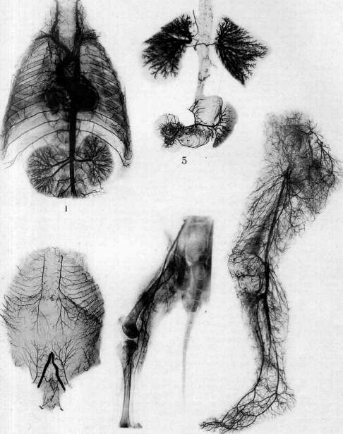
Figure 1. Large arterial trunks of the thoracic cavity of a 9-year-old child. Figure 2. Blood supply to the anterior thoracic and
abdominal wall. Figure S. Arteriogram of the femoral vessels of a living dog. Figure 4. Arterial system of the posterior lower limb. Figure J. A. pulmonalis of a newborn. Blood supply of the esophagus: participation of the a. bronchialis. (From the Laboratory of Normal and Comparative Anatomy of the State Radiological and Oncological Institute.) To the article Blood Vessels. S14 (Tandler): 5.0 of gelatin is dissolved by slight heating in 100 cm3 of water, 5.0 of potassium iodide and the necessary amount of dye are added. Besides the hot carmine mass, one can prepare a slowly setting, cold gelatin mass; immerse 6.0-10.0 of gelatin for 15 min in 100 cm3 of water. When the gelatin swells, heat it in a water bath so that the gelatin dissolves in the water. Next, 6.0-10.0 of carmine is mixed with water until a thick paste is formed; add ammonia until the carmine is completely dissolved (the liquid becomes transparent and cherry-red). This alkaline liquid is neutralized (not with acids) with 10% formalin until the smell of ammonia disappears and the smell of formalin appears; at this point, the liquid gradually turns from cherry-red to brownish-red (as if turbid). The carmine solution is added gradually to the warm gelatin solution; the resulting mass is ready for use without filtration. 1) It must be freshly prepared; 2) once set, it should not melt upon heating; 3) it should not be overheated, otherwise an insoluble compound of formalin with gelatin occurs; 4) the injected object should not be subjected to the action of acids, otherwise the carmine will dissolve and stain the surrounding tissues. As a material for injections for macroscopic and microscopic purposes, one can use a suspension of dry India ink in water (B. Ognev). The injection is performed in separate portions of mass of varying density. Portion I—1.0 of dry India ink is ground in 150 cm3 of hot water (filtered). Portion II—2.0:100 cm3 of water. Portion III—3.0:100 cm3 of water. Through arterial capillaries, venous ones are also filled during this; while under a microscope, arterial capillaries will appear as solid black lines, venous ones as if marked with a dotted line. India ink for injections is also used in a mixture with egg white (Grosser); according to Hamburger—India ink with egg white 1:1 or 2 volumes of India ink + 3 volumes of blood serum. In addition to microscopic examination of blood vessels, the method of so-called plastic reconstruction is used, which consists in the fact that the injected organ, its part, or vascular network (e.g., vessels of the liver lobule), laid out in a series of microtome sections (of a certain thickness), can be reconstructed again, but in an enlarged form. Besides the injection method for studying small blood vessels, capillaries, and initial stages of the development of the circulatory system, the benzidine staining method of I. Slonimsky can be used, based on the staining of Hb of the blood contained in the vessels; 2.0 of pure benzidine (Benzidin puriss. Merk'a) is dissolved in 20 cm3 of glacial acetic acid, 60 cm3 of distilled water and 0.5 of animal charcoal are added, shaken for 15 minutes and filtered (store away from light). As an oxidizing agent, one should use commercial hydrogen peroxide (Perhydrol Merk'a) in a 10-fold dilution (3% H2O2). To fix the Hb stain, an 8% solution of ammonium molybdate is used. The object is fixed with 80% alcohol. Using the described method, either the entire part of the organ under study is stained, or benzidine is introduced into the tissue surrounding the vessels. The study of blood vessels without injections can be performed according to the method proposed for the study of brain arteries by Stoeltzner and B. Hindze. The latter developed and applied this technique to the preparation of brain artery specimens not only of humans but also of some animals. The method consists of two main moments: 1) preparation of the arteries and 2) mounting of the prepared vessels (making a plate specimen). By preparation (under water) with tweezers, the pia mater is removed completely from the brain together with all large and smallest arteries, and the brain can either be preserved without the slightest damage (even its superficial cortex substance) or, in the process of preparation, for convenience, it is gradually dissected into separate parts. In order to obtain a complete picture of the topography of the arteries of the inner surfaces of the cerebral hemispheres, Hindze advises, before starting the removal of the pia mater, to cut off the cerebral hemispheres and remove the membrane with vessels separately from the obtained three parts of the brain, connecting them then during mounting. The pia mater extracted together with the arteries is washed and then stained with a histological dye (eosin, fuchsin, carmine). After staining, the arteries (under water) are freed from the pia mater, straightened on white paper placed on a plywood board; the arterial network straightened in this way is covered with glass, turned over, and, removing (carefully) the plywood and paper, the specimen is obtained transferred to the glass. For the purpose of studying blood vessels in vivo, namely the study of capillaries, the method of so-called capillaroscopy (see) is used; when studying the formation of collaterals and their anatomy, the experimental method is resorted to, performing ligations of arteries supplying various parts of the body on animals; after a certain amount of time, upon killing the animal, an injection of the vessels (with a solidified mass) is performed, and then they are usually examined with the help of radiography.
B. Uskov.
IV. Surgery of blood vessels. Stopping bleeding from vessels during operations. For a modern surgeon, one of the conditions ensuring a favorable outcome of operations is the ability to protect the patient from blood loss. One of the means for this is placing the patient on the operating table in various positions: for example, in a sitting position during operations on the head, especially brain operations, etc. Such measures significantly reduce bleeding. For the purpose of bloodless operating, one resorts to stopping bleeding by tamponade, digital compression of vessels, ligation above, and suturing of the operative field. Tamponade can be performed with sterile gauze balls, with which the bleeding site is pressed, or with long strips of gauze (bandage), which are held in place by hands or a dressing. This is resorted to for temporary stopping of bleeding, in order to then find and ligate the bleeding vessel, or for permanent stopping, when it is impossible to find small bleeding vessels or in the case of parenchymal bleeding. In the latter cases, the tampons remain in place until the growth of granulations; the tampon then separates either by itself or with slight effort. The final stopping of bleeding by means of tamponade in large wound cavities is best performed according to Mikulicz. The method consists of the following: a square piece of gauze in 3-4 layers is cut out of such a size that, upon filling the wound by pressing against its walls, the edges of the gauze protrude from the wound on all sides by 2-3 fingers; a thick silk or paper thread is attached to its center. This piece of gauze is taken by the middle, where the thread is tied, with a sponge forceps so that the gauze covers it on all sides like a folded umbrella; the thread is inside the bag. The bag is inserted into the wound cavity and straightened to apply it everywhere to the walls of the cavity. The bag is stuffed with strips of gauze or a bandage so tightly that the bag adheres to the walls of the wound; the thread hangs out. A dressing is applied. When removing the bag, one proceeds in the reverse [order].
Figure 17. Securing a tourniquet at the base of the leg.
according to the Keppler method: a—first moment; b—second moment. In the following order: first, the tampons are gradually pulled out; this can be started on the third day, extracting a piece of gauze or bandage at each dressing change until all tampons are removed. Then, one begins to pull on the thread of the bag itself, as a result of which it rises in its center and the tissues fill the cavity, reducing its depth. Usually, this succeeds without bleeding. To avoid the development of a putrid odor in the wound, it is better to use iodoform gauze for the bag. Digital compression of vessels requires a good knowledge of their topography; in urgent cases, this method is good but tiring. Constriction is performed with an Esmarch tourniquet, a rubber or gauze bandage. The tourniquet must be of such length that it is sufficient for at least 2 turns around the limb. It tightens very reliably, but by pressing on a small surface, it cuts deeply and easily damages vessels (subsequent thrombosis) and nerves. On the upper limbs, the Esmarch tourniquet can be used only during amputation; for other operations, it is necessary to use a rubber bandage. To hold the tourniquet in the area of the hip joint, a belt with a buckle is passed through the tourniquet and the opposite shoulder, and on the shoulder, the belt is passed through the opposite armpit (Fig. 17). Among the bloody methods of securing a tourniquet at the place required by the operation, we mention the methods of Braun [a long nail is driven into the greater trochanter (Fig. 18)], Trendelenburg (a flat steel spear, about 40 cm long, is passed from the greater trochanter inward and downward between the femur and the vessels, the end is brought out into the perineum, and to protect the latter, a removable protective sleeve is put on the sharp end of the spear, the tourniquet is applied behind the spear), and Bier (the spear is passed just above the spina anterior superior through the skin and aponeuroses of the abdominal muscles (Fig. 19); instead of a spear, a sponge forceps can be passed through a skin incision (Fig. 20)). Perthes proposed a compressor for artificial exsanguination, consisting of a hollow cuff applied around the limb. The cuff comes in four sizes; it is filled with air using a pump at a pressure slightly exceeding blood pressure. The compressor eliminates the pain caused by the pressure of a tourniquet or bandage during operations with local anesthesia. To preserve a greater amount of blood during limb amputations, the latter is previously exsanguinated as much as possible by bandaging from the periphery to the center or by elevating it. Such bandaging is inapplicable in phlegmonous processes, thrombosis, and cold abscesses. The time limit for constricting a limb with a tourniquet should be considered 2–2.5 hours; extreme caution must be exercised when constricting the upper limb, where one can easily obtain motor paralysis. Application of an Esmarch tourniquet to a limb is contraindicated in arteriosclerotics unless an amputation is intended. Momburg applied a tourniquet a finger thick and 1.5 meters long around the waist between the iliac crests and the costal arches. The tourniquet is tightened until the pulse disappears in the parts of the body below (Fig. 21). The procedure is carried out under deep anesthesia or spinal anesthesia (Bier) to overcome the resistance of the abdominal muscles. Constriction, according to Momburg, aims to stop the circulation of the abdominal aorta; therefore, it must be borne in mind that in this case, the blood flow encounters huge resistance immediately, as a result of which a significant increase in pressure occurs, to overcome which a heart of quite satisfactory strength is required. When the tourniquet is released, the pressure drops sharply due to the inclusion of the lower part of the body in the circulatory system.
an increase in pulse rate. Therefore, and
by removing the tourniquet, apply H
collapses, which are related / ''Щ
to a drop in blood pres- / Ш
sure. As a result of exces- / т
sive tightening, Burkhardt ' Щ
observed a case of peritoni- Figure 18.
tis, where the small intestine was saturated with blood in several places—crushing by the tourniquet; to avoid such a complication, it is recommended to place a cushion under the tourniquet on the abdomen. This method should be recognized as very valuable in combating bleeding, but it still cannot be considered standard, and therefore it should be resorted to only in exceptional cases when stopping bleeding by other means is impossible. Indications for exsanguination according to Momburg are as follows: 1) severe bleeding after injuries at the base of the lower limbs or pelvis; 2) extensive operations on the hip joint, especially in cases where wounds, inflammatory processes, or malignant formations are not limited only to the joint but also involve the pelvis and require rapid and radical surgery, in the absence of other means (layered ligation, ligation of the common iliac artery) to stop the bleeding; 3) aneurysm surgery
Figure 19.
Figure 20. Figure 19. Securing the tourniquet at the base of the leg using a Trendelenburg spear according to the Bier method. Figure 20. Replacement of the Trendelenburg spear with a sponge forceps according to Keppler. in the pelvic area and the upper part of the thigh; 4) interilio-abdominal exarticulation and resection of the pelvis; 5) bleeding during childbirth, especially atonic postpartum, when all other means have been exhausted; 6) situations where it is necessary to gain time during sudden severe blood loss (accidents, etc.) in the lower part of the body (vital indication). On the scalp, exsanguination is achieved by applying an Esmarch tourniquet around the head through the occiput and forehead; however, not every head shape is suitable for the method: the head of a brachycephal is suitable, and the head of a dolichocephal, where the temporal regions do not protrude at all or protrude very little, is little or not at all suitable.
Figure 21. Compression of the aorta according to Momburg.
Therefore, during surgery, hemorrhage is stopped by suturing according to Heidenhain or by clamping with special forceps (Makkas et al.). Operations on blood vessels. The methods of operating on blood vessels are numerous. The routes by which access to them is gained are also very diverse. The direct manipulations performed on them, even for the same purpose, are also diverse in technique; for example, there are 23 ways to expose the innominate artery; there are 12 main methods alone for the surgical treatment of traumatic aneurysms (Moskalenko). Likewise, to ligate the common iliac artery, one can approach it extraperitoneally or through the abdominal cavity. Operations on blood vessels are divided into two categories: 1) access to the vessel and 2) manipulations on it. Access to blood vessels is of two kinds: some, atypical, utilize the pathological-anatomical features of the case; here, it is impossible to specify any definite methods of approach to the vessel, as there will be almost as many as there are pathological changes in the circulatory system. Other approaches, typical ones, utilize the anatomical features of the location of the given vessel. Operations on vessels are reduced to the following types: 1) ligation for wounds and aneurysms; 2) suture for wounds and resections of the vessel (see Vascular suture); 3) desympathization (periarterial sympathectomy); 4) dislocation. Dislocation of vessels is currently one of the interesting experiments in vascular surgery: the brilliantly conceived experiments of Carrel—the displacement of the channel of one artery into the basin of another, for example, in stenosis of the coronary artery of the heart. The artery is transected below the narrowing; the left subclavian artery is transected, its central end is moved toward the heart and sutured to the peripheral end of the coronary artery. The blood flow is directed through the subclavian artery into the region of the heart supplied by the coronary artery (Moskalenko). Ligation of vessels is performed for the purpose of preventing hemorrhage during operations and for the purpose of immediate and final cessation of already existing hemorrhage. In modern surgical practice, ligation of a vessel is understood as the ligation of its two ends at the site of the wound (Delageniere); this is ligation at the site of hemorrhage, but it can also be performed at a selected site (along the course) of the vessel. The vessel is located in the wound and ligated immediately above and below the injury (ligation at the site of hemorrhage); if the wounded vessel is exposed above the injury and ligated there, this will be ligation at a selected site. It is preferable to perform ligation of the vessel at the site of its wound, because otherwise a large section of the vessel with its collaterals is excluded, which significantly impairs blood supply. Furthermore, ligation at a selected site is less reliable, as one can expect the resumption of hemorrhage in the wound through collaterals. In view of the above, the thought may arise that a ligature at a selected site is unnecessary; nevertheless, one has to resort to such ligation in cases of hemorrhage from large cavity wounds or during the removal of entire parts of the body, for example, ligation of the art. et venae iliacae commun. during exarticulatio interilio-abdominalis. Besides hemorrhage, there are other indications for the ligation of vessels. When one vein or another is thrombosed, then to avoid embolism or the spread of infection, ligation of the central end of the vein is required, e.g., ligation of the v. saphenae magnae at its entry into the femoral vein; wounds of large vessels or excision of a section of a vessel during the removal of a tumor or aneurysm often require ligation of the vessel. Before ligating a large vessel, one should consider the possible consequences of such ligation. Ligation of the abdominal aorta, according to Denis, always leads to death due to an excessive increase in blood pressure and cardiac fatigue. Ligation of the common carotid artery in one-third of all cases leads to death due to a disturbance of blood circulation in the brain. Ligation of the superior and inferior mesenteric arteries and veins is incompatible with life. After ligation of the main arteries of a limb, according to Wolff et al., gangrene occurs in 50% of cases of ligation of the common iliac, 20% of the femoral artery above the origin of the a. prof. femoris, 10% below it, 14 1/2% of the popliteal artery (significantly higher according to the experience of the World War), 11.2% of the external iliac, 15% of the axillary (8.3% according to later and more significant statistics by Geselevich), 4.8% of the subclavian and brachial; there are only 54 cases of ligation of the innominate artery in world literature up to 1919 (this operation was performed 7 times by Russian surgeons with 3 recoveries), with 15 cases of recovery. For the upper limb, ligation of the subclavian and axillary arteries between the site of origin of the aa. transversa scapulae et transversa colli and the beginning of the a. profunda humeri is relatively safe due to the developed collateral circulation in this section of the limb; the number of gangrenes increases with ligation of the lower segment of the brachial and ulnar arteries. Ligation of one of the arteries of the forearm or lower leg is safe. Ligation of a single femoral vein can be accompanied by severe consequences. Oppel believes that the most reliable method to protect a limb from necrosis during the ligation of large vessels (a. subclavia, etc.) is the simultaneous ligation of the corresponding vein. This procedure, in his opinion, balances the developing inequality in the inflow and outflow of blood from the given limb. This question is not yet considered finally resolved, and a number of surgeons (Küttner, Lexer, and many Russian surgeons) speak out decisively against the simultaneous ligation of the femoral artery and vein. When assessing the consequences of ligation of limb vessels, one should keep in mind not only the danger of the appearance of gangrene but also other consequences of circulatory disturbance leading to long-term dysfunction of the limb. Complete ligation of the inferior vena cava below and above the renal veins, according to the latest literature, contrary to the facts observed until now, turns out to be compatible with life. The results of ligation will vary depending on whether collateral circulation had time to form by the moment of ligation or whether it will have to develop only after the application of the ligature, as happens in wounds and other cases. The outcome of vessel ligation deteriorates sharply in the presence of wound infection. The result of ligation depends on collateral circulation; its state can be determined by the following method: the limb is exsanguinated with an Esmarch bandage; after a few minutes, the main artery of the limb is compressed, the bandage is released, and one observes how quickly reactive hyperemia of the skin appears and spreads; if the skin of the limb down to the fingers quickly assumes a normal color, then one can count on good collateral circulation (Moschcowitz's method). According to Coenen's method, the main artery is compressed at the site of its wound, the bandage is released, and reactive hyperemia is observed; if blood flows from the peripheral end of the wounded artery in a lively stream, this indicates sufficient collaterals for this artery. Korotkov advises measuring peripheral blood pressure for this purpose. Among the instruments for vessel ligation, a scalpel, scissors, surgical and anatomical forceps, instruments for passing ligatures, vessel clamps, and ligatures are necessary. For passing a ligature, a blunt Deschamp needle is used; in its absence, the ligature can be passed with forceps placed under the vessel. Among vessel clamps, Pean and Kocher clamps are most frequently used. Ligation by means of passing a ligature. The patient is placed in the appropriate position, and the operative field is prepared in the usual manner. After marking the projection of the vessel, an incision is made in the skin, penetrating to the fascia; the latter is recognized by the immobility of the layer in relation to the easily displaceable skin and subcutaneous tissue. The fascia is incised with a knife, possibly along a grooved probe, along the entire length of the skin incision. The muscles are separated bluntly. The vessel is isolated with a knife and two anatomical forceps from the vascular sheath, which is loosely connected to the artery. Upon exposure of the artery, the ligature is passed. The Deschamp needle is passed under the artery necessarily from the side of the adjacent nerve or vein to avoid wounding the latter or catching the former in the ligature. The thread is tied with a reef knot without a double first turn of the loop. Silk, catgut, and cotton thread are used for ligatures. It is best to use thin silk; it holds firmly and is well sterilized. In cases of small lateral injuries of large veins, a lateral ligation with thin silk can be performed; catgut will slip off, and the lumen of the vessel is preserved. On relatively thick bundles of tissue (adhesions, greater omentum) through which one or several vessels pass, clamps are applied before their ligation, the tissue between them is incised, and each end is sutured, and the thread is tied on both sides (Fig. 22). Such a ligature is called a ligature en masse. Small vessels usually do not need to be ligated; hemorrhage is stopped by compression with gauze sponges.
To reduce the number of ligatures, Blunk proposed a clamp in which the ends of the jaws are bent along the plane; when closed, the jaws overlap and flatten the captured vessel in the transverse direction; then the intima and the muscular coat of the vessel are twisted into two separate plugs that occlude the vessel. Blunk's clamps are applicable only on small vessels.
Figure 22.
Figure 23.
Figure 22. Ligation of a bundle of tissue. Figure 23. Compression of an artery with an aluminum plate according to Halsted. Bleeding from bone arteries is easily stopped by driving a sterilized nail into the bone canal, by pressing in sterile wax, or simply by tapping on the bone saw cut with a surgical hammer. Compression of large arteries. The definitive ligation of such arteries becomes less dangerous if their collaterals are first trained by gradual and temporary compression of the main trunk. This method was introduced into surgical practice by Jordan. Doberauer proposed a very simple method for reducing the lumen of a vessel: a drainage tube is passed under the vessel and tied outside the wound; the drain is twisted with a rod passed through the loop, as a result of which the artery is completely compressed. Halsted, Matas, and Allen use an aluminum plate for this same purpose (Fig. 23), the width of which is approximately equal to the diameter of the vessel being compressed. The plate is sufficiently strong and elastic to keep the vessel compressed and to be applied without any instrument. Removing the plate is just as easy as applying it. How safe the compression of even large vessels with such a plate is, is shown by the experiments of Halsted, in whose case the presence of the plate on the aorta of a dog for 7 months caused no changes in the vessel (Jeger). Prolonged compression of a vessel is also possible with autoplastic material, for which the fascia lata of the thigh is suitable due to its ease of procurement, elasticity, and strength; a strip of it, according to Halsted, should wrap around the vessel in a spiral. During temporary cessation of bleeding, it is necessary to take precautions against any, even minimal, damage to the vessel walls. In addition to the described methods of temporary cessation of blood circulation, several other types of gently acting clamps have been proposed for this purpose: clamps by Moore, Hopfner (Fig. 24), Carrel, and Bogoraz (Fig. 24). The first three are intended generally for work on vessels, the latter (Bogoraz) have a special purpose—parietal closure of the vessel lumen. The clamp is not always applied directly to the vessel; first, the latter is wrapped with a strip of gauze, and it is then clamped (Fig. 25). A good method is Smith's technique, in which the artery is compressed between rubber tubes. To avoid dependence on instrumentation, one can use, especially in emergency cases, all means at hand—a thin drainage tube, a strip of gauze, or a thick catgut thread folded in two—and with the help of a clamp (any kind), twist the thread or strip surrounding the artery until the vessel lumen is obliterated. Arteriotomy. Vascular suture makes it possible to temporarily open a vessel to penetrate its lumen, for example, for the purpose of removing an embolus. A brilliant example of this operation is Trendelenburg's operation for pulmonary artery embolism. To date, 6 cases of recovery after arteriotomia a. pulmonalis are known. Bleichroeder and Unger proposed a technique which may possibly have great significance for physiology and practical therapy. Bleichroeder applied this method to himself and several other voluntarily consenting people. They inserted a catheter through a large incision in an artery or vein and advanced it in the lumen of the vessel to a certain point, either to take blood from there or to introduce some medication to the corresponding site. Demarquay, and later Gaertner and Kuttner, utilized the opening of veins for experiments on introducing pure oxygen into the blood (E. Jeger). In the future, it may
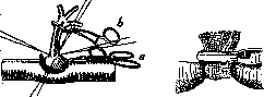
Figure 24.
Figure 1b.
Figure 24. Connection of vessels by the end-to-side method: a—Bogoraz clamp; b—Hopfner clamp. Figure 25. Compression of an artery with a strip of gauze. be possible to use this method of enriching the blood with oxygen in certain acute suffocations, for example, during obstruction of the airways by a foreign body or during closure of the trachea by diphtheritic membranes, etc. Replacement of vessel defects. Vein transplantation. The use of blood vessels to cover or reinforce sutures of nerves and tendons is well known. Vascular suture has made it possible to compensate for a deficiency in a vessel by transplanting whole segments of a vessel. A suitable material for this is a vein, especially the great saphenous vein, due to its superficial position and sufficient caliber. When suturing a vein into an artery, one must always remember the presence of valves in the vein; therefore, its peripheral end must be sutured into the central segment of the artery and, conversely, the central end into the peripheral segment of the artery. One should not take the great saphenous vein from the same limb where the transplantation will be performed. The wall of a vein transplanted into an artery thickens slightly under the influence of blood pressure. In 51 cases of vein transplantation in humans, the outcome was favorable 39 times (Wartmüller). Lexer transplanted a segment of the great saphenous vein of the same length and smaller caliber into a sixteen-centimeter defect of the iliac artery; after 5 years, the normal function of the transplant was present (Bier, Braun, Kümmel). An indispensable condition for the success of vessel transplantation is the aseptic state of the wound. Vein transplantation during war wounds should be considered inexpedient due to wound infection. The use of blood vessels to replace lost parts of the ureters, urethra, and common bile duct (d. choledochus) is interesting. Payr proposed an operation for drainage of the cerebral ventricle in hydrocephalus by transplanting a piece of vein. There are two varieties of the operation: the sinus sagittalis is exposed by a flap incision in the region of the sagittal suture; it is temporarily ligated in two places, incised, and the end of a vein or artery is implanted there; the other end is passed through a tunnel made in the brain substance by a trocar into the lateral ventricle; in this case, the vein is positioned so that its valves prevent the flow of blood from the sinus into the ventricle (Fig. 26); in the second method, a segment of vein is passed into the lateral ventricle through a small trepanation hole behind the ear and is passed under the skin to the neck, where it is connected to the external jugular vein. All the listed operations on vessels are typical, i.e., they are performed according to rules developed on the basis of the architecture of the vessels. However, they have the same peculiarity as approaches to vessels: under identical pathological-anatomical conditions, the plan of their execution in different cases may be different. The scheme of any surgical operation, including a vascular one, is built on pathological-anatomical and topographic-anatomical conditions. It is often necessary to change the plan of a vascular operation under identical pathological-anatomical conditions. Consequently, the reason for the variability of the plan of typical vascular operations in such cases must be sought in topographic-anatomical conditions, and the topography of vessels is variable; for example, the brachiocephalic artery in a narrow superior thoracic aperture lies almost along the midline, while in a wide one, it is significantly deviated to the right; the bifurcation of the aorta in a narrow and flat inferior thoracic aperture lies high—between the III-IV lumbar vertebrae, while in a uniform one, it lies lower, sometimes on the promontory. The topography of vessels predetermines the scheme of approaches, and the architecture predetermines the plan of the procedure itself on them. The architecture of vessels is no less variab
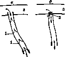
Figure 26. Payr's method: a—first method of suturing a vein; b—second method of suturing with narrowing of the implanted end (2); 1—valves; 3—sinus.
uneven distribution of vasa vasorum, inconstancy in the caliber of vessels (the deep femoral artery is sometimes equal in caliber to the femoral artery, sometimes differs sharply from it, etc.). Vascular suture yields different results depending on whether it is applied in the avascular field of the artery (healing conditions will be unfavorable) or, conversely, in the vascular field of the vessel wall. The nerve supply of the vessel is also of importance. Arteriography—obtaining an X-ray image of vessels filled with a contrast liquid [see separate table (pp. 511-512), figs. 1-5]—is performed to diagnose the patency of vessels, e.g., in gangrene of a limb or arteriovenous aneurysm (Krestovsky). A substance that blocks X-rays is injected into the artery: Lipiodol and aqueous solutions of salts. Lipiodol is injected, according to Sicard, in 1-2 cm3; but doses up to 10-12 cm3 have been used (Harvier, Lemaire); of aqueous salt solutions, Strontium bromatum (Berberine, Hirsch), Natrium jodatum, and Natrium bromatum (Krestovsky) are used. The latter is injected in a 25-percent solution in an amount of 10-12 cm3 (Egaz Moniz, dos Santos). Most authors (Singleton, dos Santos, Duval) inject the solution into the femoral artery immediately below the Poupart ligament. It is advised to inject the liquid below the deep femoral artery, e.g., in the Hunter canal. Some expose the artery, others puncture it through the skin. The artery is clamped above the needle insertion site. However, puncture of the artery through the skin in the Scarpa triangle is more common (Sicard, Singleton). The patient's pelvis is slightly elevated, the thigh is rotated outward. A long needle should be used, as for spinal anesthesia, and thin, but with a sufficient lumen, especially if viscous Lipiodol is being injected. The needle is attached directly to the syringe. When Lipiodol is injected, it is warmed, and in an amount of 1-2 cm3, it is injected all at once; with larger doses, slowly—1 cm3 per minute (Harvier). Sodium iodide or sodium bromide is injected quickly, all at once. The speed of injection is a necessary condition for the success of arteriography (dos Santos). Half an hour after the injection, one can still obtain images of the branches of the artery, but 17 minutes after the moment of injection, the vessel is no longer visible on the image; only a diffuse darkening of the tissue appears, where the contrast substance is apparently located. Lipiodol is well tolerated by the arterial endothelium; the same cannot be said for sodium solutions. Strong salt solutions (25%, not to mention 100%) cause severe pain, so one has to resort to spinal or even general anesthesia. Complications include vascular spasm in a part of the limb, increased ischemia, and general intoxication when using sodium solutions. Lipiodol seems less dangerous, but its doses are too small to obtain an image of the artery over a sufficient extent. In three cases of using a 100% solution of sodium iodide, Singleton observed a sharp deterioration in the condition of patients with senile gangrene; one died on the second day and another on the 20th day after the injection. Leibovici, in a work on arteriography, concludes that clinical methods of examination stand in the first place both in gangrene of the lower extremities and in all other cases, and only a very limited place should be assigned to arteriography in clinical methods of examination. The method must be considered risky, but in exceptional cases, it can be resorted to (Martynov). In the USSR, this method of examination was first successfully applied on a living person by Krestovsky.
Alcoholization of vessels as a method of treating various diseases has become widespread in recent years. This method is also widely used on nerves. Alcoholization is performed either in the form of an injection into the thickness of the vessel wall (Handley, Stradyn) or in the form of circular wetting of the artery over a distance of 8-10 cm (V. I. Razumovsky). The second method has undoubted advantages over the first; with injection, the destruction of nerves (which is the goal of the method) occurs in a limited space, while with "wetting" of the vessel with alcohol, the nerves are destroyed over a large distance and completely, rather than in sections, as with injection. The "wetting" method is completely harmless, which cannot be said about the "injection" method, where the possibility of a through-puncture into the lumen, as described by Handley, is not excluded. Alcoholization is performed with ethyl alcohol with a strength of no more than 80°. The procedure is indicated for various diseases: trigeminal neuralgia (carotid artery), obliterating endarteritis with its varieties (Berger, Raynaud, etc.), trophoneurosis (femoral artery) (V. I. Razumovsky), impotence (a. pudenda interna) (Vakulenko), etc. As a result of the alcoholization of vessels, as well as nerves, pain disappears and the course of the disease process improves, and perhaps function also improves (Vakulenko). Alcoholization of a vessel is analogous to desympathization: in both, the aim is to destroy the nerves on the vessels, but alcoholization does not have the disadvantages of the latter—spasm of the vessel and even its rupture. A. Sirotkin.
Related articles
Cite this page
“Blood Vessels (525).” Soviet Medical Encyclopedia. English translation of Bolshaya Meditsinskaya Entsiklopediya, 1st ed. (Moscow, 1928–1936), ed. N. A. Semashko. https://sovietmedicalencyclopedia.pages.dev/article/blood-vessels/