Capillaroscopy
Historical document, translated for reference. It reflects medical knowledge of the 1920s–30s and is not medical advice.
Summary
Capillaroscopy is a diagnostic method for examining capillaries and capillary blood circulation in the skin of a living person. This article details the technique, which involves applying a clearing oil to the skin—typically the nail fold—and observing the capillaries under magnification, and discusses the clinical significance of observing capillary morphology and circulation in various diseases.
Encyclopedia article (1928–1936)
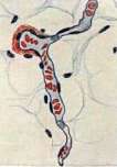
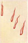
CAPILLAROSCOPY, a method for examining capillaries and capillary blood circulation in the skin of a living person. For the first time, Hitter in 1879 pointed out the possibility of examining capillaries in the mucous membrane of the human lower lip. In 1911, W. Lombard pointed out the possibility of examining capillary circulation in human skin after preliminary clearing of the epidermis with glycerin. The detailed development of the capillaroscopy method and its introduction into the number of clinical research methods belongs to O. Müller and his school. The capillaroscopy method consists of applying a drop of a good clearing oil (cedar, bergamot, liquid petrolatum or paraffin, glycerin, etc.) to any area of human skin, most preferably in the area of the nail fold of the fingers, waiting 1-3 minutes for the homogenization (clearing) of the epidermis, and then microscoping this area under strong side illumination. The nail fold is the most convenient for capillaroscopy, as the capillaries here lie superficially, are located in a horizontal plane, and are therefore clearly visible along almost their entire length; furthermore, the finger is convenient for microscoping. The latter is conducted at low magnification (20-80 times) by means of the simplest microscope or capillaroscopes specially constructed for these purposes [Müller (Zeiss), Leitz, Skulsky], which differ from ordinary botanical microscopes in that an illumination device is attached near the objective in capillaroscopes, thanks to which the need for a special setup for illumination is eliminated. When working with a simple microscope, one can use concentrated light (by means of a small magnifying glass) from a 50-100 candlepower electric lamp to illuminate the area under study; one can successfully perform capillaroscopy under sunlight (with or without a magnifying glass). When using strong illumination and during prolonged capillaroscopy, it is necessary to pass the beam of light through a cooling device to avoid thermal hyperemia. The hand being examined must be at the level of the heart, in a comfortable, completely free position. For microscopic observations, the integrity of the nail fold is necessary. Capillaroscopy of other areas of the human body's skin can be performed with the help of universal (large) capillaroscopes by O. Müller and A. Nesterov. The normal picture of capillaries in the area of the nail fold in an adult is presented in Fig. 3. The same picture in a 4-month-old child is presented in Fig. 4. As can be seen from these figures, during capillaroscopy of the first rows of the nail fold of the fingers (and toes), capillaries are visible under the microscope in the form of red (on a yellowish-pink background) elongated loops, resembling ladies' wire hairpins in shape. In each capillary loop, a narrower ascending arterial half and a wider descending venous half are distinguished; the place where the arterial half transitions into the venous one (the bend) is called the apex of the capillary loop. With thin skin, one can also see the afferent arterioles and the wider branches of the 1st subpapillary venous network under the microscope (Figs. 4 and 7). The length of the capillary loops varies depending on the length of the nail fold, but it can still be assumed that in healthy people, the length of the arterial branch of the capillary loop is 160 μ and the length of the venous one is 220 μ; when taking into account all the bends along the course of the capillary, the length of the unfolded loop (in the 1st row of the nail fold of the fingers) will be equal to 0.4 mm. In this same area, the diameter of the arterial branch of the capillary is 7.6 μ and the diameter of the venous one is 9.1 μ; the width of the entire capillary loop in its widest part is 31.6 μ; the number of capillary loops is 8 per 1 linear millimeter. In the direction from the 1st row of capillary loops toward the joint, the length and diameter of the capillaries decrease, and their more or less regular row-like arrangement is disrupted. Thanks to the application of capillaroscopy, it has been established that in healthy newborns, the capillary system of the skin is fully formed, but it appears indistinctly under the microscope, as if in a crumpled state, which is caused by the folding and maceration of the skin. The length and diameter of the capillaries of newborns are smaller than the corresponding values in adults; conversely, capillary circulation proves to be more energetic, which is dependent on the hyperemia of the skin existing in the first days after birth. During capillaroscopic examination of other areas of the skin, the capillaries appear as very irregular loops, commas, and dots, between which branches of the subpapillary venous network of varying thickness shine through. In some areas of the skin (e.g., the face), the capillaries in their shape deviate so much from ordinary loops, come into such close contact with the branches of the first subpapillary venous network, and intertwine with them so closely that one can speak of a mixed loop-network system. It has been capillaroscopically proven (Nesterov) that a pronounced flush of the cheeks, especially a pathological flush, is caused by the aforementioned approach of the first venous network to the capillary system and a significant dilation of the capillary-venous vessels. By means of capillaroscopy, more or less characteristic changes in the capillaries and capillary circulation have been established in a whole series of diseases of internal organs. Thus, during the development of cardiac weakness, one can trace the increase in signs of peripheral decompensation step by step by means of capillaroscopy. In this case, capillaroscopy confirms peripheral decompensation when it is not yet clinically determined at all. Such Figure 1. Longitudinal section of a capillary in the subcutaneous fat layer of a human: blue—endothelial nuclei; red—erythrocytes (magnification 650). Figure 2. Longitudinal section of the site of origin of two capillaries of human subcutaneous adipose tissue; capillaries—as in Fig. 1; artery with a muscular coat (magnification 650). Figure 3. Normal capillaries at the nail edge on a finger (magnification 70). Figure 4. Normal capillaries and vessels of the subpapillary plexus in a child (nail edge). Figure 5. Elongated capillaries and dilated vessels of the subpapillary plexus in an adult asthenic with vasoneurosis (nail edge, magnification 70). Figure 6. Elongated, dilated, unusually configured capillaries and dilated vessels of the subpapillary plexus in an adult with severe vasoneurosis (nail edge, magnification 70). Figure 7. Elongated and sharply dilated capillaries alongside normal ones in acrocyanosis; granular flow, stasis, narrow arterial and wide saccular venous knee (nail edge, magnification 70). Figure 8. Elongated capillaries with a narrow arterial knee; intensely colored background; hypertension (nail edge, magnification 70). Figure 9. Elongated, irregularly shaped capillaries with a narrow arterial and wide venous knee; plexus vessels dilated; acute nephritis (nail edge, magnification 70). Figure 10. Elongated, narrowed in the arterial and dilated in the venous knee capillaries, sharply deformed; subacute nephritis (nail edge, magnification 70). Figure 11. Normal and altered capillaries side by side—in vasoneurosis; flow is granular in places; continuous in places (nail edge). Figure 12. Strongly elongated, narrow in the arterial and dilated in the venous knee capillaries, with anastomoses in places; granular flow; hypertension (nail edge). Figure 13. Increased number of capillaries of normal shape with sharply granular flow; cachectic edema—nephrosis (nail edge, magnification 70). Figure 14. From a to d—increasing spasm of the venous knee of the capillary with stasis during congestion; arterial knee is very narrow (dorsum of the foot, magnification 70). Figure 15. a and b—Jolly bodies in the erythrocytes of the blood of a patient with a ruptured spleen; c and d—Cabot rings in basophilic erythrocytes; e and f—Cabot rings in orthochromatic erythrocytes (from Naegeli). Figure 16. Carbuncle. To the illustrative articles: Jolly bodies, Cabot rings, Capillaroscopy, Capillaries, Carbuncle.

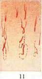
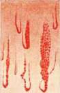
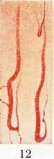

Signs of developing peripheral decompensation are: slowing, inconsistency of capillary circulation, intermittency, granularity of the erythrocyte stream, and slight dilation of the apex and venous limb of the capillary loop. In pronounced peripheral decompensation, we find a significant slowing of capillary circulation in the majority of visible capillaries, an increase in the number of functioning capillaries, their massive diffuse-uniform or locally bulging dilation, protrusion of dilated subpapillary veins, and a venous coloration of the blood in the capillaries. With the cessation of decompensation, the pathological capillary circulation disappears, but it disappears later than the clinical manifestations of decompensation. With the development of edema, the microscopic picture is outlined unclearly; the capillaries are contoured indistinctly and appear immersed in a fog; nevertheless, in the majority of cases, it is still possible to ascertain that the number of functioning capillaries (during edema) and their diameter, as well as circulation, are reduced, which is obviously dependent on the compression of the capillaries by the edematous fluid. Congestive capillary-circulatory changes in chronic decompensation occur not only in the skin but also in other organs, which is quite probable (congested lungs, liver, kidneys, etc.), and in this stable and massive dilation of the capillary system may lie one of the causes of those difficulties that are encountered in the treatment of chronic decompensation. In any pronounced decompensation (of central or peripheral origin), attention is drawn to the noticeable dilation of the subpapillary venous plexuses, their more distinct protrusion, and the peculiar, slightly bluish, cyanotic coloration of the microscopic background caused by this moment. Such "protrusion" of the subpapillary venous plexuses and the more saturated background coloration caused by them prove to be pronounced in peripheral decompensation even when neither pathological dilation of the capillaries nor, even more so, macroscopically (clinically) determinable cyanosis can yet be established. This circumstance—in connection with the fact that even in the normal capillaroscopic picture the general coloration of the background (resp. skin) is to a significant extent caused by the translucency of the rich venous subpapillary plexuses—allows one to conclude that the cyanotic coloration of the skin in peripheral decompensation is to a significant extent caused precisely by the venous vascular factor. Capillaroscopy convincingly demonstrates that the most significant disturbances in the rhythm of cardiac activity (as such) do not find any noticeable reflection in capillary circulation, which must point to the regulatory significance of the arterial, mainly precapillary, vessels. On the other hand, it has also been established capillaroscopically that a deep breath and, even more so, the Müller maneuver can lead to a short-term improvement in capillary circulation even in cases of its significant slowing. In capillaroscopic studies, objective confirmation has been given to the fact that between the magnitude of the pulse determined graphically or by palpation and the magnitude of capillary circulation, i.e., the magnitude of the blood supply to the skin (and probably other tissues), there is not only no complete parallelism, but the existence of a peripheral arterial pulse does not at all guarantee the existence of capillary circulation (Nesterov). These observations put forward the necessity of revising the methodology of clinical pulse examination, mainly from the angle of its dynamic assessment (determination of the magnitude and speed of the pulse wave roll), and also force one to admit the existence of a form of "decompensation of peripheral circulation," in the diagnosis of which capillaroscopy should occupy one of the first places (vasoneuroses, initial forms of endarteritis, spontaneous gangrene, Raynaud's disease, acrocyanosis, compensated arteriosclerosis, etc.). In pronounced peripheral arteriosclerosis in individuals with an asthenic constitution and pale coloration of the integuments, the capillaroscopic picture appears pale, with a reduced number of functioning capillaries compared to the norm. This reduction in the number of functioning capillaries does not extend to the entire field of vision uniformly, but rather in patches, in which one can sometimes notice "shadows of capillary loops," or capillaries that are very narrow and poorly functioning. Under the microscope, the capillaries are outlined distinctly in the form of elongated, sometimes stretched, sometimes pathologically twisted "elegant" loops. In the mass, the capillaries are slightly narrowed, mainly in their arterial part. The branches of the first subpapillary network stand out well. The movement of blood is less stable than in healthy individuals. Capillary-circulatory changes are localized mainly in the root of the arterial branch and have all the signs of capillary-circulatory changes of the 1st type (see Capillaries). In arteriosclerotics of digestive and hypersthenic constitutions, capillaroscopic findings are less characteristic, but it is still possible to note that the capillaries in the root of the arterial branch are narrowed, and in the venous one they are dilated, that the capillary circulation is unstable; the branches of the subpapillary venous plexuses stand out more distinctly. The capillaroscopic picture does not allow for a definite distinction between constitutional hypertension and hypertension of renal origin, but the differences between the "pale" and "crimson" types of hypertension are revealed in the capillaroscopic picture quite distinctly. In hypertension of the "pale type," the capillaroscopic picture is similar to that in arteriosclerosis, namely: the microscopic field is colored a pale pink color, the number of functioning capillaries (with sufficient cardiac activity) is reduced, their distribution in the field of vision is uneven—fields with reduced capillarization are encountered. In the mass, the capillaries are polymorphic, tortuous, elongated, deformed; the venous half and the apices of the capillary loops are dilated, the root of the arterial branch is narrowed; the blood flow is variable—from very fast or slightly slowed to periodic sharp, almost complete stops. Between these extremes is a whole gamut of transitions: rapidly or gradually slowing, intermittent, cylindrical, granular, "beaded" blood flow; changes in capillary circulation often reveal a rhythmic character. Primarily and most demonstratively, capillary-circulatory changes are revealed in the root of the arterial branch. These and other data from capillaroscopic observations in hypertension of the "pale" type quite convincingly speak in favor of the origin of this form of hypertension due to increased tone or spasm of the smallest precapillary arteries. In hypertension of the "crimson" type, in accordance with the pronounced hyperemic coloration of the skin integuments, the microscopic field is colored an intensely pink color, sometimes with a cyanotic tint. The number of functioning capillaries is increased, their orderly arrangement is disturbed, and the distribution in the field of vision is more or less uniform. In the mass, the capillaries are significantly dilated, elongated, tortuous, often deformed, and polymorphic. The number of "giant" capillaries is increased. In a compensated state, the movement of blood is distinguished by significant speed and great stability. The increase in the number of functioning capillaries, their massive dilation, and the rapid stable blood flow point with certainty to "hypercirculation" in hypertension of the "crimson" type. Such are the capillary-circulatory changes in arteriosclerosis and hypertension in a state of cardiac compensation. When cardiac insufficiency is added to these diseases, signs of congestive capillaries gradually increase in the capillaroscopic picture. An idea of the capillaroscopic picture in hypertension with circulatory insufficiency is given by Figs. 11 and 13. In acute and subacute nephritis, as Figs. 10 and 12 show, the number of functioning capillaries is reduced, the arterial branch is narrowed, the venous one is dilated; the capillaries are tortuous, deformed, and function periodically, in accordance with which spontaneous capillary-circulatory changes of the 1st type are sharply pronounced (see Capillaries). A portion of the capillaries periodically becomes completely empty, deprived of blood, due to which the microscopic picture appears paler than in the norm. In the period of increasing inflammatory changes in the kidneys, the number of non-functioning capillaries reaches 1/3 and even 1/2 of all visible capillaries. With the cessation of the inflammatory process in the kidneys, the number of non-functioning capillaries gradually decreases, the periods of their exclusion from circulation shorten, and the capillaries dilate; however, as systematic observations show, capillary-circulatory changes appear earlier than the clinically well-determinable signs of nephritis and disappear later than their elimination. In chronic glomerulonephritis, the capillaroscopic picture approaches that in hypertension of the "pale" type. In clinically pronounced nephrosis with edema on the extremities, the capillaroscopic picture appears foggy, the capillaries are outlined indistinctly, in a slightly narrowed form.
A greater intermittency of blood flow than normal is noted. With the disappearance of edema, the capillaroscopic picture quickly returns to normal. Thanks to the application of capillaroscopy, it has been possible to study the group of vasoneuroses in more detail. In particular, it was possible to quite clearly outline a specific vasoneurotic anomaly conditioned by constitutional and external causes. In this anomaly, in the majority of cases among representatives of an asthenic constitution, with primary complaints of high excitability and easy fatigability, reduced working capacity, chilliness and sweating of the hands and feet, migraine-like and neuralgic pains, periodic or constant constipation, palpitations, skipped heartbeats, shortness of breath during rapid movements, irregularities of menses in women, etc., we find the following picture under the microscope. In the area of the nail fold of the skin, corresponding to the cyanotic coloration of the skin of the extremities, the general background of the microscopic picture appears more intensely colored than normal, which depends on an increase in the number and diameter of capillaries filled with blood, on the significant protrusion of subpapillary venous plexuses, and the venous coloration of the blood in the indicated vessels. Many capillaries significantly exceed normal ones in their dimensions and therefore can be called giant; the number of such giant, as well as generally tortuous, dilated, and deformed capillaries, is increased compared to the norm. The dilation is more pronounced in the apex and the venous branch of the capillaries. In most cases, this dilation is not expressed uniformly, but in patches. Depending on the extent of the dilation, its magnitude, and the character of the increase and decrease in diameter, the dilation has the appearance of either nodularity, varicose swellings, or true capillary aneurysms. The size and number of such deforming dilations vary from case to case and, in the same case, from capillary to capillary. With a combination of significant deforming dilations and tortuosity, the most bizarre, sometimes fantastic figures are obtained. Figures of simple and complex eights, complex loops, rosettes, etc., are a common phenomenon in vasoneurotics. Some capillaries turn out to be so dilated (50-60 μ) that they are already noticeable to the naked eye. However, it should be admitted that, no matter how significant the deformations of the capillary loops in vasoneurosis are, it is not possible to find any deformations strictly characteristic specifically for this disease. In most cases, the blood flow is slowed down, sometimes very significantly, due to which it becomes intermittent (cylindrical), granular. In no less than 60% of all cases, capillary-circulatory changes in vasoneurosis proceed slowly, sluggishly, are localized predominantly in the apex and the venous half of the capillary loop, and in their appearance very much resemble peristaltic waves (capillary-circulatory changes of the 2nd type). The calculations performed speak with certainty to the fact that the described changes in vasoneurosis turn out to be an almost constant finding in the above-indicated clinical symptom complex and therefore can be called characteristic. The most probable cause of capillary-circulatory changes in vasoneurosis is a primary spasm of the precapillary arteries and the subsequent dilation of capillaries and capillary veins. As for the nature of the changes in the capillaroscopic picture in vasoneurosis, in most cases, we are dealing with undoubtedly far-advanced functional (trophic) disturbances in the tone of the capillary wall, although in severe chronic forms, the participation of some organic changes cannot be excluded. Probably, the peculiar dilations and protrusions of capillaries, which are known by the name of "capillary aneurysms," found by many authors, should also be attributed to the manifestations of vasoneurosis. According to the observations of Moos, these capillary aneurysms can most often be found in the skin of the shoulder, and they are encountered much more often in women (74%) than in men (36%); they were not found at all in newborn children; with age, the number of these findings increases; in the maximum quantity, these "aneurysms" were found by Moos in pregnant women. Capillaroscopic observations allow us to conclude that a significant part of such aneurysmal dilations has a temporary, transient, probably functional character, and only in a certain part of cases, where these deformations were tracked over a long period of time (up to 10 days), is it natural to think about the participation of organic changes in the capillary wall. Figures 5, 6, and 7 provide an idea of the microscopic picture in vasoneurosis by O. Müller. The capillaroscopic picture in acrocyanosis has much in common with that in the vasoneurotic anomaly (Fig. 9). Figure 14 provides an idea of the granular blood flow due to significant skin edema. In the capillaroscopic picture, undoubted dynamic (functional) or morphological (organic) changes in the capillary system of the skin are revealed in pulmonary tuberculosis, syphilis (stage III), diabetes, Basedow's disease, ulcerative and hemorrhagic diatheses, bronchial asthma, blood diseases, severe infections (typhus, influenza, sepsis, rheumatism, diphtheria, meningitis), in spontaneous gangrene of the extremities, cirrhosis of the liver, organic lesions of the central and peripheral nervous system (neuritis, syringomyelia), eclampsia, etc. Thus, in pulmonary tuberculosis II-III B-C, the capillaries turn out to be dilated, tortuous, sometimes very significantly; capillary circulation in the majority of cases (85%) is slowed down, unstable. In the period close to the lethal outcome, the dilation of the capillaries increases significantly, the circulation slows down, and the blood acquires a dark color (a manifestation of asphyxia) (Fig. 8). In syphilis, in the II and especially the III period, the appearance of true capillary aneurysms was noted, which, under the influence of successful therapy, decreased in size and number, and sometimes disappeared completely. An idea of the morphological changes in capillaries in diabetes mellitus can be obtained from a comparison of the normal capillaroscopic picture [see separate table (art. 511-512), Fig. 5] and the capillaroscopic picture in this disease [see separate table (art. 511-512), Fig. 6]. In bronchial asthma, the capillaroscopic picture has much in common with that in the vasoneurotic anomaly and arteriosclerosis. In anemias, the capillaries have a yellow-orange color and are often dilated. The diagnosis of leukemic leukocytosis is fully accessible to capillaroscopic examination, in which we find under the microscope a pale color of the field of view with dilated capillaries, along which a highly characteristic "variegated" stream of erythrocytes moves slowly (or with deceleration). In polycythemia, the capillaries are very sharply dilated and, as it were, clogged with a dense mass of erythrocytes. From the observations of E. Koch on the determination of the duration of the complete circulation of blood in the human organism in various diseases, it follows that the movement of blood in leukemia and polycythemia turns out to be slowed down in general (up to 30"-50" instead of the normal 20.9"), which is why the slowing of capillary circulation in the skin noted in capillaroscopic observations should be considered only a partial manifestation of the decrease in the speed of circulation in the entire organism. The capillaries of the skin turn out to be changed in hemorrhagic diatheses. Thus, according to the observations of P. Lukomsky, in the majority of cases of Werlhof's disease, the capillaries turn out to be narrowed, deformed, with a slowed granular blood flow. In scurvy, the capillaries turn out to be polymorphic, in part significantly dilated, with varicose swellings and hemorrhages (Nesterov, Lukomsky). There are still very few capillaroscopic observations on this issue, and for now, one can only draw one essential conclusion: that in hemorrhagic diatheses, morphological changes undoubtedly take place. Capillaroscopy has established extremely interesting changes in blood circulation in capillaries during pregnancy, during childbirth, in the menstrual period, depending on the time of day and year, the nature of nutrition, etc. A number of capillaroscopic observations have established the influence on capillaries and capillary circulation of solar, light, simple, mineral, and carbon dioxide baths of various temperatures, the mercury-quartz lamp, diathermy, galvanization, faradization, Bergonié's method, d'Arsonvalization, X-ray treatment, etc. Capillaroscopic observation states quite definitely that Quincke's capillary pulse depends on the pulsation not of the capillaries, but of the smallest arteries. Capillaroscopy has provided a number of valuable data for the anatomy and physiology of capillaries and capillary blood circulation. Thanks to capillaroscopy, we can study capillary blood circulation, which has been little accessible to us until now, under completely physiological conditions. In the clinic, capillaroscopy has the significance of an auxiliary method that complements and deepens our knowledge obtained with the help of other clinical methods of examination.
A. Nesterov. Capillaroscopy in children. The capillary system in children presents certain peculiarities depending on age. The development of the cutaneous capillary network begins in the second half of pregnancy. First, the deep network of capillaries—the subpapillary layer—develops; the latter is accessible to capillaroscopic observation in the first two months after birth; it is visible in the capillaroscope in the form of a disordered network. The superficial network of cutaneous capillaries begins to develop towards the end of pregnancy, later than the deep subpapillary network; it is distinguishable in the capillaroscope only in an infant from the 3rd month of life; the capillaries of the papillary layer of the cutaneous capillary network have a curved, hairpin-like shape. With age, with the manifestation of constitutional peculiarities in the child, the capillary network acquires morphological features. In an asthenic constitution, the capillary loops are narrow and few in number; in a lymphatic one, the capillary loops are numerous and wide; in a neuropathic one, they are short and tortuous. The methodology of capillaroscopy in children is the same as the methodology in adults. The capillaroscopic picture of the cutaneous capillary network acquires special interest in acute childhood infections that sharply affect the state of the cardiovascular apparatus. In typhoid fever in children, the capillaries are strongly tortuous; in vasoneurotics with a labile capillary apparatus, in a severe clinical picture of the disease, there is an atonic state of the cutaneous capillary network. In measles infection, there is hemorrhagic imbibition around the capillaries. The paresis characteristic of severe infections is absent. All capillary loops are dilated, increased in quantity, and arranged in small groups [see separate table (col. 511-512), fig. 7]. In scarlet fever in the acute period, there is an alveolar arrangement of the capillary loops, and they are uniformly dilated. In the recovery period, the capillary loops present a picture of a spastic-atonic state. In nephritis, in the 3rd-4th week, a spasm of the capillaries sometimes occurs some time before the appearance of renal symptoms. Due to the special electivity of the scarlet fever toxin for the vascular system, the latter remains vulnerable for a long time, as the capillaroscope shows; upon the end of the disease, the capillaries remain in a state of uniform dilation with a slowed flow [see separate table (col. 511-512), figure 8].
Related articles
Mentioned in
Cite this page
“Capillaroscopy.” Soviet Medical Encyclopedia. English translation of Bolshaya Meditsinskaya Entsiklopediya, 1st ed. (Moscow, 1928–1936), ed. N. A. Semashko. https://sovietmedicalencyclopedia.pages.dev/article/capillaroscopy/