Capillaries
Historical document, translated for reference. It reflects medical knowledge of the 1920s–30s and is not medical advice.
Summary
This article from the 1928–1936 Great Medical Encyclopedia examines the anatomy, histology, and development of blood and lymphatic capillaries, detailing their endothelial structure, pericytes, and physiological roles.
Encyclopedia article (1928–1936)
CAPILLARIES (from the French capillaire - hair-like), or hair-like vessels, are microscopically small vessels with the thinnest wall. 1. Blood capillaries, connecting the branches of arteries and veins into a single closed system [see separate table (p. 223-224), fig. 1 and 2], represent the vessels of the smallest caliber, forming networks between the terminal branches of arteries (arterioles) and the initial branches of veins (venules). In living objects, as well as with conventional fixation and staining methods, capillaries have the appearance of thin transparent tubes with smooth and even edges; elongated nuclei protruding into the lumen are visible in the wall (fig. 1). The diameter of the thinnest capillaries (muscles, nerves, retina) is 4.5 - 6.7 μ; medium ones (skin, mucous membranes) - 6.7 - 11 μ; wide ones (glands, bones) - 9 - 13 μ, reaching up to 18 - 22 μ (Ebner). Capillaries were considered for a long time to be intracellular channels,
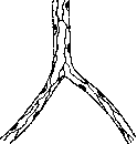
Figure 2. Capillary treated with silver nitrate. Cell boundaries in the form of black lines.
until Hoyer (1865), injecting them with weak solutions of silver nitrate (1/4-1/2%), discovered boundaries between individual cells in the form of black lines (a method still used today). This proved that the capillary wall consists of a single layer of flat epithelium (endothelium; figure 2). Endothelial cells are always more or less elongated along the length of the vessel; their contours may be smooth, but more often have the appearance of jagged, tortuous lines. In thin capillaries, the lumen is bounded by only two cells, the nuclei of which are located on different sides, alternating along the length of the capillary; in wider ones - by three or four. Textbooks usually list capillaries in which silver plating could not reveal cell boundaries: liver capillaries, membranae hyaloideae of the frog, choriocapillaris, Malpighian corpuscles; from this the conclusion is drawn about their syncytial nature; however, in the hyaloid membrane and glomeruli they were successfully impregnated (Zimmermann, Nussbaum). On silver-impregnated preparations, black spots of round or angular shape often appear along the cell boundaries, especially frequently at the confluence of several cells. They were explained by the accumulation of silver in the openings between cells (Arnold) and the smaller openings were called stigmata (spots), and the larger ones - stomata (stomata); through such preformed passages in cases of diapedesis, erythrocytes pass and leukocytes can emigrate. Subsequently, injections with organic silver salts with certain precautions led to the conviction that the spots are accidental precipitates (Alferov, Renaut); however, the real character of the openings in some cases cannot be denied (Prenant). This follows from the fine structure of the endothelium elucidated by Ranvier and Kolosov: an endothelial cell consists of a cuticular plate facing the lumen and a deep protoplasmic part with a nucleus; the plates of neighboring cells are cemented by intermediate substance, and silver is deposited between them during impregnation; the deep parts are connected by means of protoplasmic bridges so that narrow slits remain between them. Upon irritation, the cells round up, move away from each other, and openings appear between them, which are thus not preformed, but facultative. A number of scientists attribute to the capillary endothelium the capacity for phagocytosis (dyes, bacteria), inflammatory proliferation, hematopoietic function, etc. Outside the endothelium in some capillaries, a thin structureless membrana propria is described; it is clearly visible on larger capillaries and directly continues into the intima of arterioles and venules (Kolliker).

Figure 3. Rouget cells embracing a capillary with their processes.
Most capillaries are built of a single endothelium, but in some places they also have an outer cellular sheath, adventitia capillaris, perithelium, the structure and significance of which have not yet been clarified. Rouget described branching cells around capillaries, the nuclei of which are located along the length of the capillary, and the processes go across, embracing it with rings; he considered them contractile, muscular elements. Almost simultaneously, such "contractile cells" were described by Eberth as connective tissue cells of the perithelium (fig. 3). Since then, many works have been devoted to them, and some authors are inclined to recognize them as connective tissue. Zimmermann, who proposed calling them pericytes, did not find transverse processes in human capillaries. Between the elements of the endothelial and adventitial layers, there is the closest genetic relationship: together with the endothelium, the capillary-adventitial cells probably constitute elements of the reticulo-endothelial system of each organ. As a product of mesenchyme, adventitial cells are subject to rather significant quantitative and morphological fluctuations depending on the organ taken, its functional state, the nature and strength of irritations, etc. Capillaries almost always branch, forming networks of various forms in different organs: in muscles and nerves - elongated along the length of fibers, in membranes - of a more regular polygonal form, in glands - in the form of baskets enveloping the cells, and so on. On one hand, capillary networks pass into arterioles, on the other - into venules; preliminarily they merge into capillaries of larger diameter, on which the membrana propria and individual muscle cells become noticeable (pre-capillary vessels). - In some organs, the number of capillaries is extremely limited, and sometimes they are completely absent, e.g., in some parts of the cerebral cortex (Pfeiffer), on the skin of the face. In these cases, one also speaks of arteriovenous anastomoses or "deriving channels." Depending on the anatomical structure and function of the organ taken, the capillary system is represented in the form of: 1) simple or complex loops (capillaries of skin papillae and synovial villi), 2) looped network (capillaries of intestinal villi), 3) vascular glomeruli (vascular glomeruli of the kidneys), 4) lacunae (cavernous bodies of the penis, spleen and placenta) and 5) networks. The latter form is the most frequent. In capillary networks, the shape of the capillary loops, their size, number and direction turn out to be different in various tissues and organs. It is undeniable that the shape of the capillary network, the shape, size, number and direction of the loops constituting it represent a fully expressed adaptation to the structure of the organ and the function it performs. Thus, in the papillae of the tongue and skin, a loop system of capillaries is encountered, in muscles, tendons and ligaments - a network with a longitudinal direction of loops. In organs with intensive metabolism and wide "functional accommodation" (for example, in the lungs, muscles, liver), the number of capillaries is greater, the networks are denser, their loops are narrower; conversely, in functionally inactive cartilages, ligaments and tendons, the capillary network is poor, its loops are wide and rare. Development of capillaries. Primary development of capillaries occurs at very early stages of embryonic life in connection with the development of the circulatory system in general, which at the moment of origin has simple endothelial walls. Secondary development proceeds from the already existing capillary network and is observed in all growing organs; it can be observed in life in the tail of a tadpole (Golubev). On the wall of the capillary, a pointed outgrowth appears due to the proliferation of the protoplasm of the endothelial cell (fig. 4); it continues to grow until it meets with a similar outgrowth coming to meet it, or with the wall of another capillary, and then their fusion occurs. The new trabecula of the capillary network arising in this way, at first solid, gradually acquires a lumen, and the nuclei formed from karyokinetic division of the neighboring endothelium pass into it. For the first time, a young capillary represents a syncytium without cell boundaries; its division into cells occurs subsequently. The development of capillaries from special vasoformative cells (cellules vasoformatives Ranvier) was not confirmed, and the cells themselves are now considered remnants of a degenerating capillary network.
v. Karpov. Physiology of capillaries. The arterial and venous systems, connected in their narrowest (from the point of view of the sum of the lumen of the entire vascular bed in a given place) part by means of the heart cavities, in the widest part, i.e., in the tissues, close by means of blood capillaries. Therefore, blood capillaries are the widest and most intimately adjacent part of the cardiovascular system to tissues. In blood capillaries, the closest anatomical and biochemical

Figure 4. Development of capillaries in the tail of a newt: 1 and
contact between blood and tissues. Thanks to this contact and the special histological and physico-chemical structure of the capillary walls, the capillary system carries out the necessary metabolic processes between living tissue and blood, blood and air, blood and chyme in the intestinal wall, etc. Capillaries are thus not only one of the transport sections of the circulatory system, but also that section of it where the exchange of substances takes place between blood and tissues, and between blood and the external environment. Blood capillaries are invisible to the naked eye and therefore were not known in science prior to the invention of the microscope. The credit for discovering capillaries belongs to Malpighi (M. Malpighi), who in 1661 established their presence in the mesentery, urinary bladder, and lungs of the frog. The first observations of capillary circulation in warm-blooded animals belong to Leeuwenhoek (1695) and then Cowper (1704). Circulation in human capillaries was first described by ophthalmologists in various parts of the human eye. The best objects for studying capillary circulation are the thin and transparent parts of various cold-blooded and warm-blooded animals (mesentery, tongue, urinary bladder, lungs, swimming web of the frog, tail of the tadpole, fish fins, lungs of the triton, mesentery of the guinea pig, rabbit, cat, rat, flying membrane of the bat, pancreas of the rabbit, incubated chicken eggs, etc.). Capillary circulation in the most diverse animals has a completely definite similarity, which undoubtedly reflects the identity of the basic function of capillaries in various animals. The differences relate mainly to the speed of blood movement and the stability of the circulation. In all examined animals, blood moves through the capillaries in a continuous stream, without pulsatory accelerations, which cease already in the smallest arteries with a diameter of about 0.25 mm (Hürthle). With a significant dilation of the supplying small arteries and a small number of their branches (terminal arterioles), as well as in the case of a sharp slowing of cardiac activity (with a large pulse amplitude), pulse-synchronous accelerations of circulation can penetrate into the capillaries and cause the so-called capillary pulse. With significant difficulty in venous outflow, and with stasis of central or local-vascular origin, a capillary pulse can also arise, which will be expressed in extremely characteristic jerk-like advancements of blood from the capillaries into the veins. Observing capillary circulation for several minutes, one can make sure that it does not remain constant all the time, but undergoes a series of changes under the influence of internal causes. Depending on the dilation of the supplying arteries (under the influence of thermal, mechanical, electrical, nervous, and other irritations), capillary circulation increases, and capillaries dilate; upon constriction of the supplying artery (under the influence of cold, electrical stimulation, adrenaline), capillary circulation decreases, and capillaries constrict. Blood sometimes passes quickly and evenly through the capillaries, sometimes its course slows down, circulation decreases, capillaries constrict, and some of them are completely deprived of blood and disappear from view. After a few minutes, the empty capillaries begin to function again, but other neighboring capillaries become empty, and so on. The shift of these "working" capillaries with "reserve" capillaries occurs either quickly or slowly, depending on the organ being studied, internal and external influences. At present, based on the studies of A. Krogh, Nesterov, Tanneberg, Richards, Kylin, and others, it can be recognized that in a resting organ only a part of the capillaries functions, while another part of them is as if in reserve in case of increased activity of the organ. Thus, Krogh, by irritating a dog's muscle, obtained an increase in the number of capillaries from 5 (resting muscle) to 195 in 1 mm2. In other dog muscles, he found the number of usually functioning capillaries to be equal to 30-40% of the total number of capillaries. Nesterov, by inducing reactive-stagnant hyperemia of the skin in humans, obtained an increase in the number of functioning capillaries by an average of 17.4%, but in individual cases this percentage reached 34. Experience shows that the same capillaries can be either "working" or "reserve"; there are no specially "working" or specially "reserve" capillaries. The mechanism of the opening of "reserve" capillaries and the closing of "working" capillaries is still unknown, but most likely it is based on metabolic, physico-chemical changes in tissues, of which capillaries are essentially an integral part (F. Kraus, R. Virchow). In wide capillaries, erythrocytes and leukocytes have an unequal arrangement in the moving blood: erythrocytes, being specifically heavier, occupy a central, axial position; leukocytes, being lighter, are located in the peripheral layers (Donders, Schklarewsky, and others). The "rolling movement" of leukocytes near the capillary wall is explained by the influence on them of the faster moving central layers of fluid. The existence of a parietal plasma layer, almost free of formed elements, finds its explanation in the ratio of fluid layers moving in the capillaries at different speeds. In narrow capillaries, the layered arrangement of formed elements and plasma is disrupted. The extremely pronounced plasticity and elasticity of erythrocytes and leukocytes guarantee their free passage through the narrowest capillaries (Fig. 5), their most intimate contact with the capillary wall, which is necessary for metabolism, and relatively low blood friction. The latter ensures fast circulation at a relatively low pressure. To determine the resistance that the moving blood encounters in the capillaries, the following formula of Poiseuille has been proposed: v = \frac{(P_1 - P_2) \pi r^4 t}{8 \eta l}, where \pi is a known value; P_1 is the hydrostatic pressure at the beginning, P_2 at the end of the tube; r is the radius, l is the length of the tube; t is time, and \eta is a certain iov

Figure 5. Normal blood flow in the lung capillaries of a frog, filmed cinematographically: E — erythrocytes; A — alveolar epithelium; L — leukocytes.
constant value of internal friction. As can be seen from this formula, the amount of fluid flowing out of the end of the tube per unit of time must be proportional to the fourth power of the radius of this tube. In view of the fact that blood capillaries are much narrower than the tubes with which Poiseuille's formula was verified, the diameter of capillaries is generally non-constant and subject to fluctuations over time, capillaries are distensible, anastomose with each other, are located in various planes, while the circulating blood represents a heterogeneous mass, Poiseuille's formula cannot be used for accurate calculation of intracapillary friction. Some idea of the magnitude of the obstacle overcome by the heart when advancing blood through the capillary system can be obtained by taking into account the total extent and total lumen of the capillary system. - The length of capillaries, as is known, turns out to be different in various tissues and organs, and in the same organ during the rest period and during the work period. On average, according to Tigerstedt and Champans (R. Tigerstedt, Champans), the length of capillaries in the human body is 0.2-0.5 m; according to Dieter and Cheng (Dieter, Ch.-S.-Cheng), the length of capillaries in the nail fold of fingers is 0.16-0.4 mm, and according to the latest research by A. Nesterov, 0.4 mm. The number of capillaries per unit surface or volume of tissue is known only for muscles and human skin. Krogh assumes 2,000 capillaries per 1 mm2 for human muscles, and the total extent of all capillaries in human musculature (whose weight is 50 kg) is 100,000 km, i.e., such a "capillary thread" could wrap around the globe 2½ times, and the total surface of all capillaries would be equal to 6,300 m2. According to Nesterov's calculations, 1 mm of human skin contains about 55 proper capillary loops, and the length of all capillaries of human skin is 30.8 km. If we assume that the calculations performed indicate only the order of those quantities with which we are dealing in reality, then even in this case it should be recognized that the capillary system presents enormous resistance to the moving blood, resp. to cardiac activity. This conclusion is also confirmed by the calculation of the total number of capillaries and the total lumen of the capillary system. It is known that the velocity of blood flow in each segment of the circulatory system is inversely proportional to the area of its cross-section. Since the velocity of blood movement in capillaries is approximately 200 times less than the velocity of its movement in the aorta (Suter), it can be considered that the entire capillary system is almost 200 times wider than the aorta. If we further assume that the cross-sectional area of the aorta of an adult is equal to 8 cm2 (Suter), then the area of the entire capillary system of the systemic circulation according to the conditions just indicated will be equal to 1,600 cm2, or 160,000 mm2. Dividing the total area of the capillary system by the cross-sectional area of a single capillary, which with an average capillary diameter of 10 µ will be equal to about 0.00008 mm2 (more precisely, 0.000079 mm2), we will obtain the number of all capillaries of the systemic circulation; this number will be equal to 2 billion. This figure is confirmed by another calculation. At 70 heart contractions per minute during each systole, the left ventricle ejects 69 cm3 of blood into the arteries, resp. into the capillary system; at 60 heart contractions per minute, the minute volume of blood entering the arteries, resp. the capillary system, for 1 second will already be equal to 80 cm3, or 80,000 mm3. Assuming that in a capillary with a cross-sectional area of 0.00008 mm2 blood moves at a speed of 0.5 mm per 1 second and that the amount of blood flowing through it for 1 second will be equal to 0.00004 mm3, we get 80,000 : 0.00004 = 2,000,000,000, i.e., a total of 2 billion capillaries of the systemic circulation. It is completely obvious that the advancement of blood through such a mass of capillary tubes encounters great resistance. - Taking into account the resistance in the capillaries proper, one must also keep in mind the very large resistance in the capillary arteries and veins. According to B. Lewy, for example, the resistance in precapillary arteries is no less than in capillaries proper. Contractility of capillaries. Not a single question of the capillary problem has contributed to such a deepening of our knowledge of capillaries in general and capillary physiology in particular as the question of the independent contractility of capillaries. The doctrine of capillary contractility has been developed in various directions. Corresponding to the main forms of changes in the capillary lumen and capillary circulation, the entire doctrine of capillary contractility is divided into 3 sections: 1) spontaneous changes in the capillary lumen (under normal and pathological conditions); 2) changes in the capillary lumen under the influence of nervous and haematogenic stimuli; 3) changes in the capillary lumen under the influence of various external physical and chemical agents. - 1. The first indications regarding spontaneous changes in the capillary lumen and capillary circulation are found in S. Stricker, who worked with excised parts from young tadpoles and the nictitating membrane of a frog. Subsequently, Ch. Rouget, Vimtrup, Ch. Tannenberg, and others confirmed and expanded Stricker's point of view. With the spread of the capillaroscopic method (see Capillaroscopy), the number of observations noting spontaneous contractions of capillaries is rapidly increasing. Both under normal and especially under pathological conditions, spontaneous contractions of capillaries are very diverse in strength, duration, prevalence, and character; however, their main forms can be placed into 2 main groups. Capillary contractions of the 1st type proceed quickly, are localized mainly at the root of the arterial branch, very frequently exhibit a rhythmic character, and are often accompanied by narrowing of capillaries and their complete or partial emptying; rapid onset and quick resolution are characteristic. Contractions of capillaries of the 2nd type proceed slowly, are localized mainly at the apex and venous half of the capillary loop (in human skin), and in character often resemble the picture of sporadic local constricting (tonic) or peristalsis-like contractions of the capillary wall; slow initial and final periods are characteristic. The number of capillaries captured by contraction can be very different: from 1-2 capillaries to capillaries of the entire field of vision; capillary diameter and capillary circulation change from barely noticeable and transient fluctuations to the complete disappearance of capillaries from the field of vision and complete cessation of circulation. Many authors consider spontaneous contractions of capillaries to be an expression of active capillary contractility. However, if we take into account that the capillary wall possesses an extremely limited number of those cellular elements to which a contractile function could be attributed, that capillary contractions of the 1st type proceed very quickly, as if synchronously with the blood flow, are detected primarily at the root of the arterial branch, i.e., in the immediate vicinity of the arteries, sometimes have a pulsating rhythmic character, clearly reflecting fluctuations in the inflow of arterial blood, and are "washed away" by the accelerated forward and reverse blood flow, then it will be more correct to recognize that their main cause is circulatory changes in the system of small and smallest arteries. Part of the contractions of capillaries of the 2nd type has a peristalsis-like character and is therefore considered by many authors under the name of capillary peristalsis. On the other hand, an objective analysis of the conditions under which peristalsis-like contractions of capillaries of the second type are observed leads Krogh, Nesterov, and others to deny peristaltic contractions of the capillary wall. As for the origin of the entire group of contractions of capillaries of the 2nd type, here, in addition to the indicated circulatory changes in the system of supplying precapillary arteries, one must also take into account: slowing of capillary circulation of various origins, irregularities of the capillary lumen and irregularities of the inner surface of the capillary wall, disturbance of equilibrium in the distribution of fluids between blood and tissues, sedimentation and agglutination of erythrocytes, grouping and filtering out of erythrocytes, as well as active changes in the shape and even arrangement of endothelial and adventitial cells (their swelling, emigration, and immigration). Such changes in the shape, arrangement, and number of endothelial and adventitial cells—depending on the strength and nature of stimuli—can now be considered proven and therefore the existence of active, independent changes in the capillary lumen, based on these peculiar properties of endothelial and adventitial cells, can also be considered proven. 2. The influence of nervous stimuli on capillaries. The presence of nerve endings in the capillary wall can now be considered fully proven. Meanwhile, the purpose of these nerves still remains unclear. Applying electrification to certain nerves, M. Sergeyev, Steinach, and Kahn obtained isolated or combined with arteries narrowing or expansion of capillaries. A number of authors come to recognize the motor innervation of capillaries on the basis of experiments with chemical stimuli, on the basis of observations on the course of the local skin reaction, on the course of the reaction after transection of peripheral nerves or posterior roots. A number of other authors deny the motor innervation of capillaries.
If we take into account the discrepancy in the statements of authors and the assertion of the most authoritative researcher on this question, Krogh, that "our acquaintance with the innervation of capillaries is extremely imperfect," we must recognize that the question of the motor innervation of the capillary wall at the present time remains open. Bearing in mind the undoubted importance of the capillary wall in the process of metabolism between blood and tissues, as well as the great reactivity and mobility of the cellular elements composing it, one can assume with full right that the pericapillary nerves have a regulatory significance for these basic functions of the capillary wall. Of the various hormonal influences on capillaries, the effect of adrenaline has been best studied. The literature data on this issue are very significant, but extremely contradictory. Thus, in the observations of one group of authors, the capillary lumen under the influence of adrenaline decreased very significantly and moreover throughout its entire extent; other authors note contraction mainly in the system of precapillary arteries and in the arterial part of the capillaries. Heimberger observed the contraction of capillaries with intra- and pericapillary administration of adrenaline. Finally, a third group of authors in their experiments obtained an expansion of capillaries or the absence of definite reactions under the influence of adrenaline. Based on the literary material, as well as recent observations (Nesterov), it can be recognized that capillaries can react to adrenaline both by a decrease and by an increase in the lumen, which depends on the dose of adrenaline taken, the pre-experimental state of the capillaries, precapillary arteries and the entire blood circulation as well, the species of animal, its nutritional state, experimental and other conditions. In the vast majority of cases, circulatory changes in capillaries (in experiments on living objects) under the action of adrenaline to one degree or another reflect circulatory changes in the system of the smallest supplying arteries and draining veins, and therefore do not provide direct and exact evidence in favor of active contractions of the capillary wall. 3. To prove the active contractility of the capillary wall, a very large number of clinical and experimental observations have been carried out using various types of electricity (galvanic, faradic currents, d'Arsonval), heat, cold, X-rays, quartz lamps, sunlight, arterial and stagnant hyperemia, cessation of arterial inflow and venous blood outflow, Müller and Valsalva experiments, mechanical irritations, etc. The main conclusions that can be drawn from this extremely rich scientific material are presented in the following form. 1. Capillary circulation is extremely mobile and reacts easily, i.e., can change dramatically under the influence of the most insignificant external and internal, physiological and pathological causes. Thus, a slight heating is enough to obtain hyperemia under the microscope when it cannot be noticed with the naked eye; a slight agitation (in humans) is enough for the capillary circulation in the skin of the fingers to completely stop for a few moments and then recover again within a few seconds to normal, etc. 2. Circulatory changes in capillaries in most cases are a consequence of circulatory changes in the system of the smallest precapillary arteries, which perfectly fulfill the role of the finest regulator of local circulation ("mechano-transport diaphragm between the heart and capillaries"). The speed of blood movement in capillaries. By virtue of known laws of hydrodynamics in a system of communicating vessels, the speed of fluid movement is inversely proportional to the cross-sectional area of a given part of the system. In the cardiovascular system, the widest part is the capillary system, and therefore a priori one can conclude that it is in the capillaries that the speed of blood movement will be the least. Experience fully confirms this conclusion. To determine the speed of blood movement in capillaries, some authors determined the time of movement of a column of blood (or individual erythrocytes) along a precisely measured segment of the capillary; others used for this purpose the entoptic picture of blood circulation in the capillaries of the retina, others compared the movement of erythrocytes with the movement of an object whose speed was easily determined; finally, Gürtle and Basler used cinematographic filming of capillary circulation for this purpose. The values of its speed found by various authors are compared in Table 1 (see page 243). Thus, the speed of capillary circulation is very small; it fluctuates within 0.12-1.8 mm per 1 sec. To characterize the speed of blood movement through capillaries, the calculations of Stewart and Zoth are interesting, namely, they found that for the passage of 1 mm3 of blood through capillaries with a diameter of 10 µl, it will take about 6-7 hours, and for the passage of 1 cm3—about 250 days. Such slow movement of blood through capillaries guarantees maximum utilization of blood as an internal exchange environment. Blood pressure in capillaries. The normal movement of blood from arteries to veins through capillaries indicates that capillary pressure is less than arterial, but greater than venous. Like the speed of capillary circulation, capillary pressure is very variable and depends on many causes. Ceteris paribus, it increases with an increase in the arterial inflow and a decrease in the venous outflow of blood. However, there is no direct relationship between arterial and venous pressure on the one hand and capillary pressure on the other. Thus, fluctuations in capillary pressure when changing the position of the studied hand relative to the level of the heart do not reflect the changes occurring in hydrostatic pressure in the arteries. With arterial hypertension, capillary pressure can be normal and even below normal, etc. The cited and a number of other facts compel us to recognize that the magnitude of capillary pressure is determined not only by the state of arterial inflow and venous blood outflow, but also by causes of another kind. Among them, we must keep in mind mainly the regulatory activity of precapillary arteries, fluctuations in the physico-chemical state of the colloids of the capillary wall and surrounding tissues, the state of lymph outflow, etc. The exact determination of capillary pressure is extremely difficult due to the microscopically small size of capillaries and their hidden position deep in the tissues. Of the methods proposed for this purpose, some can be called direct, others indirect. Direct, or bloody methods for determining capillary pressure consist in determining the pressure at which the outflow of blood from a tissue incision stops (Basler, Weiss), or the pressure in a capillary into which, under the control of a microscope, the finest glass tube connected at the other end with a manometer is inserted (Carrier, Rehberg, Landis). Indirect, or compression methods take into account capillary pressure by the pressure necessary to change the color of the skin or to change the capillary circulation determined under the microscope. The values of capillary pressure obtained by different authors and by different methods are compared in Table 2. Taking into account all the difficulties in determining capillary pressure, we can say that the most perfect methodology was used and therefore the most accurate results were obtained by: Basler, Goldman, Kraus, Hill, Kilin, Nesterov, Nevermann, Britanisky and Weissman, Rominger and Török-Raika. According to the data of these authors, capillary pressure is very low; for the skin of the hand, for example, it fluctuates within 4-9 mm Hg, i.e., it very slightly exceeds the blood pressure in the median cubital vein (according to Moritz and Taboga—0.7-6.6 mm Hg, on average—3.8 mm Hg).
Table 1. Animal and organ of study. Found speed in mm/sec. Author. Frog, abdominal muscle ........ 0.28. Gels. Tail of tadpole ........ 0.57. Weber. Swimming membrane ........ 0.51. Valentin. Gills of larvae of sa... ........ 0.25. Volkmann. Tail of tadpole ........ 0.40. Vierordt. Fish fins ........ 0.12. Bühler. Mesentery of young ........ 0.80. Schleiach. Swimming membrane ........ 0.36. Zeller. Retina of human ........ 0.6-0.9. Nesterov. Retina of human ........ 0.5. Gürtle. Conjunctiva of human ........ 0.5-0.8. Basler. Conjunctiva of human ........ 0.7-1.8. ... Finger skin of hand ........ 0.5-1.8. ... Mesentery of frog ........ 0.2-1.2. ... Sartorius muscle ........ 0.24-1.7. ...
Table 2. Place of determination of capillary pressure. Pressure in mm Hg. Author. Skin of fingers of human hand ..... Skin of human hand. Swimming membrane of frog ...... Swimming membrane of frog ...... Hand skin .....
Table 2 (continued).
(in adults) . . . Frog...... 37.7 70.5 7.3-11 14.7-44 20-42 25-30 52.2 35-45 6.6-8.8 17-25 6-9 8-14 4-6.5 5.5-11 6.6-8 18-22 8-9 1.5-9.5 3.3-5.5 20-30 Kris Nathanson Roy and Brown Lapinsky Rotermund Busch Recklinghausen Schiller Danzer and Hooker Lombard Basler and Goldman Landerer Kraus Gill Keilin and Zecher Nesterov Neverman Britanisky and Weisman Boas and Frant Rominger Vigevani Terek-Raika Lioesni Landis Participation of blood capillaries in metabolism. The delivery to tissues of necessary nutritional materials (organic crystalloids, colloids, O2, salts, water) and the removal of metabolic products are carried out by means of blood that has reached the capillary system. Metabolism between blood and tissues naturally takes place at the site of greatest anatomical contact between them, i.e., in the capillary system. This participation of the capillary wall may be conceived as a mechanical factor, a permeable membrane located between the blood—cumulatively adapted to nourishing tissues—and the finely differentiated parenchymal cells of various organs in their metabolism. It can be considered established that the process of gas exchange between blood and tissues through the capillary wall proceeds according to known laws of diffusion without the participation of special cellular forces. To account for the values of tissue consumption of O2 and release of CO2, one can utilize a comparison of the content of these gases in arterial and venous blood. This accounting can be conducted by determining absolute changes (in volume percentages) in the gas content of arterial and venous blood or by determining the percentage utilization of arterial oxygen in tissues, respectively in capillaries. Table 3 gives an idea of the absolute gas changes in venous blood. From the table, it is evident that the gas content in the blood of closely related species of animals turns out Experimental animal Dog . . . on average Dog . . . on average Horse . . . on average Sheep . . . Ram . . . Chicken . . Duck . . . Human 4 . Content in 100 cm3 of venous blood (in cm3) O2 5.5-16.6 11.9 11.9-17.3 14.5 5.9-9.4 6.7 6.5 5.4 4.1 5.2-9.0 13.37 13.60 CO2 38.8-47.5 44.3 48.5-51.5 50.1 48.5-81.6 55.9 48.3 55.5 67.5 38.4-55 48.3-60.4 100 cm3 of blood in capil. lost gained O2 CO2 7.3 5.5 7.9 5.9 7.3 6.5 6.3 8.7 6.5 5.8 Author Schaffer Bohr-Henriques Zuntz-Hagemann Preyer Shchelkov Jolyet » Loew and Lundsgaard Harrop to be very different, wherein for the dog, for example, venous blood is richer in O2 and poorer in CO2 than in the horse, ram, and birds. It also follows from the table that the values of the absolute loss of O2 and addition of CO2 in 100 cm3 of venous blood lie close to each other. The relations are different when determining the relative percentage changes in the gas composition of arterial blood. Table 4 (borrowed from Loewy) gives an idea of the "percentage utilization of arterial oxygen at rest." Experimental animal Dog. Horse Human Utilization by tissues of arterial oxygen in % Saturation of venous blood with oxygen (relative to total saturation from atmospheric air) in % 51.64 Maximum-75.8 Minimum-34.56 20-34 57.7 43.7 63-80 Various factors exert an influence on the change in the gas composition of blood as it passes through capillaries, among which work, temperature, and venous congestion are of particular importance. During the work of an organ, the gas composition of the blood circulating in it changes under the influence of increased metabolism (increased absorption of O2 and greater production of CO2) and accelerated circulation (increased influx of O2). Zuntz and Hagemann, studying the gas composition of blood (from the heart) in a horse at rest and after exercise, found a decrease in O2 content from 6.66 volume % (during rest) to 4.36% after work; Levy and Lewandowski in 15 experiments on 8 humans found on average after work a decrease in blood O2 content by 6% (by volume), which in % of arterial O2 utilization yielded 31.5%. Significant deviations of temperature from the average normal lead to significant changes in capillary circulation and at the same time to changes in the gas composition of blood. In experiments by Barcroft and Nagahashi upon cooling the arm, the blood taken from the basilic vein showed a significant decrease in O2 content (down to 1/3). Congestion leads to definite changes in the gas composition of blood. Thus, Levy, studying the gas composition of blood obtained from the arm vein before and after congestion, found a drop in O2 content from 13.3% (by volume) before congestion to 6.99% after half a minute of congestion. The capillary wall is in a different position when non-gaseous nutritional materials pass through it. Experience shows that in some organs, certain substances pass through the capillary wall into the tissue of the organ even in cases where the concentration of these substances in the blood is less than in the organs' tissues. Thus, according to Heidenhain's observations, the calcium concentration in cow's milk reaches 1.7 g per 1 liter, whereas in the blood from which the mammary gland receives calcium, its content is equal to only 0.18 g per 1 liter. It is known that the molecular concentration of urine, and in particular the concentration of urea in urine, is higher than in blood, meanwhile the excretion of nitrogenous metabolic products from the blood by the kidneys continues uninterruptedly. It is entirely obvious that for the movement of calcium ions in the first case and urea molecules in the second, some energy must be expended, without the participation of which, by virtue of physical laws of osmosis alone, this movement could not be accomplished. This energy can be provided only by living cells and partly probably by the endothelial cells of the capillary wall. This energy may be called "osmotic energy" (Siebeck). The intimate mechanism of the conversion of chemical energy of cells into osmotic energy (by analogy with muscle cells, where chemical energy is converted into energy of movement) is unknown to us, but it is obvious that it is closest of all to those processes which we designate by the concepts "secretion" and "resorption." The nature and intensity of these processes are determined, as is known, by the sum of internal and external moments, to the influence of which the "secretory" activity of the endothelial cells of the capillary wall is also evidently subject. Of the internal moments, cell and plasma structure exerts a decisive influence on this function—precisely those properties that have been extremely poorly studied in capillary endothelium. Of the external moments that exert an absolute influence on the exchange-transport function of the capillary wall, one should point out the hydrostatic pressure under which blood (together with nutritional materials dissolved in Table 4. Remarks Directly obtained Quantity of O2 calculated from O2 tension it) moves through the capillaries; the osmotic pressure of the blood and of that fluid which constantly bathes capillaries from the tissue side; osmotic pressure undoubtedly exerts a significant influence on the course of the metabolic process between blood and tissues (through the capillary wall), and, as experience shows, not only the absolute magnitude of osmotic pressure is of importance, but also the type and mixture of dissolved particles; thus, the ratio of the most important cations (Na+, K+, Ca++), the concentration of H+ and OH- ions, as well as the physical-chemical properties of the solution dependent on the indicated moments (surface tension, viscosity, stability, "swelling pressure," and other properties of lyophilic colloids) play a major role here. Besides the influence on the capillary wall in the exchange process of the just-indicated "physical coordinates" (Siebeck), there is also known the influence of hormones and nervous influences closely related to them ("chemical coordinates" of Siebeck). Unfortunately, in this complex chain of dependencies of the exchange-transport function of the capillary wall on a whole series of known and still little-known moments, only some of them have found a more or less definite place (osmotic and hydrostatic pressure). The study of this question is greatly complicated thanks to the existence of the closest anatomical and functional connection between the capillary wall and the tissues surrounding it, which gives the right to consider capillaries with their surrounding tissues as a single functional whole. From this point of view, both a very sharp isolation of the capillary wall to the value of a functionally isolated "secretory" unit and the other extreme—the identification of the capillary wall with an inactive permeable, e.g., gelatinous membrane—should be considered incorrect. An exceptionally important role in the metabolic process belongs to the mesenchymal apparatus.
Hence follows the significance for this process of the capillary wall, which is constructed of mesenchymal elements and to which, therefore, a very definite and apparently active role must be ascribed in the process of metabolism between the blood and tissues. As regards specific information concerning the passage through the capillary wall of individual representatives of the group of nutritional materials of interest to us, in experimental and clinical observations the capillary wall has proved to be easily permeable to water and crystalloids and little (Krogh, Lewis) or completely impermeable (Schulemann, Volhard) to colloids. Under pathological conditions, the permeability of the capillary wall can undergo significant changes [experiments by Krogh, Tanneberg, and others with the injection of various dyes, experiments by Denecke, Morawitz, and others]. As experiment and clinical observation show, the capillary wall is also permeable to various substances foreign to a given animal (poisons, drugs, etc.). The capillary wall is extremely reactive and mobile, moreover to varying degrees and in various forms depending on the organ and its functional state. Thus, it is known that under normal conditions the capillary wall is easily permeable to formed elements of the blood in the bone marrow and spleen. Under pathological conditions, the capillary wall becomes easily permeable to these same elements already in a wide variety of organs and to a very varying degree. When tissue integrity is impaired, capillaries not only dilate and thus increase the influx of nutritional materials, but also increase significantly in number. With the formation of a scar, this process goes in the reverse direction, and the number of capillaries in scar tissue becomes smaller than in the original tissue. The described process once again emphasizes the proposition that capillaries and the tissues surrounding them constitute a single functional whole. A. Nesterov. Pathology of Capillaries. Abnormalities in the development of capillaries can be expressed in the excessive development of the capillary network, which, with the simultaneous dilation of vessel lumens, gives the picture of so-called telangiectasias. Processes of angiomatosis of tissues and organs (see Angioma) are adjacent to this same category of phenomena. However, phenomena of this kind can also develop during life in connection with changes in the structure and function of the capillary walls themselves, their innervation, and during various pathological processes in the surrounding tissues, especially those accompanied by the loss of tissue turgor, for example in cirrhosis of the liver. Prolonged dilation of capillaries (e.g., in cavernous angiomas, during the development of collateral circulation) entails hypertrophy of their walls; capillaries sometimes turn into thick-walled trunks resembling veins and even arteries. There are indications that on the basis of partial underdevelopment of capillary walls, the development of their localized aneurysms is possible (especially in the branching region); thus some authors try to explain the origin of miliary aneurysms of the brain. In connection with the doctrine of so-called "reserve" and "working" capillaries (see above), the concept of anemia and hyperemia of a particular tissue or organ changes somewhat in the sense that these terms denote not only a decrease or increase in the mass of blood in the existing dilated "working" capillaries, but also a decrease or increase in the number of functioning capillaries at the expense of "reserve" ones. It is possible that a massive, all the more universal dilation of the capillary bed at the expense of reserve capillaries, creating a significant increase in the passive "blood depot," creates in some cases a difficulty for the central organ, i.e., the heart, causing its decompensation due to the insufficiency of blood inflow to the heart, or due to too small a pulse volume of blood. Blood stasis, which frequently arises in capillaries, has a mechanism of development that is not yet fully elucidated; reference is made to the importance of changes in the innervation of precapillary arterioles and the capillaries themselves, as well as to changes in the properties of erythrocytes themselves, e.g., an increase in their agglutinability, volume, etc. On the basis of the same circulatory difficulties, thrombi arise in capillaries, usually bearing the character of so-called hyaline thrombi. In connection with changes in the capillary walls and the properties of the blood itself (up to now both of these components cannot be clearly differentiated), capillary hemorrhages per diapedesin easily arise; sometimes (especially in the brain) these hemorrhages are ring-shaped (so-called Ringblutungen of German authors). Various embolisms (bacterial, tissue, air) can also lead to hemorrhages. With thrombotic closures, and especially with bacterial embolisms, degenerative-necrobiotic changes, desquamation, disintegration of the endothelium, as well as inflammatory processes, usually with the participation of nearby pericapillary elements (so-called perithelium) and frequently of parenchymal cells of the corresponding organ (e.g., neuroglia in the nervous system), arise in the capillary walls. Such inflammatory processes can be accompanied by complete destruction and resorption of capillaries. Among degenerative and infiltrative processes in capillaries, hyaline and amyloid degeneration of their walls is frequently observed, leading to a significant thickening of the walls and a narrowing of the lumen. Deposition of fatty substances in capillary walls is a very frequent phenomenon; however, it is hardly correct to interpret this phenomenon always as degenerative: a purely functional absorption (adsorption) of fat droplets and grains can take place here, especially with an increase in their content in the blood and tissue fluids. Indeed, in all more or less pronounced lipemias (infections, especially relapsing fever, sepsis, diabetes mellitus), lipoid infiltration of capillaries is observed (liver, brain, skeletal muscles). Deposition of calcium and iron compounds is also not uncommon, and in both cases capillaries are stained dark violet, almost black, by hematoxylin. Deposition of iron, up to complete impregnation of capillary walls, is especially frequent in the brain, e.g., around foci of hemorrhages, around dead cysticerci, etc. One may also note the deposition in capillaries of malarial pigment, silver salts (see Argyria), and coal dust. In some infectious diseases, pathogens of the disease can be found in the capillary walls, for example in their endothelium. They can also sometimes be detected in large quantities in the capillary lumens (malarial plasmodia, meningococci, etc.).
Related articles
Mentioned in
Cite this page
“Capillaries.” Soviet Medical Encyclopedia. English translation of Bolshaya Meditsinskaya Entsiklopediya, 1st ed. (Moscow, 1928–1936), ed. N. A. Semashko. https://sovietmedicalencyclopedia.pages.dev/article/capillaries/