Artery
Historical document, translated for reference. It reflects medical knowledge of the 1920s–30s and is not medical advice.
Summary
This article from the 1928–1936 Soviet Great Medical Encyclopedia discusses the anatomical classification, types of branching, structural architecture, and variability of arteries in the human body.
Encyclopedia article (1928–1936)
ARTERY, arteria (from Greek aer-air and tereo-contain), a very old, but still used term denoting 1) the windpipe, art. tracheia (Latin aspera arteria) and 2) blood vessel, which the ancient Greeks believed contained air (pneuma)-art. leia (Latin levis-smooth) with a smooth surface, later by the Arabists-vena pulsatilis or saltans (dancing). -Types of arteries. Arteries constitute a part of the vascular system, representing a network of tubes through which blood flows from the pumping apparatus-the heart-to the periphery. In the arterial system, there are two extreme types (Shevkunenko): the main (magistral) and the dispersed, and a third-the transitional, or mixed. The first is characterized by the presence of a well-expressed main trunk, gradually decreasing in caliber, from which secondary branches depart progressively along its entire length. The dispersed type differs in that the main trunk is short and rapidly breaks up into the entire mass of secondary branches. The mixed type possesses features of the extremes: the main trunk is short, the secondary branches retain some sequence in their origin, proper to the main type, but the places of their origin are concentrated in a more or less short section. According to the laws of hemo- and hydrodynamics, a bed constructed according to the main type presents fewer obstacles to the movement of fluid through it. From a comparative anatomical point of view, the main type, found in humans in 65% of cases, is more perfect. Secondary branches of arteries depart at various angles-acute or obtuse (retrograde). The former is more characteristic of the main type and from the standpoint of hemodynamics is the most advantageous.
In the construction of the arterial system in the same individual, both the main and dispersed types are encountered. Two circumstances are important here: 1) the differentiation of organs-such organs of primary vital importance as the brain and heart are more abundantly supplied with vessels of the main type; parenchymatous organs and muscles-in 55%; 2) the benefits of blood distribution: for example, the main trunk of the superior mesenteric artery in 80% is built according to the main type, and the terminal branches according to the dispersed type, forming a series of arcs of the 1st, 2nd, and 5th orders. By this, it is achieved that the blood stream directed along the main bed approaches the terminal ramifications under the most favorable conditions. The unfavorable conditions, from the standpoint of hemodynamics, of the dispersed type of terminal ramifications of the superior mesenteric artery are expedient as they provide the intestine with the most developed collateral network, which is truly necessary if one takes into account its ability to displacement, looping of folds, etc. (Kupriyanov). Of essential importance in the conditions of blood flow is the internal architecture of arteries: the form, direction, arrangement of the ostia of collaterals, and the crest-spur located at the site of origin of secondary branches (Lisitsyn). -By the method of further and final branching, arteries can be divided into two types: some arteries, before passing into terminal branches, form more or less significant and numerous anastomoses, allowing blood to flow freely to a given area even during blockage or compression of the shortest path (for example, arteries of the skin); in other cases, arterial trunks right up to capillary vessels do not form more or less noticeable collaterals, and the cessation of flow through such a small trunk immediately deprives the entire area supplied by it of nutrition-these are terminal arteries (such are the arteries of the gastric mucosa, spleen, cerebral cortex, and some others).
Like the entire circulatory system in general, arteries appear very variable both in the topographical sense (numerous anomalies of the course) and in the sense of the individual existence of a given trunk depending on the conditions of blood circulation; for example, in the case of transection or blockage of a given arterial trunk, collateral, often very small, vessels can form fairly large vessels that fully replace the excluded part of the main pathway; with the development of tumors, with increased function (mammary glands during pregnancy), from capillary vessels, just as in the early stages of development, arterial trunks can develop, sometimes very powerful; on the other hand, under changed mechanical conditions or during the reverse development of an organ (pregnant uterus, mammary glands, etc.), not only capillaries, but also arterial trunks can become obliterated and atrophy to complete disappearance. Structure of the arterial wall. Arteries are derivatives of the mesenchyme. Initially, arteries are thin tubes whose walls consist of only a single endothelium; later, from the same mesenchymal elements, circular muscle fibers, elastic elements, and adventitia are formed. The arterial wall in type consists of three layers: inner (intima), middle (media), and outer (adventitia). All these layers can be clearly distinguished in any artery, but according to histological structure, considerable differences are observed in them depending on the location and, in particular, on the caliber of the vessel, and therefore all arteries can be divided into three types: small-caliber arteries-muscular type, large-caliber arteries-elastic type, and medium-caliber arteries-transition type.
Figure 1. Small artery (A) and arteriole (B) from the pia mater of a dog: 1-nuclei of endothelial cells at the edge and middle of the lumen; 2-nuclei of smooth muscle cells; 3-nuclei of connective tissue cells of the adventitia; 4-capillaries.
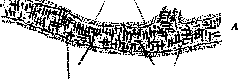
consists of three layers: inner (intima), middle (media), and outer (adventitia). All these layers can be clearly distinguished in any artery, but according to histological structure, considerable differences are observed in them depending on the location and, in particular, on the caliber of the vessel, and therefore all arteries can be divided into three types: small-caliber arteries-muscular type, large-caliber arteries-elastic type, and medium-caliber arteries-transition type.-In small arteries (see figures 1 and 2), the inner layer consists of a layer of endothelial cells covering the inner surface of the vessel with a smooth layer and preventing blood clotting; immediately beneath the endothelium lies the internal elastic membrane (membrana elastica interna); from the contraction of the middle muscular layer, it forms longitudinal folds and on the cross-section of the vessel appears
Figure 2. Small artery of the muscular type: 1 and 2-adventitia (1-longitudinal muscle bundles, 2-connective tissue); 4-media, circular smooth muscle fibers; 5 and 6-intima (5-membrana elastica interna, 6-endothelial layer); 3 and 10-perivascular connective tissue; 7 and 9-blood corpuscles in a clot; 8-clotted blood plasma.
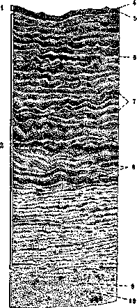
as a more or less thick shiny strip, to which the nuclei of endothelial cells visible in profile tightly adhere. Behind the internal elastic membrane goes the middle tunic, consisting of a powerful layer of circularly arranged muscle fibers (smooth)-the thickest section of the arterial wall; in some cases (sites of arterial branching, splenic artery) one can also see thin bundles of smooth muscle fibers running longitudinally and lying closer to the internal elastic membrane. Externally, the connective tissue tunic-adventitia-adheres to the muscular tunic, consisting of bundles of collagenous fibers having a predominantly longitudinal direction and a network of elastic fibers, which in larger trunks often forms a thickening at the border with the middle tunic-membrana elastica externa.-The wall of large arterial trunks (aorta, subclavian artery, internal iliac artery) presents significant differences from the type just described, concerning all three layers of the wall (see figure 3). The intima is no longer formed by endothelium alone, but between it and the internal elastic membrane there is a more or less significant layer consisting of thin bundles of collagen fibers and thin elastic networks having a predominantly longitudinal direction; between
Figure 3. Large artery of the elastic type: 1-intima, 2-media, 3-adventitia; 4-endothelium; 5-fibrous layers of the intima; 6-elastic plates; 7-bundles of smooth muscle fibers; 8-nuclei of smooth muscle fibers; 9-elastic fibers of the adventitia; 10-bundles of collagen fibers.
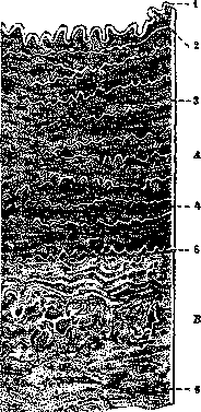
Fig. 4. Transitional artery (medium caliber) A - media B - adventitia; 1 - internal elastic membrane; 2 - endothelial layer; 3 - elastic fibers; 4 - bundles of smooth muscle fibers; 5 - external elastic membrane; 6 - vasa vasorum. Among the fibrous elements lie irregularly branched cells, anastomosing with their processes and forming a cellular network - Langhans cells. The internal elastic membrane usually splits into two layers, of which the inner one often consists of small fibers merging with the network of fibrous layers of the intima. The middle layer (media) is characterized by the presence of a large number of elastic fibers and terminal plates, which are interspersed with relatively few smooth muscle fibers (at the beginning of the aorta, smooth muscles are usually completely absent). The adventitia of large arteries contains thin branches of blood vessels (vasa vasorum) in its thickness, the capillary branches of which also extend into the outer part of the middle layer. Between these two extreme types of arteries, there is a gradual transition in vessels of medium caliber (e.g., a. brachialis, a. femoralis), which approach either the muscular type or the elastic type, and for this reason are called arteries of the transitional type (see Figure 4). In addition to the division of arterial vessels into the three mentioned types, it is necessary to single out the finest arterial trunks that pass into capillary vessels, the so-called arterioles; the muscular layer of such a vessel consists of a single row of circular muscle fibers, which near the artery lie closely next to each other, while near the capillaries they are spaced farther apart at considerable distances from each other; the internal elastic membrane of the arteriole is very thin and may be visible only after special staining for elastic tissue; the endothelial layer passes directly into the endothelium of capillaries; the adventitia of the arteriole is very poorly developed and passes into a thin meshwork of delicate bundles of collagen fibers surrounding the capillaries. The wall of arterial vessels contains a large number of nerve fibers, forming around the vessel a dense plexus containing small ganglion cells; into this plexus enter both sympathetic nerves and cerebrospinal nerves (Maliantovich). The nutrition of the arteries themselves (of large caliber) occurs through vasa vasorum (see above); in addition, nutrition is also maintained from the lumen of the vessel itself. Macroscopic study of arteries is performed by dissection after preliminary filling with hardening masses (gypsum, gelatin, wax+colophony+turpentine, Teichmann's mixture, etc.) and radiologically, after injection of radiopaque (not allowing X-ray) masses. Pathology of Arteries. Developmental defects of the aorta (and everything concerning the aorta) - see Aorta, as well as Heart. In the rest of the arterial system, isolated deviations from the normal type of development are often encountered, which mostly belong to the so-called variations and have no pathological significance. As for the pathological processes of degenerative-atrophic, hyperplastic and even inflammatory nature observed in the arterial system, most of them represent only individual components of that extensive and extremely rich in content pathological form, which is called arteriosclerosis (see), and only a few of them can be attributed to a greater or lesser degree of independent significance. Among the latter are: 1) Limited necroses of the wall (mainly of the middle, less often of the inner shell) of various arteries (spleen, heart, skin, brain, extremities), which often arise under the influence of various infectious and toxic effects, e.g., in abdominal and typhus, scarlet fever, diphtheria, cerebrospinal meningitis, septic diseases, extensive skin burns, and finally, experimentally with intravenous injections of adrenaline to rabbits (in the latter case, there is a tendency to rapid successive calcification). In addition, Jaffe described a case of primary multiple focal necrosis of the middle shell of the pulmonary artery in a newborn, histologically picture resembling experimental adrenaline necroses of mediae of animals. 2) Hyaline degeneration is observed independently of arteriosclerosis in small arteries of many organs under various conditions. Thus (according to Herxheimer), in the spleen, hyalinosis of the pulp arteries and follicles (but not trabecular arteries) is often found - especially in persons over 10 years old - without any specific diseases, almost as a physiological phenomenon, increasing in frequency and intensity with age. The same change is found in the arterioles and capillaries of tumors and various organs with nutritional disorders, occurring either in connection with local inflammatory processes, or with peculiarities of function (e.g., in the uterus after pregnancy), or with atrophy of the organ (e.g., in the ovary), or finally, without determinable causes. In addition, there are cases of primary hyalinosis of small arteries of many organs (mostly with predominant involvement of the digestive tract, especially the tongue) as an independent peculiar disease. In all these cases, the process of formation of hyaline masses in the arteries begins subendothelially (rarely under the internal elastic membrane, leaving the entire intima completely untouched) and spreads towards the periphery, with the lumen usually narrowing to a greater or lesser degree, and sometimes completely disappearing. The muscular membrane in larger vessels remains for a long time in the form of an unevenly thinned strip located outside the hyaline ring, while in small arteries it is quickly involved in the process: it loses nuclei, homogenizes and becomes indistinguishable. 3) Amyloid degeneration of arteries occurs as a particular manifestation of general amyloidosis and affects, predominantly, small and medium arteries; large arterial vessels are affected by it very rarely and only to a slight degree. The deposition of amyloid masses usually begins with the muscular membrane, and in the smallest branches it proceeds so rapidly that the vascular wall as if immediately in toto turns into glassy amyloid substance. In larger trunks, the first deposits may sometimes occur in the adventitia. See also Amyloid degeneration. 4) Fatty degeneration of the inner shell belongs to the changes of the arterial system that develop unusually early and are more common than all others. According to the data of Saltikov and Zinserling, the beginning of this process should be attributed to the 3-6th month of life, and from the age of 8 it is determined on section in all without exception cases. Most often the changes are found in the aorta, but in addition, they can also be observed in large branches arising from the aortic arch, especially in the carotid arteries, in the iliac arteries, as well as in the pulmonary artery. In contrast to this, as if physiological, process of fatty degeneration of vessels occurring under the influence of certain specific poisonings (phosphorus, alcohol), infections (especially in relapsing fever) or severe anemic conditions, small arteries and capillaries are more often affected, with fatty degeneration involving, mainly, the endothelium and muscle fibers of the media. 5) Deposition of lime as the most common finding is observed in arteries as a manifestation of secondary dystrophic calcification of various pathological products, for example, fat breakdown, necrotic areas, foci of hyaline degeneration, etc. As for primary lime deposits, here first of all deserves mention the calcification of the middle shell, which (as shown by Mohckeberg, Faber and others) occurs regularly, starting in the pelvic arteries and arteries of the abdominal cavity and then gradually passing to the aorta and much less frequently affecting other arteries. The first signs of this process in the pelvic arteries can sometimes be found already in children in the 1st year of life, and by the age of 10-20 years it is determined in almost 100%. Its essence consists in the appearance in the middle shell of the smallest grains of lime (often detectable only with the help of the reaction with AgNO3), located in arteries of both elastic and muscular type, between muscle cells along the course of elastic fibers, as if dusting the latter and only occasionally collecting in somewhat larger clusters. The internal elastic membrane can also participate in the process, to a lesser extent - the intima, even less - the adventitia, and in the latter two, as well as in the media, lime is deposited, mainly on the elastic fibers (perhaps, in the ground substance between them). It is interesting that these changes are observed just as regularly in horses and in cattle, starting approximately from the 3-4th year of life. The deposits just described under normal conditions are never significant. If they exceed physiological limits, they usually give the picture of changes that is already considered as an independent disease and amounts to the coarse petrification of the middle shell of many arteries of the muscular type, especially of the arteries of the extremities (especially a. femoralis).
Here, as in the previous case, the muscle cells themselves are not directly calcified, but rather die gradually under the pressure of accumulating calcareous masses. The process begins in the middle layers of the media and then spreads outward and inward within it, sometimes also involving the internal elastic membrane. The deposits are distributed unevenly along the vessel wall, but in separate areas, mostly in the form of circularly running bands, as a result of which the Artery in pronounced cases resembles a goose trachea. Such a vessel may be subject to fractures, and in the healing of which a connective tissue thickening forms on the wall—something like a callus. It is interesting that in those places where the Artery bends frequently (popliteal fossa, groin flexure), it as a rule remains free from changes. Another form of pathological petrification of the Artery is the calcification of the internal elastic membrane, which is either isolated or serves as the beginning of more extensive calcareous deposits, subsequently sometimes occupying the entire vascular wall. The first can be observed not infrequently outside of any specific diseases in individual small arteries of many organs, especially the thyroid gland; the second occurs in cases of extensive calcareous metastases, as well as in that rare pathological form which is called universal calcinosis or calcareous gout. For these latter conditions, on the one hand, the extreme extent of the process is characteristic, which sometimes involves almost all arteries of medium and small caliber; on the other hand, the particularly severe involvement of the blood vessels of the lungs, stomach, and kidneys is characteristic, which is presumably due to the excretion of acidic products (carbon dioxide, urine, gastric juice) by these organs and the corresponding increased alkalinity of their tissues. As for the further fate of the calcareous deposits, they can remain unchanged (or gradually increase in size) for an indefinitely long time; often, however, their resorption by giant cells and proliferating connective tissue is observed, sometimes with the subsequent formation of large bony areas in their place.-6) The deposition of iron in the walls of vessels apparently occurs much more frequently than was previously assumed. It is localized on the internal elastic membrane (with possible subsequent spread to the media), often combining with petrification, from which it is indistinguishable on ordinary preparations and is revealed only with the help of appropriate microchemical reactions. It arises in connection with local or general disorders of iron metabolism (around resorbing foci of hemorrhages, in internal organs in hemosiderosis and hemochromatosis), but sometimes without quite definite causes (for example, in the thyroid gland in myxedema, some goiters).-Summarizing all the data presented regarding regressive processes in the arterial wall, it should be noted that in terms of quantity and volume of almost physiological age-related changes (hyalinosis, fatty degeneration, petrification), the vascular, in particular arterial, system among other systems of the organism apparently stands in first place, being (probably under the influence of the constant and significant mechanical work it performs) one of the most vulnerable. Among the elements making up the arterial wall, the connective tissue elements (especially the ground substance and fibers) are the least resistant, while the muscle cells prove to be significantly more durable. As for the progressive changes of the Artery, besides the physiological thickening of the intima of all arteries, beginning from birth and continuing with interruptions until mature age, and functional hypertrophy (e.g., in the uterus during pregnancy), they almost entirely belong to the so-called arteriosclerosis (see) and therefore most of them will only be mentioned briefly here. These include: 1) Compensatory proliferations of the intima, arising under the influence of prolonged changes in blood pressure in a given area (e.g., under the influence of increased pressure in hypertensions, nephrosclerosis or decreased pressure, as occurs in the umbilical vessels and the ductus arteriosus after birth), as well as due to violation of the normal properties (distensibility, contractility) of the vascular wall. The latter is most pronounced in areas of significant calcification of the middle or internal elastic membrane.-2) Processes of pathological organization in thrombosis and embolism and regenerative proliferations occurring in the healing of wounds. In the case of small punctured or punctured-cut through wounds, the puncture channel is quickly filled with thrombus. Subsequently, the organization of this thrombus occurs, which proceeds, mainly, from the superficial layer of the intima and much less from other membranes. Thus, a connective tissue thickening (vascular callus) is obtained on the vessel wall, which gradually scars. Over time, new formation of elastic fibers occurs in it, but neither elastic membranes nor muscle cells appear. This process differs in no essential way from the one just described, which takes place in the area of the vascular suture. In more extensive damage to the wall, if fatal bleeding does not occur, aneurysms form (see). In case of a complete transverse rupture or incision of a healthy artery, first of all, there occurs a very sharp longitudinal and transverse contraction of the segments of the media, as a result of which the intima folds into numerous folds, and bleeding in small and even medium-sized arteries almost always stops. Further healing proceeds by the formation of thrombi in the segments with subsequent proliferation of the intima and organization.-3) Inflammatory non-specific proliferations of the intima (endarteritis productiva s. obliterans) often occur in vessels passing near or through foci of various kinds of chronic inflammations, but sometimes develop independently (for example, in chronic lead poisoning) and can lead to gangrene of the extremities or intermittent claudication. This latter group forms a transition to the true inflammatory diseases of the arterial system, among which there are both acute and chronic forms. Acute inflammatory changes of arteries of various calibers (arteritides) are encountered in severe cases of many infectious diseases (scarlet fever, sepsis, pneumonia, typhoid fever, typhus, diphtheria, influenza). They consist, on the one hand, of cellular infiltration of the wall, developing partly due to emigration of cells from vasa vasorum, and partly due to proliferation of adventitial elements, and on the other hand, of more or less destructive changes of the elastic and muscle fibers. Often, mural thrombosis is added to this. Of particular importance is the purulent inflammation of arteries, which can arise either by the spread of the process from surrounding tissues, or due to the primary fixation of the causative agents on the inner membrane or in vasa vasorum (mycotic embolism). Besides purulent infiltration of the wall, thrombus formation always occurs here (in mycotic embolism the process begins with this), which subsequently often undergoes purulent melting. Necrosis and purulent melting of the wall itself lead to the development of ulcers, ruptures, perforations, or aneurysms. The pathological importance of the process lies in the ease of development of multiple emboli due to purulent melting of the thrombus and in the possibility of profuse (often fatal) bleeding from the destroyed artery. In small arteries, with a slower course of the process (especially when it spreads to the vessel from surrounding parts), fairly early obliteration of the lumen is possible due to inflammatory proliferation of the intima. A special form of inflammation of small arteries is given by typhus. The most characteristic features of it are, on the one hand, focal destructive-necrotic changes of the inner and especially the middle membrane with mural thrombosis, and on the other hand, proliferation of adventitial elements leading to the formation of characteristic cellular nodules. Depending on the stage and severity of the disease, changes of the first or second kind may predominate. Among the chronic inflammatory processes in the arterial system, besides the above-mentioned non-specific productive endarteritis and so-called periarteritis nodosa (see Periarteritis), it is necessary to note the specific lesions of arteries in tuberculosis, actinomycosis, and syphilis.-In tuberculosis, the changes in arteries in the initial stages are expressed in the form of specific peri- or endoarteritis. The first is very common and can be observed in almost every large or small tuberculous focus when the process spreads from surrounding tissues to the outer membrane of the artery. Usually, due to the rather slow rate of development of changes, the reactive proliferation of the intima (by itself or in connection with subsequent thrombosis and organization) manages to lead the vessel to obliteration before the other vascular membranes are involved in the area of specific granulations.
Only in large vessels or with rapid development of the process and a tendency of the tissue to caseous disintegration does the vessel become perforated and bleeding occur, before which a hernia-like aneurysm usually forms on the affected area of the wall due to the bulging of the intima under pressure through the site of destruction of the adventitia and media. Another, much rarer form of tuberculosis of the arteries is characterized by the development of tubercles on the inner membrane, which occurs when infectious material (e.g., caseous masses) enters the bloodstream. The fixation of bacilli on the intima is greatly facilitated by any changes in it (e.g., atheromatous)* that make its surface uneven. The development of a tubercle on the intima is always accompanied by more or less significant bulging into the vessel lumen, which in small arteries easily leads to obstruction, and if they are also terminal vessels, then to infarction (kidney, bones). At the site of the latter, tuberculous granulations usually develop rapidly. Tubercles of the intima are of much greater importance in the spread of the process, since in caseous necrosis and disintegration their infectious material much more easily and frequently enters the bloodstream, causing infection of the entire branching area of the given artery. Everything said regarding tuberculosis can be fully applied to actinomycotic lesions of the vessels. The latter, besides its much greater rarity, differs only in the structure and further fate (purulent melting) of the specific granulomas. Syphilitic changes of the arteries, with few exceptions, belong to the picture of tertiary syphilis and are most often found in the areas of development of gummatous nodes. Small arteries passing through the area of a gumma always participate in the process, and the caseous necrosis of the syphilitic granuloma is undoubtedly largely due to the condition of its vessels. The latter is characterized by small-cell infiltration of the outer membrane and, in places, the middle layer, and proliferation of the intima, which in general leads to a sharp thickening of the wall and narrowing, and then to complete closure of the lumen. With necrosis of the gumma, the vessels also undergo necrosis, and their elastic tissue is preserved in the necrotic mass for a very long time, thanks to which they can easily be detected there with appropriate stains (sometimes important for differential diagnosis from tubercle). Isolated lesions in syphilis, besides the aorta (see), are most characteristic of cerebral arteries. Here, in the initial stages, the matter also comes down to focal small-cell infiltration of the outer membrane and reactive non-specific proliferation of the intima. With a more severe process, the cellular accumulations in the adventitia become denser and spread around the entire circumference of the vessel, extending to the periadventitial tissue as well as to the middle membrane. Lymphoid cells may be mixed with giant cells and polymorphonuclear leukocytes, which are more often found near the foci of necrosis that arise here, located mainly at the border with the media. Subsequently, specific infiltrates with necroses appear in all layers of the middle membrane and, especially, in the thickened inner membrane, destroying the elastic tissue as they develop. The vessel lumen is often closed either by cellular masses or a thrombus, causing corresponding consequences (areas of softening or even death). In cases of regression of the process, fibrous transformation of the affected areas of the wall occurs with the new formation of elastic fibers. In typical cases of syphilis, the arteries of the brain base appear thickened, dense, cylindrical in shape, and brilliantly white or slightly yellowish in color (resembling "boiled macaroni"). Unlike arteriosclerosis, here calcification is mostly absent (at least significant), while necrotic foci, if visible to the naked eye, are usually located in the outer layers of the vessel wall. Among tumors developing from arterial vessels, only angiomas of the corresponding type (angioma arteriosum) develop, the starting point of which is most often the arterial branches of the scalp or face (see Angioma). Treatment of arterial diseases comes down to eliminating the corresponding causes or treating the underlying diseases (tuberculosis, syphilis, etc.). Operations are performed on arteries: ligation (ligatura), freeing the artery from constricting scars (arteriolysis), incision of the artery wall and opening of its lumen with the aim of extracting an embolus or foreign body, suture of the artery, resection of a segment of the artery with subsequent replacement of the defect with a vein, plastic operations on the trunk and collaterals with the aim of maintaining continuity of blood flow, desilatization of the artery, anastomosis between an artery and a vein, and operations for aneurysms (Hurtes, Anel, Brasdor-Vardroop, Antyllus, Philagrius, Matas).
Related articles
Cite this page
“Artery.” Soviet Medical Encyclopedia. English translation of Bolshaya Meditsinskaya Entsiklopediya, 1st ed. (Moscow, 1928–1936), ed. N. A. Semashko. https://sovietmedicalencyclopedia.pages.dev/article/artery/