Arteriosclerosis
Historical document, translated for reference. It reflects medical knowledge of the 1920s–30s and is not medical advice.
Summary
This article from the 1928–1936 Soviet Great Medical Encyclopedia defines arteriosclerosis as a collective term for various pathological arterial changes, including age-related thickening, syphilitic mesaortitis, calcification, hyalinosis, and atherosclerosis. It traces the historical evolution of the concept from Virchow's inflammatory theory to Marchand's degenerative-hyperplastic model, emphasizing the clinical significance of atherosclerosis.
Encyclopedia article (1928–1936)
ARTERIOSCLEROSIS. Contents: Pathological anatomy of arteriosclerosis . . . 261 Etiology and pathogenesis . . . . . . . . . . . . . . 266 Symptomatology and clinical forms . . . . . . . 271 Diagnosis . . . . . . . . . . . . . . . . . . . . . . . 283 Prognosis . . . . . . . . . . . . . . . . . . . . . . . 284 Prophylaxis and therapy . . . . . . . . . . . . . . 284 Statistics . . . . . . . . . . . . . . . . . . . . . . . 290 Pathological anatomy of arteriosclerosis. Arteriosclerosis (arteriosclerosis), sclerosis of the arteries, is a chronic disease of the arteries, expressed as a thickening of their walls—either more diffuse or in the form of separate plaques. Corresponding to the altered areas, the arterial wall hardens, loses its elasticity, and the arteries acquire a tortuous course. The term arteriosclerosis reflects only the external features of the altered arteries. It was first introduced by Lobstein (1833), who also gave the first rough anatomical description of arteriosclerotic changes. The essence of arteriosclerosis was first described in detail by R. Virchow (1879); however, his point of view was subsequently abandoned. Virchow classified arteriosclerosis into the group of "parenchymatous" inflammations. He believed that this process begins with "cloudy swelling" of the inner lining of the arteries due to increased nutrition of the intima, resulting partly in degeneration and partly in hypertrophy. Virchow distinguished two types of arterial changes: a purely passive process of fatty degeneration without a preliminary stage of parenchymatous inflammation ("fettige Usur") and arteriosclerosis proper, i.e., an inflammatory process resulting in degeneration or hyperplasia. Considering arteriosclerosis an inflammatory process, Virchow gave it the name endarteriitis chr. deformans. Virchow's doctrine of arteriosclerosis persisted for decades, though it underwent various modifications. Thus, in connection with Cohnheim's doctrine of inflammation, arteriosclerosis began to be viewed as a proliferative inflammation, and the deposition of fat began to be interpreted as a secondary degeneration of the inflamed, thickened inner lining. Thus, the main change in arteriosclerosis began to be seen as a connective tissue thickening of the intima (A. nodosa and A. diffusa). Thoma (1914) spoke out categorically against the inflammatory nature of arteriosclerosis. According to his doctrine, arteriosclerosis always begins with a weakening of the middle layer of the arteries (angiomalacia), as a result of which the lumen of the arteries expands, and the speed of blood flow through them decreases. The latter circumstance causes a compensatory thickening of the inner lining, thanks to which the lumen of the arteries becomes the same as before. Thus, Thoma first tried to explain the essence of arteriosclerosis by the mechanical conditions of blood circulation. In his view, connective tissue thickening of the intima represents the main change in arteriosclerosis. Jores studied in detail the origin and structure of the thickened intima in arteriosclerosis and distinguished two main groups of such thickenings—hyperplastic and regenerative. In Jores's opinion, arteriosclerosis is based on hypertrophy of the intima, occurring initially due to the development of the so-called elastic-hyperplastic layer. This layer easily undergoes fatty degeneration, and as a result of the latter, new connective tissue is formed and a so-called regenerative layer appears. Further refinement of the concepts of the essence of arteriosclerosis was made by Marchand, who came to the conclusion that arteriosclerosis is not an inflammatory process, but a degeneration of the vascular wall, closely connected with its hyperplasia; hence the name proposed by Marchand for a more precise designation of arteriosclerosis—atherosclerosis, i.e., a combination of atherosis (fatty decay) with sclerosis. At the basis of arteriosclerosis, in Marchand's opinion, lies a disturbance in the nutrition of the arterial walls; however, Marchand did not give a more precise definition of what this nutritional disturbance consists of. With further study of the pathology of the arteries, a number of independent processes were isolated within the group of arteriosclerosis that, in essence, have nothing in common with each other. Thus, at the present time, it can be said that arteriosclerosis is, in essence, a collective term for several types of pathological changes in the arteries. This concept partly includes such changes in the arteries that should be separated into a special group as normal age-related thickenings of the arterial walls. They consist of a thickening of the inner lining of the arterial walls that begins in childhood and gradually progresses with age (Aschoff, Jores, Maslov, and others). This process is most sharply expressed in the coronary arteries of the heart (Volkova). It is characteristic not only of humans but also of many animals. In addition to the thickening of the inner lining, age-related changes in the arteries also consist of an increase in the amount of intermediate chromotropic substance in the middle layer (Solovyov). The thickened inner lining of the arteries, when fully developed, consists of three typical layers: muscular-elastic, hyperplastic, and connective tissue. The second type of pathological changes in the arteries, which can be separated from the group of arteriosclerosis, constitutes syphilitic mesaortitis (see Aorta and Syphilitic aortitis), syphilitic and other endarteriitis (see Artery). The next (third) process, separated from the group of arteriosclerosis into an independent nosological unit, is the process of calcification of the middle layer of the arteries (Monckeberg, 1903). The fourth form of arterial wall lesion, separated from the general group of arteriosclerosis, is hyalinosis of the arteries, which, as a rule, affects the small arteries of internal organs (see Artery). Finally, the fifth, most important independent form of arterial lesion belonging to the group of arteriosclerosis is atherosclerosis (Marchand). This process is especially important in clinical terms. It leads to extremely sharp disfigurement of the arterial walls, causes their narrowing or even rupture, and is an extremely frequent cause of death in old age. Macroscopic changes in the arteries in atherosclerosis begin with the appearance on their inner surface, in certain typical places, of yellowish streaks and spots (especially at the sites of the origin of lateral branches). The latter, in more severe stages of the development of atherosclerosis, become elevated, dense, whitish, protrude in the form of plaques into the lumen of the arteries, and sometimes ulcerate due to the transformation of their central part into "atheromatous decay" (see figure). Yellowish streaks and spots on the inner surface of the aorta and on the bicuspid valve are, as a rule, encountered already in early childhood. In one-year-old children, they are present in almost all cases. These changes in children are localized in the initial part of the aorta above the valves and gradually spread further, especially often affecting the sites of the origin of lateral branches. Significant disagreements have arisen on the question of what relationship the currently indicated changes in the aorta in children have to the picture of atherosclerosis in adults. Most authors at the present time recognize that yellowish streaks and spots represent, both in children and in adults, only early stages of the development of more sharply expressed changes in the form of plaque-like thickenings of the inner lining, characteristic of atherosclerosis.
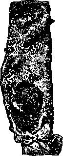
Severe atherosclerosis
of the aorta; large atheromatous ulcers (according to Aschoff). (Ribbert, Aschoff, Saltykov, Lubarsch, Zinserling). Thus, atherosclerotic changes in the arteries begin as early as early childhood. Subsequently, they either develop more or less rapidly and present the picture of adult atherosclerosis, or they halt in their development and, likely, may even disappear. Atherosclerotic changes always begin in the aorta, in its initial part. From there, they spread toward the periphery, primarily affecting the large elastic-type arteries branching off the aorta (carotid, subclavian, iliac), but also spreading to muscular-type arteries. In general, the process spreads systemically, i.e., it usually involves an entire system of branches of a given artery, weakening in intensity from the center toward the periphery. Smaller muscular-type arteries are usually not affected by atherosclerosis. In different cases, even in old age, very different degrees of development of atherosclerosis are encountered. Furthermore, often, along with more or less sharply pronounced atherosclerosis of the entire arterial system, a particularly sharp lesion of the arteries of one specific area is discovered, for example, the arteries of the brain, heart, etc. Microscopically, atherosclerosis is characterized by the accumulation of lipoid substances in the inner lining of the arteries, among which cholesterol is particularly abundant, as well as by a thickening of the inner layer. The view that prevailed until recently, that atherosclerosis represents a primary thickening of the inner layer of the arteries with secondary degeneration, must be abandoned. In recent years, mainly on the basis of experimental studies, it has been possible to prove that the process of atherosclerosis begins with the appearance of lipoid substances in the intima, and the hyperplasia of the latter represents a secondary phenomenon (Anichkov). Similar indications were obtained when studying the earliest stages of the development of atherosclerosis in humans. The cited observations shifted the entire question of the pathogenesis of atherosclerosis onto a completely new plane. Since atherosclerosis begins with the deposition of lipoids, this means that this factor must have the most important significance in the pathogenesis of this process. The same conclusion follows from the results of chemical and microchemical studies of the walls of atherosclerotic-altered arteries. It turned out that they always contain a huge amount of lipoids, in particular, cholesterol (Windaus, Lemoine, Schönheimer). In 1913, it was first possible, by feeding rabbits cholesterol, to induce in them typical atherosclerotic changes of the arteries, quite similar to human ones (Anichkov, Wacker, Hueck, and others). The successful attempts to obtain atherosclerosis in rabbits, made by previous authors (Ignatovsky, Saltykov), should be explained by the fact that they fed their experimental animals food containing cholesterol. The study of the development of experimental cholesterol atherosclerosis, as well as parallel systematic studies of human material (Zinserling), have to a significant extent clarified the formal genesis of atherosclerosis.
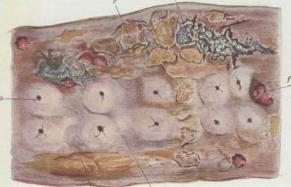
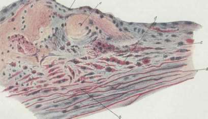
Fig. 1. Atherosclerosis of the thoracic aorta of an elderly woman (natural size): a - spotted lipid deposits on an almost normal intima; b - sclerotic plaques at the sites of origin of the intercostal arteries; c - ulcerated plaques; d - atheromatous foci covered by the intima; e - atheromatous ulcers; f - calcified foci. Fig. 2. Atherosclerosis of the aorta, beginning atherosclerosis in a child (staining: orcein, Sudan, hematoxylin; magnification 80x): a - muscular and elastic layer; b - fatty-degenerated muscle cells; c - intima; d - proliferation of intimal cells; e - foci of lipoids; f - endothelium, partially damaged (according to Aschoff). It turned out that in atherosclerosis, the issue is not about the degeneration of a hyperplastically thickened inner lining, but about the primary infiltration of the latter with lipoids, among which cholesterol is of particularly important significance. Lipoids penetrate into the inner lining of the artery directly from the lumen along with the flow of tissue lymph (Ribbert, Aschoff, Anichkov). If the latter contains a large amount of lipoids, they are deposited in the intima, in its interstitial substance. Further stages of the process consist of reactive changes arising following the deposition of lipoids. These changes consist partly of the accumulation of wandering cells—macrophages—and partly of the development of fibrous and elastic tissue. Thus, the process proceeds in a manner completely opposite to how it was previously imagined: there is not a primary thickening of the inner lining with subsequent fatty degeneration, but a primary infiltration with lipoids with subsequent reactive tissue hyperplasia. From this, it is evident that, from the point of view of formal genesis, atherosclerosis must be classified as an infiltrative process. The factors leading to the emergence of atherosclerosis are very numerous. However, without the presence of primary cholesterol deposition in the arterial walls, this process does not arise. Thus, in its etiology, a disturbance of cholesterol metabolism has very important significance. Furthermore, mechanical moments play an essential role in the emergence of atherosclerosis. Their significance is evident from a consideration of the localization of atherosclerotic lesions of the aorta: they always develop in places subjected to the most severe mechanical influences. The latter particularly includes increased blood pressure. Mechanical moments, as, likely, a whole series of other factors, can predispose the arterial wall to the deposition of cholesterol in it, mixed with other lipoids, as a result of which atherosclerosis begins. Thus, in the etiology of atherosclerosis, there are, firstly, general factors in the form of a disorder of cholesterol metabolism and, secondly, local factors that create particularly favorable conditions in the arterial walls for the deposition of lipoids there. Previous views on the etiology of atherosclerosis suffered from the defect that they sought to find one general "cause" for atherosclerosis in the form of, for example, mechanical, toxic, or nervous influences. However, this could not be confirmed experimentally. At the present time, the "lipoid" or "cholesterol" theory of atherosclerosis is the best-substantiated experimentally, although it also assigns a prominent place in the etiology of this process to the other factors just mentioned. As a result, this theory is also called the "combination" theory of atherosclerosis. It views atherosclerosis as a manifestation of a disease of metabolism, in particular, lipoid (cholesterol) metabolism. The factors leading to the disorder of lipoid and especially cholesterol metabolism, as well as the essence of this disorder, however, remain unclear to this day. Atherosclerotic changes in the arteries are encountered not only in humans but also in some animals (for example, in old dogs, in pigs). Experimentally, they can be obtained (by feeding cholesterol) in rabbits, guinea pigs, and chickens, as well as in rats and mice—in the latter case, however, only with the help of certain additional factors [feeding with protein, castration (Loewenthal)]. Arteriosclerosis represents, in essence, one of the manifestations of the process of lipoid deposition in the fibrous-elastic tissue of the organism in general. Similar infiltrative phenomena (lipoid deposition) are observed in the heart valves, tendons, in the sclera and cornea of the eye (senile arcus—gerontoxon), etc. The consequences of arteriosclerosis for the organism can be very serious. These include: 1) impairment of the blood supply to organs (heart, brain) due to the narrowing of the lumen of the arteries supplying them, and also sometimes due to accompanying thrombosis (consequence—atrophy and necrosis of organs); 2) ruptures of small arteries, especially in the brain—senile apoplexy; 3) dilation of the arteries (see Aneurysm); 4) disorders of general blood circulation due to a decrease in the elastic properties of the arterial walls. Thanks to the rigidity of the arteries in arteriosclerosis, their auxiliary function in blood circulation is weakened, and the work of the heart increases. The latter circumstance, along with other factors, leads to hypertrophy of the heart and to hypertension, sometimes with secondary developing heart failure, especially with simultaneous involvement of the coronary arteries.
N. Anichkov. Etiology and pathogenesis. The term "arteriosclerosis," as indicated above, currently unites several different pathological processes. The differentiation of these various types of arteriosclerosis in relation to etiology, influence on the function of arteries, symptomatology, etc., is an immediate task of clinical medicine. From a clinical point of view, only those arteriosclerotic changes which lead to a disturbance in the function of the arteries of the corresponding organs are discussed here. Clinical medicine has until now classified arteriosclerosis only by its localization and has distinguished on this principle arteriosclerosis of the aorta, coronary, cerebral, pulmonary, and renal arteries, arteries of the gastrointestinal tract, pancreas, extremities, etc. In relation to some localizations of arteriosclerosis, this division coincides with the above-cited pathological-histological division: thus, arteriosclerosis of the aorta, pulmonary artery, and coronary vessels is atherosclerosis; of the cerebral and renal arteries (medium-sized)—atherosclerosis, (small-sized)—hyalinosis and fatty degeneration; of the arteries of the extremities—calcification of the tunica media. A general uniform involvement of all arteries by arteriosclerosis is encountered relatively rarely; usually, one is dealing with the presence of clinically manifesting arteriosclerosis of one or another of the above-indicated localizations or, at least, with a more or less pronounced predominance of arteriosclerosis in one or another vascular region, in one or another organ or organs. Therefore, for the most part, in clinical practice, one is dealing with arteriosclerosis.
with the disease of a specific organ and most often with arteriosclerosis of the heart, aorta, brain, and kidneys. When arteriosclerosis affects, predominantly and more or less diffusely, the small, precapillary arteries—arterioles—one speaks of arteriolosclerosis; by its histochemical character, this is always hyalinosis and fatty degeneration of the arterioles. From the arteriolosclerotic processes, clinical practice singles out the particularly significant arteriolosclerosis of the kidneys, corresponding, according to previous terminology, to chronic interstitial nephritis (see). Pathological anatomists often encounter arteriosclerosis of the spleen as well, and more rarely of the pancreas, but no definite clinical picture corresponds (as yet) to the arteriosclerosis of these organs. A general, more or less widespread arteriosclerosis of all organs and tissues [the previous concept of arterio-capillaro-fibrosis (Gull, Sutton) and the modern concept of Munk, Munzer, and others] has not yet been established pathologically and anatomically. The concept of arteriosclerosis is usually associated with the idea of increased arterial pressure. Initially, it was believed that these two phenomena were inseparably linked causally, with arteriosclerosis being the primary factor. At the present time, it can be considered established that 1) arteriosclerosis is far from always accompanied by hypertension (according to Romberg-Sawada, only in 12.3% of cases), and 2) that cases of hypertension are very often encountered where arteriosclerosis is weakly expressed or even absent, and the kidneys are not changed at all. As a result, the concept of hypertension as an independent pathological functional state was created (see Hypertension). Hypertension is undoubtedly very often combined with arteriosclerosis, especially with arteriolosclerosis of the kidneys, but their interrelationship has not yet been sufficiently clarified. Perhaps, in some cases, arteriosclerosis is a primary phenomenon and causes hypertension (reflex arteriosclerosis of the kidneys, brain); in other cases, on the contrary, hypertension causes arteriosclerosis or, at the very least, contributes to its development. Finally, it is admissible that under other conditions, hypertension and arteriosclerosis are the consequence of one common pathogenetic factor or a combination of factors. Clinically, arteriosclerosis is a disease of old age. It is rarely detected before the age of 45 and, in general, manifests itself more frequently and is more sharply expressed the older the person is. However, there is no complete parallelism between age and the degree of arteriosclerosis, and cases are not rare where arteriosclerosis is not detected by any pathological phenomena in very old age and, conversely, sometimes arteriosclerosis already produces severe consequences in relatively young people in the fourth and even the third decade of life (Jaffe, Sternberg). In autopsy material, Saltykov found arteriosclerosis in men in 78.27% and in women in 76.53% (1,200 cadavers). Schubert, when examining autopsy material, found more pronounced arteriosclerosis more often in men than in women. Clinical material also gives the impression of a more frequent disease in men and, moreover, with more severe forms of arteriosclerosis (more frequent chronic poisoning with alcohol and tobacco in men, more active participation in the struggle for existence, constitutional predisposition). The significance of age in the etiology of arteriosclerosis can be imagined as follows: the longer certain harmful influences can act on the vessels, all other conditions being equal, the more pronounced changes in the vessels they must cause. But, in addition to the duration of harmful influences, the speed and degree of development of arteriosclerosis are also determined by the intensity of these influences and the resistance of the arterial walls. The latter can vary depending on individual, or constitutional, factors. On the basis of clinical experience, there is no doubt that arteriosclerosis belongs to diseases in the etiology of which hereditary predisposition plays a large role, and the impression is created that, in addition to a congenital general predisposition to arteriosclerosis, there is also a congenital, or familial, predisposition to arteriosclerosis of certain vascular regions. The influence of constitution on the development of arteriosclerosis can be imagined as one or another influence on the vessels of constitutional anomalies of metabolism (e.g., cholesterol). Quite definite is the special predisposition to arteriosclerosis—perhaps, more accurately, to atherosclerosis—of individuals with a hypersthenic constitution. In such individuals, chemical analysis of the blood reveals a higher level of cholesterol and uric acid in the blood (Myasnikov, Grotel). A hypersthenic constitution predisposes, as is known, to obesity, gout, diabetes, and stone disease, and apparently links these diseases with arteriosclerosis. French clinicians group this set of diseases (and some others) under a special diathesis—arthritism—and believe that this diathesis also predisposes to arteriosclerosis. As for other etiological factors and influences determining the development of arteriosclerosis, they can be divided into mechanical and chemical or physico-chemical. Mechanical factors can be imagined as excessive passive and active work of the vessels: passive—in the form of intensified, excessive fluctuations in the tension of the arterial walls as a result of too frequent, rapid, and strong fluctuations in arterial pressure; active—under the influence of excessively frequent and intensified contractions and relaxations of the arteries, caused by the frequently and sharply changing blood supply to the corresponding organs during their excessive, too frequently and sharply changing and fluctuating work. This is supported by the observations of pathological anatomists, who often find early or intensified development of arteriosclerosis, in particular atherosclerosis, where, due to physiological conditions or pathological causes, the arterial wall is subjected to intensified mechanical influences, either in the form of excessive fluctuations of tension or in the form of prolonged, intensified tension under the influence of increased pressure. Some clinicians (Baeumler, Romberg, and others) point to more frequent arteriosclerotic involvement of the peripheral arteries in people engaged in physical labor, with men having it in the arteries of the upper extremities (to a greater degree in the right than in the left), and women in the arteries of the lower extremities. The impression is created that in the urban population, especially in individuals leading a sedentary lifestyle with an intense expenditure of mental and emotional energy, arteriosclerosis is localized predominantly in the arteries of the brain, heart, in the aorta, and in the arteries of the abdominal viscera. But these data are not the result of precise studies, but of clinical and polyclinical impressions. With this reservation, it is necessary to point out the influence of emotional distress on the development of arteriosclerosis. It gives the impression that in individuals whose lives are restless due to their profession, character, or unfavorable external conditions, as well as in those who suffer from increased excitability of the nervous system and various psychoneuroses, arteriosclerosis develops earlier and more severely. Also deserving of mention are the indications of some authors, e.g., Pavinsky, regarding the predominant development of coronary atherosclerosis under the influence of emotional distress. Especially in individuals distinguished by increased excitability of the vasomotor nervous apparatus and suffering from so-called vasomotor or vascular neuroses (Oppenheim, Pal), arteriosclerosis apparently develops more frequently and at an accelerated pace. That a disturbance in the function of the vasomotor nerves can indeed contribute to the development of arteriosclerosis is supported, among other things, by the cases of Botkin and Huchard, who observed intensified development of arteriosclerosis on the side of the body where, due to a unilateral lesion of the spinal cord, a disturbance of the normal function of the vasomotor nervous apparatus was observed. Moreover, the disturbance of innervation may, perhaps, have in the etiology of arteriosclerosis not only the significance of a factor determining the incorrect mechanical work of the vascular wall, but also a factor disrupting its nutrition (“trophic” influence of innervation). Heavy physical labor, especially in connection with deprivation and distress, also contributes to the development of arteriosclerosis. An illustration of this is the particularly frequent finding by pathological anatomists of pronounced arteriosclerosis (and, predominantly, also of the coronary vessels) in relatively young participants of the imperialist war of 1914–18 (Mönckeberg, Kohlhaas, Jaffe, Sternberg). Great significance in the etiology of arteriosclerosis, as already indicated, belongs to hypertension. Therefore, Basch at one time called hypertension “latent arteriosclerosis,” and Huchard “presclerosis.” On the basis of accumulated clinical material, at the present time, it cannot be accepted that hypertension always and quickly leads to arteriosclerosis, but these terms illustrate perfectly the close connection that exists between hypertension and arteriosclerosis. Among the influences causing arteriosclerosis or contributing to its development, on the basis of clinical observations, one could place chronic alcohol poisoning; on the other hand, there are observations (mainly by pathological anatomists) denying such a connection. Tobacco smoking undoubtedly predisposes to spastic contractions of the arteries, complicating arteriosclerosis in the form of attacks of angina pectoris or abdominal angina, so-called intermittent claudication, etc. Regarding lead, there is no doubt that chronic poisoning with it inevitably leads to the development of arteriosclerosis. In addition to generally accepted clinical experience, one can also cite the observations of Friedrich, who established that arteriosclerosis in those working with lead and lead compounds develops both more frequently and earlier.
Among workers dealing with lead, he found arteriosclerosis in 23%; among workers of the same factory not dealing with lead and of the same age—in 4.1%. The influence of infectious diseases in the etiology of arteriosclerosis is controversial; some authors attribute the greatest significance to them (e.g., Saltykov, Faber), because they found arteriosclerotic changes in those who died at a young age much more often in cases where an infectious disease had preceded. Others (Anichkov) do not consider this proven. Clinicians point to the frequent acceleration and intensification of the development of arteriosclerosis following infectious diseases. Among chronic infectious diseases, syphilis is not only a cause of specific aortitis and arteritis but may also contribute significantly to the development of ordinary arteriosclerosis. Only some pathologists point to tuberculosis as a significant cause of arteriosclerosis (Faber). One of the earliest changes in the function of the arterial wall under the influence of arteriosclerosis is a decrease in its ability to actively change its lumen depending on the needs of the corresponding organ for blood supply, and since increased blood supply is required primarily during the work of an organ, a decrease in its performance results (Potain's meiopragia). Furthermore, with arteriosclerosis, a decrease in the elastic distensibility of the arteries develops—they approach rigid tubes. With the intermittent pumping of blood by the heart into the aorta, insufficient elastic distensibility of the arteries reduces the blood supply to organs, because with an intermittent flow, an elastically distensible tube conducts more blood per unit of time than a rigid one. In addition, the blood supply to the corresponding organ is reduced if, as a result of diffuse arteriosclerosis of smaller arteries, a narrowing of the lumen of a significant number of these arteries occurs, or if a thrombus forms in a larger branch at the site of a sclerotic change in the wall. Then the work of the given organ becomes insufficient even during its complete or relative rest—e.g., cardiac activity due to sclerosis of the coronary arteries becomes insufficient even during complete rest of the entire organism. This reduced, to a greater or lesser degree, performance of the corresponding organs and the changes caused by their insufficient blood supply lie at the basis of the main clinical manifestations of arteriosclerosis. Sclerotic changes in the arteries themselves are not clinically determined, with the exception of changes in peripheral arteries accessible to examination and the aorta. Thus, regarding the recognition of the most important clinically relevant localizations of arteriosclerosis, we have only indirect signs.
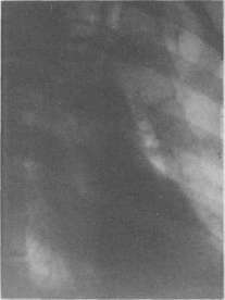
Symptomatology and clinical forms of arteriosclerosis. Atherosclerosis of the aorta, when more strongly developed, leads to its elongation and dilation; in this case, in the area corresponding to the sternal end of the 2nd costal cartilage and the 2nd intercostal space, dullness appears, extending to the sternum and to the right, extending beyond the edge of the sternum by 1-2 finger breadths. The intensity and size of this dullness vary depending on the degree of elongation and dilation of the aorta; all transitions are observed from barely noticeable only with weak percussion and only when compared with the left side, to almost complete dullness. Pulsation in the area of this dullness is usually observed only with stronger aneurysmal dilation of the ascending aorta, which, as a consequence of atherosclerosis, is very rare. Much more often, atherosclerosis of the aorta is detected by palpation (retrosternal pulsation). Both the aforementioned dullness and retrosternal pulsation are usually considered signs of aortic dilation. In reality, in the origin of both phenomena, the predominant role is usually played not by dilation, but by the elongation of the aorta, which is more characteristic of atherosclerosis. The fact is that its ends are fixed to a certain extent, therefore, with the elongation of the entire aorta, its arch must stand higher, and since the aorta is located in the chest cavity in a direction from front and right, to back and left, then with its elongation, the ascending part of the aorta must press more closely against the anterior chest wall. Auscultatorily, atherosclerosis of the aorta is usually manifested by a systolic murmur over the aorta and an intensification (accentuation) or metallic tint of its 2nd tone. A systolic murmur can be caused by 1) rigidity of the aortic wall (relative narrowing of the aorta during the ejection of blood into it by the left ventricle), or 2) changes, i.e., thickening and rigidity of the aortic valves due to their involvement by the same atherosclerosis, or 3) dilation of the initial part of the aorta, or 4) significant irregularities of its inner surface. In cases where there is no systolic murmur over the aorta, it sometimes appears if the patient puts their hands behind their head (Sirotinin, Kukoverov). The intensification of the 2nd tone and its metallic character are most often caused by an increase above the norm, by the beginning of diastole, of the difference between the pressure above and below the semilunar valves of the aorta. This increase in the difference occurs due to an increase in blood pressure in the aorta, where it is higher at the end of systole due to the rigidity of its walls than in the normal state (see below regarding the increase in the amplitude of pulse fluctuations of arterial pressure in atherosclerosis). The cause of the metallic character of the 2nd tone of the aorta is, in addition, considered to be changes in the aortic valves and aortic walls in the sense of their greater density, due to the development of connective tissue and calcification. The indicated auscultatory phenomena in atherosclerosis of the aorta are sometimes detected more clearly if one listens with a stethoscope placed, if possible, parallel to the body axis in the jugular notch. Due to the high position of the arch of the atherosclerotic aorta, the vessels branching from it are also raised. This is especially noticeable in the subclavian arteries and, in particular, in the right one, as it is located more superficially. The arch of the subclavian artery rises 1-2 cm above the 1st rib and becomes significantly more accessible to palpation than in the normal state, when it lies directly on the rib. At the same time, the subclavian arteries show sharp pulsation in atherosclerosis of the aorta (Trunecek's symptom). Among the signs of atherosclerosis of the aortic arch, one can partly include the difference in the pulse magnitude of both radial arteries (p. differens), since this difference depends most often on sharp atherosclerotic changes of the aorta at the site of origin of either the brachiocephalic trunk or the left subclavian artery. Radioscopy reveals in atherosclerosis of the aorta its elongation and dilation and an intensification of its shadow. Elongation is manifested during dorso-ventral fluoroscopy or radiography not only by the elongation of the shadow of the large vessels but also by its widening, with a sharper protrusion, both to the right—of the arch of the ascending aorta, and to the left—of the upper left arch formed by the aortic arch; this left upper arch forms in these cases the so-called beak-like protrusion of the aortic arch. This widening of the shadow of the large vessels during dorso-ventral fluoroscopy to the right and left is the result of the displacement, due to the elongation of the entire aorta, of its ascending part to the right and forward, and the descending part to the back and left. If there is also dilation of the aorta, it is determined better during fluoroscopy or radiography in the first (see table, fig. 1) and second oblique directions (using Frick's technique), because in this case, the ascending and descending aortas are seen more isolated from other large vessels (pulmonary artery, superior vena cava). During fluoroscopy in the second oblique direction, the entire aortic arch is visible, from the ascending aorta to the initial part of the descending one, and by the width of the lumen between them ("aortic window") one can judge the degree of aortic elongation. The atherosclerotic aorta gives a denser shadow than a normal one, but it is very difficult to determine correctly, taking into account all the individual characteristics of a given patient and the conditions of fluoroscopy, whether the shadow of the aorta in a given patient is denser than normal. It is more correct to judge the intensification of the aortic shadow by whether the descending aorta is visible in the first oblique direction of the rays, and the arch of the aorta itself in the second oblique direction. Normally, in these directions of fluoroscopy, these parts of the aorta are not visible, as their shadow disappears against the background of the light lung fields. In atherosclerosis of the aorta, in the first oblique direction, the descending aorta is sometimes visible along its entire length. It must be emphasized that the determination of aortic dilation and elongation by radiological methods requires great experience. Very often they, and even "aortic aneurysms," are diagnosed in elderly people.
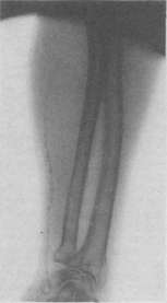
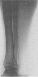
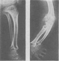
1 - atherosclerosis of the aorta (in the I oblique direction). 2 - arteriosclerosis of the arteries of the forearm (calcification of the tunica media). 3 - arteriosclerosis of the arteries of the lower leg (calcification of the tunica media). 4 and 5 - limbs in achondroplasia. To the articles Arteriosclerosis, Achondroplasia. in such cases where there is only the usual physiological age-related dilation and elongation of the aorta. More accurate results are given, of course, by orthodiagraphic measurement of the aorta in various directions of X-rays and by the Kreuzfuchs method, or corresponding teleradiographs (see fig.).
Orthodiagrams of the heart and large vessels in atherosclerosis (various types in terms of the predominance of elongation or dilation). Small drawings depict the measurement of aortic width according to the Kreuzfuchs method. Atherosclerosis of the aorta, in terms of subjective manifestations, often proceeds completely latently; those subjective sensations, mostly pain and shortness of breath, with which it may be accompanied, are caused primarily by subsequent phenomena: narrowing of the vessels branching off from the aorta and weakening of cardiac activity. Narrowing of the coronary ostia contributes to the development of typical attacks of angina pectoris. In addition, sometimes pains are observed that are not entirely characteristic of angina pectoris; they are similar to angina pectoris in localization, as they are most often felt behind the sternum in the form of distension, pressure, less often burning, and spread to the upper extremities, to the neck, and to the upper part of the abdomen, but their paroxysmal character is less pronounced, they are prolonged, sometimes felt for days and weeks, with periodic intensifications that are vaguely demarcated (aortalgia). In the region of the heart, in the axillary cavity, along the inner surface of the upper extremities, predominantly the left, hyperesthesia is sometimes determined. In the upper extremities, especially the left, numbness, a crawling sensation, and similar paresthesias are observed. Sometimes pain is felt only in the upper extremities, predominantly the left, sometimes only in the shoulder joints, and due to their prolonged nature, they are mistaken for "rheumatic" pain. All these painful sensations can appear at complete rest, but are sometimes caused or intensified by emotional distress and physical exertion; it is often difficult to distinguish them from angina pectoris. Apparently, these painful phenomena are not connected with damage to the coronary vessels, and one can only assume that all these sensations are the result of irritation or changes in those nervous (sympathetic) elements that are embedded in the wall of the aorta and surround it (plexus cardiacus and plexus aorticus) and should be explained by viscero-sensory reflexes. For pain in the upper extremities accompanied by palpitations, Huchard sees the cause in the irritation of the plexus brachialis by a highly situated and intensely pulsating arteria subclavia. Dysphagia, sometimes observed in atherosclerosis of the aorta even in the absence of dilation pressing on the esophagus, is also explained by a reflex from the periaortic nerve plexuses. Narrowing of the ostia of the innominate artery and the left carotid artery due to atherosclerosis of the aortic arch can, by causing insufficient blood supply to the brain, manifest as tinnitus, dizziness in the form of attacks (especially when moving from a horizontal to a vertical position), and fainting. Narrowing of the ostia of the same innominate and left subclavian arteries can manifest (in addition to the already mentioned pulsus differens) as a sensation of weakness in the arms, their rapid fatigability, various paresthesias, and pain appearing after brief work (analogous to pain in Charcot's intermittent claudication). Narrowing of the ostia of the intercostal arteries is the cause of pain in the back and sides, resembling intercostal neuralgia, but depending to a greater extent on corresponding movements. Finally, narrowing of the ostium of the arteria coeliaca can be the cause of "abdominal angina"—attacks of pain resembling angina pectoris in character. The clinical picture of atherosclerosis of the aorta also includes shortness of breath, caused by difficulty and subsequent weakening of cardiac activity and appearing either under the influence of increased movement (dyspnee d'effort) or only in the form of attacks of so-called cardiac asthma; attacks of suffocation are sometimes combined with painful sensations of an anginal character. Consequences of atherosclerosis of the aorta can be its thrombosis, ruptures, and aneurysms. Atherosclerosis of the abdominal aorta, which is often more pronounced in cases of aortic involvement than atherosclerosis of the thoracic aorta, can be determined only directly by palpation, which is possible, of course, only with thin abdominal walls. In this case, it is sometimes possible to palpate an enlarged, often curved, abdominal aorta. Due to the curvature and relatively small pulsatory expansion, it is sometimes mistaken for a tumor. Atherosclerosis of the pulmonary artery often develops as a phenomenon accompanying certain diseases of the heart and lungs, namely, stenosis of the left venous orifice, certain congenital heart defects (e.g., ductus arteriosus Botalli persistens), pulmonary emphysema, kyphoscoliosis, and, consequently, such pathological states in which the pressure in the pulmonary artery is strongly and prolonged elevated; but the development of sclerosis of the pulmonary artery is observed, although very rarely, even in the absence of reasons for an increase in pressure within it and even at a relatively young age. In these cases, preceding severe infectious diseases are sometimes noted. The following clinical signs are considered characteristic of pulmonary sclerosis: 1) dullness at the left edge of the sternum (due to dilation of the pulmonary artery) and the 2nd arch of the left border of the X-ray shadow of the heart protruding strongly to the left; 2) strong increase in cardiac dullness to the right and a diffuse impulse of the dilated and hypertrophied right ventricle; 3) a systolic, and sometimes diastolic, murmur over the pulmonary artery due to changes in its valves and ostium by the arteriosclerotic process; 4) strong cyanosis with relatively weakly expressed shortness of breath and in the absence of edema; 5) repeated severe pulmonary hemorrhages; 6) attacks of pain in the region of the base of the heart, radiating into the lung and accompanied by an increase in cyanosis and a feeling of anxiety and fear (Posselt). All these signs have much in common with the signs of those diseases of the heart and lungs (for example, stenosis of the left venous orifice) in which atherosclerosis of the pulmonary artery is most often observed, therefore its diagnosis in these conditions appears difficult, as it is hard to assess to what extent some symptoms belong to the underlying disease and to what extent to pulmonary sclerosis. Cyanosis deserves special attention as a sign of pulmonary sclerosis if it is strongly expressed and its degree does not correspond to other manifestations of the underlying suffering, as well as attacks of pain, which are, however, similar to attacks of angina pectoris, but accompanied not by pallor of the skin, but by strong cyanosis. Atherosclerosis of peripheral vessels accessible to the eye and palpation (arteria temporalis, brachialis, radialis, dorsalis pedis, etc.) is sometimes revealed by the fact that they become more visible, as, due to the greater rigidity of their walls and due to the increasing tortuosity resulting from elongation, they stand out more sharply in the surrounding softer tissues. With the passage of each pulse wave, arteriosclerotic arteries are displaced more strongly and faster due to greater tortuosity, less elasticity, and due to a sharper rise in pulse pressure. This is especially clearly visible in the arteriae brachialis and cubitalis. To determine the presence of atherosclerosis of peripheral arteries by palpation, it is necessary to examine, if possible, all accessible arteries, as sometimes some of them (e.g., arteries of the lower extremities) already present strong changes, whereas others (e.g., arteries of the upper extremities) are still normal to the touch. If one palpates a sclerotic artery, e.g., the radial, pressing it with a finger so strongly against the underlying bone as to squeeze the blood out of it, it seems to be a rounder and denser cord than a normal artery. However, it is necessary to remember that even strongly contracted arteries, as well as arteries with hypertrophied elastic and muscular tissue, but still without arteriosclerotic changes, can give a similar sensation. Only if irregularities are felt when sliding fingers longitudinally along the artery is an arteriosclerotic change in the arterial wall certain. At high degrees of atherosclerosis, the arteries turn into cords to the touch, uneven like a goose trachea or rosary beads. Upon palpation of the pulse of a sclerotic artery, a sensation of a faster and more energetic rise of the pulse wave is obtained. Sphygmograms fully correspond to this sensation, differing by a steep rise of the wave. This steep rise depends on the fact that, due to the rigidity of the aortic walls, the arterial pressure naturally rises much faster with each systole of the left ventricle than with a more compliant elastic vessel wall. The steep rise is often followed by a rounded or flat peak—also an expression of the reduced elasticity of the arterial wall. The secondary waves are fewer the stronger the atherosclerosis and the lower the blood pressure. Dicrotism is always weakly expressed. In general, the sphygmogram of peripheral arteries in atherosclerosis approaches the configuration of the aortic sphygmogram. A more significant change appears to be the change in pulse tension, but precise study of pulse tension by measuring arterial pressure with special instruments has shown that it is not as frequently elevated in atherosclerosis as was previously thought, and that it is often not elevated at all or only to a slight degree.
In atherosclerosis, arterial pressure depends, mainly, on whether it is accompanied by an increase in the tonic contraction of the arteries of the whole body (or, at least, the region innervated by the celiac nerve), i.e., hypertension. Atherosclerosis of the aorta does not cause a significant rise in the average level of arterial pressure, but it increases the maximum and slightly lowers the minimum pressure. Instead of the norm of 70 and 120 mm of mercury for the minimum and maximum pressure, for example, 60 and 150 mm are determined. The amplitude, therefore, will be increased to 90 instead of 50. The degree of increase of this amplitude can, in the absence of any signs of heart failure, and taking into account the pulse rate, be used as an indicator of the degree of atherosclerotic rigidity of the aorta. The elasticity of the aorta and large vessels can also be judged to a certain extent by the steepness of the rise of the sphygmographic curve and by the speed of propagation of the pulse wave. Regarding the determination of the functional capacity of peripheral arteries, the methodology is even less developed. The ability of arteries in arteriosclerosis to increase or decrease the tonic contraction of circular musculature and change their lumen can be determined with the help of a plethysmograph by including a limb in it and irritating the skin, for example, with cold or heat. A decrease in the volume fluctuations of the limb under the influence of these stimuli, compared with the norm, corresponds to the degree of arteriosclerosis of the supplying artery (Romberg, O. Müller). Despite this clinically and experimentally established insufficiency of the reactive contractility of sclerosed arteries, there are clinical grounds to assume that arteries affected by arteriosclerosis, compared with normal ones, are distinguished by increased excitability of the musculature in the sense of a tendency to spastic contractions. This tendency manifests itself, apparently, especially sharply if one more factor is added—an increase in the excitability of the nervous system, specifically the vasomotor one. In these cases, arteries sometimes respond to stimuli requiring dilation for an increased influx of blood (e.g., when the corresponding organ begins to work) with the opposite movement—spastic contraction, or at least they do not dilate appropriately. Such insufficient or incorrect reactions of the arteries, apparently, lie at the basis of some attacks of angina pectoris in arteriosclerosis of the coronary vessels, often appearing precisely when the heart is required to increase its work; these arterial reactions, apparently, cause intermittent claudication in sclerosis of the arteries of the lower extremities, attacks of abdominal angina occurring during intestinal digestion, etc. Sometimes these local vascular spasms cause and are accompanied by an increase in the tonic contraction of the arteries of the whole body, which causes, of course, a sharp rise in arterial pressure (vascular crises of Pal). The change in the arteries is the fundamental primary phenomenon in arteriosclerosis—all the following painful phenomena in arteriosclerotics are secondary. In terms of frequency and significance, the first place among these secondary phenomena belongs to phenomena from the heart. In arteriosclerosis, the heart can suffer in three ways: 1) due to arteriosclerosis of the coronary arteries, 2) due to overstrain because of the difficulty of blood flow in sclerotic arteries, and 3) in the case of arteriosclerotic damage to the heart valves with the development of one or another defect. The difficulty in the work of the heart due to the hypertension so often accompanying arteriosclerosis, strictly speaking, does not relate to heart damage from arteriosclerosis. As for coronary arteriosclerosis, it affects almost exclusively the main trunks and larger and medium branches of the coronary vessels and leads to diffuse or, more often, focal degenerative changes of the heart muscle or to focal necrosis with the subsequent development of scars. Due to these changes, the heart muscle weakens to a greater or lesser extent, and a picture of heart failure with all the consequences gradually develops. In the beginning, this insufficiency manifests itself only when increased demands are placed on the heart in terms of work—Potain's meiopragie. The indicated scars (and perhaps foci of degenerative changes), localizing in the conduction system of the heart, can cause all types of arrhythmia: the most frequent of them is extrasystole, less often—"atrial fibrillation" (pulsus irregularis perpetuus); sometimes—a disturbance of coordination in the activity of the atria and ventricles up to complete block with corresponding phenomena of the Adams-Stokes symptom complex (see) or without them, and, finally, pulsus alternans. But most characteristic for damage to the coronary vessels is angina pectoris in all its numerous variations (see Angina pectoris). Still, there is by no means a complete parallelism between the degree of arteriosclerosis of the coronary arteries and the presence and degree of angina pectoris. Often, arteriosclerosis of the coronary arteries, through narrowing and the formation of thrombi, leads to occlusion of the coronary arteries, to the formation of cardiac infarcts, and to death or the development of cardiac aneurysms (see Aneurysm of the heart). All lesions of the heart, or rather the heart muscle, which are a consequence of arteriosclerosis of the coronary arteries, can be combined under the name of atherosclerotic cardiopathy or myocardiopathy—in short, cardiosclerosis (see). This diagnosis must replace those diagnoses of "myocarditis" that are usually made in cases of damage to the heart muscle due to arteriosclerosis. Myocarditis is an inflammation of the heart muscle, whereas in cardiosclerosis there are, in any case, no inflammatory phenomena as a primary process. The second way in which arteriosclerosis of the arteries causes a change in the heart is the difficulty of its work due to a decrease in the extensibility of the arteries, in particular the aorta. The blood entering the aorta during the contraction of the left ventricle stretches it, and part of the energy of the heart contraction passes into the tension energy of the aortic wall. This energy, during diastole with the contraction of the aorta, due to the elasticity of its walls, is again converted into the kinetic energy of blood movement. Thanks to this, the flow of blood in the arteries during diastole does not stop but continues with great energy, and the next contraction of the left ventricle has to overcome the force of inertia of the blood mass to a comparatively small degree. Therefore, the more rigid the aorta, the more energy the heart has to spend to overcome the force of inertia of the blood. The heart can compensate for the loss of the function of elastic extensibility of the arterial wall only through increased work. It hypertrophies; first and mainly—the left ventricle, and when it begins to weaken, then the left atrium and the right ventricle as well. But arteriosclerosis itself usually progresses, and the increase in blood pressure and the increase in pulse fluctuations so often accompanying arteriosclerosis contribute to this. Due to this, the difficulties of blood circulation in the arteries grow, and the heart muscle itself often moves into unfavorable working conditions due to the gradually developing arteriosclerosis of its vessels. Therefore, the heart must sooner or later turn out to be unable to compensate for the ever-increasing difficulties with increased work; despite hypertrophy, it overstrains, weakens, and dilation of its cavities occurs (first again the left ventricle), and signs of heart failure gradually appear and develop. The first sign of this is usually shortness of breath during strenuous movements (dyspnee d'effort) and nocturnal attacks of suffocation (cardiac asthma), often accompanied by Cheyne-Stokes respiration. The third way in which arteriosclerosis causes a change in the heart (the least important) is atherosclerotic damage to its valves, most often the aortic ones, sometimes leading to their insufficiency and even more rarely to stenosis. Of all cases of aortic valve insufficiency (Romberg's material), only in 5.5% was the cause arteriosclerosis. Sometimes arteriosclerosis also localizes on the aortic cusp of the mitral valve, causing its thickening and shortening, usually leading to valve insufficiency or—very rarely—to stenosis of the left venous orifice. In general, the importance of arteriosclerosis in the etiology of heart valve defects was previously overestimated; according to Romberg's material, only 3.7% of all valve defects are of arteriosclerotic origin. In terms of its significance, arteriosclerosis of the brain is in second place. The clinical pictures of arteriosclerosis of the brain are extremely diverse. One can distinguish general and local manifestations. General ones can depend on arteriosclerosis of the large cerebral arteries or on diffuse arteriosclerosis of the small ones. Manifestations of a general nature are a decrease in the functional capacity of the brain, expressed in the weakening and qualitative change of the psyche. Local manifestations of arteriosclerosis of the brain are different depending on the place of the lesion, the degree, and the direction of the process. The Cheyne-Stokes type of respiration is usually considered a sign of arteriosclerosis of the medulla oblongata. In a weakly expressed degree, i.e., without complete cessation of breathing, only with periodic weakening of it, it is sometimes observed in arteriosclerotics for months or even years.
Stronger degrees of Cheyne-Stokes respiration, i.e., with periodic cessation of breathing, towards the end of which consciousness is sometimes lost and cyanosis or (more rarely) epileptoid twitching in the face and limbs appears, depend on a more severe impairment of the blood supply to the respiratory center and have a prognostically extremely unfavorable significance. However, in the origin of these more severe forms, an accompanying, for the most part, weakening of cardiac activity plays a significant role. More severe manifestations of arteriosclerosis of the cerebral arteries include rapidly disappearing paralysis, blindness, deafness, loss of speech, and, finally, seizures of general convulsions with loss of consciousness, resembling epileptic ones. At the basis of these phenomena, "spastic" contractions of the corresponding arterial branches are assumed, analogous to the above-mentioned spasms of the coronary vessels, arteries of the lower extremities, etc. Other local manifestations of arteriosclerosis of the cerebral arteries, specifically of the brain stem, have great practical significance, namely: 1) closure of the arterial lumen due to thickening of their walls and thrombosis and, even more often, 2) rupture of the walls of the arterial branches. In the first case, due to the cessation of blood flow to a certain area of the brain, necrosis and softening of it develop; in the second, hemorrhage into the brain tissue; clinically, this is expressed by the picture of an apoplectic stroke (see Brain Apoplexy). Depending on the location and extent of the focus of softening or hemorrhage, rapid death occurs, or after the subsidence of the signs of apoplexy, a picture of the loss of certain brain functions remains: monoplegia, hemiplegia, aphasia, hemianopsia. At the present time, some authors consider hemorrhage into the brain due to arteriosclerosis as a consequence of a focal lesion of the brain tissue with necrosis of the walls of small arteries in the given focus (Rosenblath) or as a consequence of a more prolonged spasm of the corresponding arteriole, leading to necrosis of its wall and rupture after the cessation of the spasm (Westphal). This latter point of view establishes a connection between cerebral hemorrhages and the above-mentioned clinical manifestations, which are considered as a manifestation of angiospasm. Of the sense organs, the eye suffers most often in arteriosclerosis. Sclerosis of the retinal arteries is detected during ophthalmoscopy in the form of tortuosity and pallor of the arteries; these changes can, however, also be caused by spasm of the retinal arteries; more typical for arteriosclerosis is a whitish reflex (double contour) along the sides of the arterial blood column; hemorrhages into the retina, often multiple, are considered a characteristic manifestation of arteriosclerosis. At the present time, they are rightly considered a manifestation of arteriolosclerosis or retinal arteritis, observed as a characteristic concomitant phenomenon in the so-called malignant, or diffuse, form of renal arteriolosclerosis. Thrombosis and embolism of the retinal arteries or hemorrhagic glaucoma are rarely observed. Manifestations of arteriosclerosis of the arteries of the middle and inner ear in the form of tinnitus and slowly progressive hearing loss are also not infrequently observed. Arteriosclerosis of the renal arteries belongs to the most frequent localizations of this process, which can be explained both by the fact that the renal arteries apparently have a greater workload in terms of alternating contraction and dilation than most others, and by the fact that there must be a relatively higher pressure in the renal arteries and arterioles, since the blood flow in them has to overcome obstacles in the form of two capillary networks. Changes in all arteries are observed in the kidneys, starting from the art. renalis to its smallest branches inclusive, and between cases with focal involvement of only individual large arteries and cases with involvement of all small and smallest ones, there exist all transitional degrees. Arteriosclerosis of the renal arteries, due to the development of rigidity of the arterial walls and narrowing of the smaller branches, leads to chronic insufficiency of the blood supply to the kidney. But with the unevenness of the process in the arteries, with their focal involvement, this insufficiency does not lead to anemia of the tissue of the entire kidney, but only of its individual areas. If this insufficiency of blood supply reaches a significant degree, it leads to atrophy of that area of renal tissue which was supplied with blood through the narrowed or obliterated artery. Depending on which arteries are subjected to arteriosclerosis, these atrophic areas will be of greater or lesser size and located either densely or sparsely. The scars developing in these areas due to anemic atrophy will vary accordingly in size and number, causing either coarse or fine nodularity or granulation of the renal surface. In view of the fact that renal tissue is distinguished by a large reserve of functional capacity, atrophy of even a significant part of it does not lead to insufficiency of renal function. Therefore, in the majority of cases, arteriosclerosis of the kidneys is not accompanied by either edema or uremic phenomena, but manifests its existence, perhaps, only by nocturia in those cases where the daytime work of the kidneys is already insufficient for the removal from the blood of all substances subject to excretion, and usually by slight cylindruria and albuminuria. In those cases where there is diffuse sclerosis of the small renal arteries, the entire organ, due to densely scattered foci of anemic atrophy, decreases in volume, and its surface appears fine-grained. This is the so-called arteriolosclerosis of the kidneys or, according to the old nomenclature, primary contracted kidney (nephrocirrhosis genuina Jores); it was previously incorrectly considered the result of chronic interstitial inflammation; during life, arteriosclerosis of the kidneys is accompanied by a very strong increase in blood pressure and corresponding hypertrophy of the heart. Death occurs—in more chronically progressing cases or with less uniform involvement of the arterioles—much more often from gradual weakening of cardiac activity or due to cerebral hemorrhages long before the manifestation of signs of renal insufficiency (arteriolosclerosis renum s. nephrosclerosis arteriolosclerotica lenta s. benigna). Diffuse, more rapidly progressing sclerosis of the renal arterioles, in particular of the afferent arteries of the glomeruli, often leads to a picture of severe renal insufficiency, respectively, azotemic uremia (nephrosclerosis arteriolosclerotica progressiva s. maligna s. gravis). Arteriosclerosis of the mesenteric arteries, specifically the superior one, sometimes causes severe pain (angina abdominalis, dysbasia angiosclerotica intestinalis). These pains sometimes occur daily during digestion, usually at a certain hour, especially after the intake of large amounts of food; in some cases, they last for periods of several days in a row, continuing for several hours, and sometimes they appear in the form of separate, more severe seizures. The pains are more often localized in the upper part of the abdomen, sometimes accompanied by severe flatulence and constipation. In the origin of these pains, apparently, the same angiospastic phenomena play a role as those that cause seizures of angina pectoris and intermittent claudication. As there, these spasms occur especially easily precisely at the moment when the rigid vessels must dilate for a greater inflow of blood into the organ beginning to work (paradoxical reaction of vessels). Glycosuria, which is often observed in arteriosclerotics, is most often, apparently, a consequence of sclerosis of the arteries of the pancreas, leading to atrophy of the islets of Langerhans. But, in addition, glycosuria in arteriosclerosis can also be of central nervous, hepatic, and renal origin, when, due to changes in their arteries, among other functions, the function of these organs in relation to sugar metabolism, respectively, sugar excretion, also suffers. Clinical picture in arteriosclerosis of the peripheral arteries. Manifestations of arteriosclerosis of the peripheral arteries supplying the skin, subcutaneous tissue, muscles, and fascia are emaciation, thinning, and dryness of the skin, so typical for old age, severe chilliness, constantly cold extremities, and various paresthesias in them. Pains in the back, lower back, and extremities observed in old age, especially at the beginning of any movement, e.g., when standing up, are also caused by changes in the corresponding arteries. In cases of unilateral or bilateral severe pain, e.g., in the back, one can assume narrowing of the intercostal arteries, the orifices of which in the aorta are, as is known, a favorite site for the localization of atherosclerotic foci in the aorta. Arteriosclerosis of the arteries supplying the muscles of the extremities lowers their working capacity, causing rapid fatigue. Very characteristic are pains in the legs appearing during walking, sometimes only in one leg, sometimes in both (depending on the degree of involvement of the corresponding arteries). These pains force the patient to limp, to stop, but they quickly cease upon rest; upon resuming walking, they soon appear again (dysbasia angiosclerotica intermittens, claudicatio intermittens). These pains are usually accompanied by cooling and pallor of the legs, sometimes by paresthesias, and are apparently caused by the fact that sclerotic arteries react to the demands for increased blood flow not by dilation, but, on the contrary, by spasmodic constriction. Much more rarely, similar phenomena are observed in the upper extremities.
It should be noted, however, that the narrowing of the arteries of the lower extremities does not necessarily lead to the above-described pains in the legs, since strong narrowing of these arteries is also observed without any pain; for these pains to appear, as has already been repeatedly pointed out, one more factor must be added—increased excitability of the corresponding vessels in the sense of a tendency toward intensified tonic contractions. Intermittent claudication is often a harbinger of a more serious consequence of atherosclerotic narrowing of the arteries of the extremities, namely, a harbinger of necrosis, gangrene—spontaneous and senile gangrene (gangraena spontanea, gangraena senilis). In these cases, arteriosclerosis usually leads to a persistent narrowing of the lumen of the corresponding arteries; on the corresponding leg or on both, the pulse in the posterior tibial artery and in the dorsalis pedis artery is felt with difficulty, or more often is not felt at all. Necrosis is also observed almost exclusively in the lower extremities and almost always begins with the toes. Usually, the immediate cause of necrosis is some, in itself insignificant, superficial inflammatory process caused by damage to the skin; this inflammatory process serves as a cause for the development of gangrene—either due to the spread of inflammation to the arterial wall and the formation of a thrombus blocking the lumen, or because the inflammatory tissue, requiring a greater influx of blood than healthy tissue, dies more easily. Gangrene in these cases is usually moist; more rarely, namely in those cases where there was no inflammatory process, mummification occurs. The clinical picture of intermittent claudication and, so to speak, spontaneous gangrene described here manifests itself with pathological-anatomical changes in the arteries of the extremities, which are classified by the majority of authors as arteriosclerotic, and by a minority are considered a separate disease—obliterating endarteritis. The manifestation of this disease often at a relatively young age and the typical localization in the lower extremities speak in favor of it being a separate disease. Recently, a new point of view has been put forward—the anatomical changes of the artery in this disease are considered as the result of frequent and intensified spasms, which are the primary process. This point of view explains the clinical picture and the relationship of this "disease" with arteriosclerosis more satisfactorily. Above, it has been repeatedly necessary to point out the tendency of arteries in arteriosclerosis to spasm. From the point of view of the combined influence of angioneurosis and arteriosclerosis, all variants of this form are more understandable. The clinical picture of arteriosclerosis is composed of the indicated phenomena. All kinds of variations and combinations are observed, since the spread of the process in the arteries is distinguished by great diversity, and a complete lack of parallelism between the degree of arteriosclerosis of the arteries of various organs and various parts of the body is possible. Diagnosis. As noted above, when diagnosing arteriosclerosis of the internal organs, one should not attach decisive importance to the presence of signs of arteriosclerosis of the peripheral arteries. In particular, one should not attach great importance to the visibility and tortuosity of the temporal artery, since it is often visible (with thin skin) and tortuous even normally. With any increase in arterial pressure and with the dilation of peripheral vessels (after intense physical movements), it stands out even more clearly, and its tortuosity increases even in the complete absence of sclerotic changes in it. When diagnosing arteriosclerosis by the presence of signs of angina pectoris, intermittent claudication, and other angiospastic phenomena, it must be remembered that all these spasms, as already indicated, are the result of at least two factors, of which one can be very small if the other is large; it is highly probable that similar angiospasms can occur even with completely unchanged arteries (regarding the differential diagnosis of hypertensive manifestations and manifestations of arteriosclerosis, see Hypertension). Difficulties are also encountered in the differential diagnosis of arteriosclerosis and cardiovascular neurosis. If one is guided by the objective symptoms of the first disease and the greater or lesser possibility of one or the other of them (age, state of the nervous system, etc.), then in pure cases finding the correct solution to the question is, for the most part, relatively not so difficult; however, in cases of combinations of one or the other disease, as in general in cases of such combinations of organic disease and neurosis, clarifying what belongs to one and what to the other is extremely difficult. The question in the differential diagnosis of arteriosclerosis of the kidneys and hypertension boils down, mainly, to a decision—is there a significant impairment of kidney function, which will be indicated by a distinct impairment of the concentration ability of the kidneys, azotemia and an increase in the Hibbard constant (see), albuminuria, hematuria, retinal hemorrhage, or the so-called albuminuric retinitis. Among other manifestations of arteriosclerosis, angina pectoris and abdominal angina sometimes present diagnostic difficulty (for the diagnosis of the former, see Angina pectoris). Recognizing abdominal angina is not easy, since usually its only distinct manifestation is pain, and the localization of this pain does not present anything typical, and abdominal angina can be confused with stomach diseases and hepatic colic. The following will speak in favor of abdominal angina: 1) the absence of objective data from the corresponding organs (stomach, liver, gallbladder); 2) the presence of arteriosclerosis; 3) strong abuse of tobacco and alcohol in the anamnesis. (For the differential diagnosis between arteriosclerosis of the aorta and luetic aortitis, see Syphilitic aortitis.) Prognosis. The prognosis in pronounced arteriosclerosis is always relatively unfavorable, firstly, in view of the tendency to progress, and secondly, due to the possibility of complications from the most important organs for life, primarily from the brain and heart. As for heart failure in arteriosclerosis, if it is caused by overwork of the heart due to an increase in arterial pressure, the prognosis with proper treatment is relatively favorable. If, however, this insufficiency is the result of changes in the heart muscle (or coronary arteries), then the prognosis is unfavorable. An increase in blood pressure in arteriosclerosis must be considered a prognostically unfavorable phenomenon, since with it the atherosclerotic process progresses faster, and the possibility of complications, especially cerebral hemorrhages and heart failure, is greater. Prevention and therapy. The prevention of arteriosclerosis follows from its etiology and pathogenesis and, insofar as excessive mental and emotional overstrain, chronic poisoning, etc., are etiological factors, it does not require special exposition, since it coincides with the requirements of hygienic living conditions. With a hereditary and constitutional predisposition to arteriosclerosis, individual prevention should be carried out in the sense of observing a dietary regimen and lifestyle (see below). Preventive treatment with iodine preparations in such cases is hardly appropriate, since the mode of action of iodine in arteriosclerosis is not sufficiently clear and since the harmful effect of iodine during long-term intake without the presence of the changes on which it is supposed to act cannot be excluded. Arteriosclerosis in old age, when it manifests itself clinically, in all probability, never undergoes regression; consequently, treatment has the goal only of: 1) stopping or slowing down the further development of the disease; 2) eliminating complications depending on functional changes and 3) combating the consequences of arteriosclerosis. To stop the atherosclerotic process, it is necessary to eliminate everything that causes it and that supports its development. Of the etiological moments, functional increase in blood pressure is remediable if it is caused by too abundant nutrition, habitual intake of too large quantities of food at once, too heavy physical exertion, mental agitation, overwork from mental labor; chronic poisoning with alcohol and tobacco is also remediable, and irregularities of metabolism—obesity and gout—are also remediable to a certain extent. Nutrition should be regulated in terms of quantity so that, with the patient's excessive weight, he gradually loses weight to the approximate norm determined by his height and physique; but the weight loss should be gradual, no more than 50 g (approximately) per week per 16.5 kg of weight. Conversely, with insufficient nourishment, it is necessary first of all to ensure that the patient's weight does not fall and, if possible, even increases. It is necessary to choose food substances that are known to be harmless in terms of their effect on the cardiovascular system: milk and dairy products (cream, sour milk, butter, sour cream, cottage cheese), vegetables (with the exception of spicy ones), fruits and berries, all flour and so-called sweet dishes, all porridges, all types of bread and cookies. As for the significance of protein and special animal-protein food (meat, fish, poultry) in the etiology of arteriosclerosis, it is necessary to admit that there is no strict proof of the harm of this food in arteriosclerosis yet, but clinical experience convinces one of the existence of such harm. It has been established that animal proteins possess the specific property of strongly increasing metabolism ("specific dynamic action" of proteins).
This property depends on the fact that they act as irritants on the cells of the entire organism; clinical observations indicating the harm of a protein-rich diet in diseases of the blood vessels and heart provide grounds to assume that the breakdown products of proteins act as particularly strong irritants on the cells of the cardiovascular nervous system specifically. On the other hand, it is firmly established that the consumption of 30-50 g of protein daily with food is sufficient to maintain full working capacity. Such an amount of protein is always present even in a strictly vegetarian diet. Therefore, it must be recognized that protein-rich food substances should be limited or even completely excluded when choosing a diet for an arteriosclerotic patient. The latter is appropriate in cases of significant elevation of blood pressure, a strong tendency toward angina pectoris or abdominal angina, and in the presence of symptoms of cardiac weakness. In other cases, animal food may be permitted in limited quantities. It is best to allow the patient no more than one dish of meat, fish, or poultry per day, while meat stocks (bouillon, fish soup) are better excluded entirely, as they contain almost nothing nutritious and can be harmful due to their content of extractive substances and a large amount of salts, which contribute to the intake of excess fluid. Boiled meat, or fish, is less harmful than fried. The difference between dark and white meat in the sense of the aforementioned unfavorable influence is not so great as to attach significance to it. Alimentary hypercholesterolemia has been proven only with the intake of such large quantities of cholesterol that are practically never encountered (after the introduction of 10 eggs per day, and even then, not always). Therefore, in this sense, it is sufficient to warn patients not to introduce an excess of the corresponding food substances (eggs, animal fats). All salted, canned meat and fish products should definitely be excluded from the diet in arteriosclerosis due to animal protein and table salt. The latter, in significant quantities, acts unfavorably on cardiac and renal activity. Therefore, it is necessary to consume as little salt as possible and reduce to a minimum all sharp seasonings, such as pepper, horseradish, mustard, vinegar, etc., in view of their harmful effect on the kidneys and liver. The maximum amount of fluid for drinking is 6-7 glasses per day (counting all liquid dishes). Strong restriction of the amount of water introduced, so-called dry eating, is unnecessary. Only in cases of edema of cardiac origin can the amount of fluid be reduced to 3-4 glasses per day. Tea and coffee are, in all probability, harmful only in strong solutions and large quantities. 1-2 cups of weak coffee and 2-3 glasses of weak tea (not too hot) per day are permissible. With high sensitivity to these drinks, it is advisable to replace coffee with its substitutes—barley, acorn, etc.—and tea with strawberry leaf tea. Cocoa can be considered harmless. A predominantly milk diet is definitely beneficial in arteriosclerosis, as it is in heart diseases. An exclusively milk diet is used only in severe cases of cardiac weakness. The amount of work permitted to patients should depend on the state of cardiac activity, and the amount of mental work also on the presence of arteriosclerosis of the cerebral vessels. In cases of cardiac insufficiency, the most important remedy is complete rest, both physical and mental. If there are no signs of cardiac weakness, then the patient should be guided by the basic rule for all physical movements: physical movements are harmless and even useful if they do not cause any significant shortness of breath or fatigue. Thus, patients can gradually accustom themselves to physical exercises, for example, to walks, which is highly desirable, especially in cases of obesity. Mental work is desirable only in such an amount that it does not cause any signs of overwork—headaches, dizziness, poor sleep. Mental work that is associated with anxiety, agitation, tension, etc., is absolutely undesirable, and in general, all psychic irritations and excitations are undesirable as they cause sharp fluctuations in blood pressure and increase cardiac activity. Tobacco smoking should be absolutely forbidden if there is any expressed tendency toward spasmodic contractions of the arteries. There is no doubt that tobacco contributes significantly to their development. In other cases, tobacco is still undesirable, since arteriosclerosis itself causes a tendency toward angiospastic phenomena, and therefore it is irrational to add a second factor acting in the same direction. The above prescriptions are the most essential part of the therapy for arteriosclerosis. Physical methods of treatment can be considered as an addition to dietary and hygienic measures, promoting more energetic circulation in the corresponding organs through cautious exercise of the arteries, and consequently, metabolism. Cautious hydrotherapy is quite appropriate as long as heart function is satisfactory. In cases of incipient and generally weakly expressed insufficiency of cardiac activity, carbonated—carbon dioxide or oxygen—baths are most appropriate. Elevated arterial pressure is not a contraindication to the use of carbon dioxide baths. Of course, it is better not to allow carbon dioxide baths to patients in the presence of other data indicating the possibility of a cerebral hemorrhage. Showers, cold baths, hot steam and dry-air baths, and all hydrotherapeutic procedures that are more energetic in terms of temperature and strong pressure are not safe in arteriosclerosis. Gymnastics, especially active, and massage have a beneficial effect on the subjective manifestations of arteriosclerosis, but can be used only with satisfactory cardiac activity. A warm and dry climate is very favorable in arteriosclerosis. The altitude above sea level should not be more than 500-1,000 m. It is advisable to send arteriosclerotics living in the north to the south for the entire cold season. Among the medicinal agents used against arteriosclerosis, iodine preparations are the most popular; however, the actual success of treatment with iodine, based on the majority of competent reviews, is in any case not so great and relates primarily to the functional and subjective manifestations of the disease (blood pressure, dizziness, headaches, etc.). Some attribute to iodine a resorptive effect in relation to the sclerotic process itself, others—an improvement in blood circulation by reducing the viscosity of the blood (Romberg), others—a lowering of blood pressure (Huchard) due to the dilation of small vessels by weakening the tone of the arterial musculature and reducing its tendency to spastic contractions. Finally, in relation to experimental alimentary arteriosclerosis in animals, there is an observation that the removal of the thyroid gland contributes to it. Since there is reason to believe that iodine stimulates the function of the thyroid gland, its beneficial effect in arteriosclerosis can presumably be explained in this way. In any case, long-term intake of iodine apparently contributes to the lowering of pathologically elevated pressure with a simultaneous increase in pulse rate (Rutkevich). A very large number of iodine preparations are used; their value is usually determined by: 1) their iodine content, 2) the ease, or speed, of iodine cleavage in the organism and its excretion from the organism, and 3) the property of causing side effects (iodism). The iodine content in the most commonly used preparations is as follows: 1 g of KI corresponds (in grams) to Preparations J (in %): 47.5, 80, 10, 0.95, 7.6, 3.04, 7.6, 3.07, 7.6, 3.8, 1.6, 5.066, 1.04, 0.95, 7.6 JNa... Tinct. jodi... Sajodin... Thiodin... (Cognet). One can, however, doubt whether the strength of the therapeutic effect of the preparations is proportional to their iodine content. Since iodine produces one effect or another in the organism, apparently, only if it appears in it in the form of a free ion (in statu nascendi?), then perhaps that preparation will be more effective from which free iodine is more easily cleaved. The phenomena of iodism depend, apparently, on the fact that from the iodine compounds circulating in the blood and excreted by the mucous membranes, free iodine, which acts as an irritant, is cleaved within the thickness of the membranes or on their surface. What causes this cleavage has not yet been established. The mucous membranes of the upper respiratory tract possess this ability, apparently, to a greater extent than the mucous membranes of the digestive tract. There is no unity in the choice of iodine preparations and dosage in the treatment of arteriosclerosis. Some prefer iodide salts, others organic protein and fat compounds of iodine, others give iodine tincture, etc. Usually, potassium or sodium iodide is prescribed in an amount of 0.5-1.0 per day in an aqueous solution, with sodium bicarbonate, immediately after meals, in milk, or in alkaline water. Iodine tincture is prescribed in amounts from 2 to 10-20 drops twice a day in milk, in gradually increasing doses. Others prefer organic iodine preparations: Sajodin, Jodeigon, Jodglidin, etc., which are all, for the most part, prescribed in an amount of 2-4 tablets per day. In general, the dose used is such that it contains less pure iodine than in the usual doses of potassium iodide.
It is customary to administer iodine for a very long time and periodically, for example, during the first 10-20 days of each month; every fourth month is skipped, sometimes even 3-4 winter months entirely. The predisposition to iodism varies extremely individually. Among the factors contributing to it, one can name: 1) difficulty in the excretion of iodine in the urine in certain forms of nephritis; 2) a predisposition to catarrhs of the respiratory tract; 3) gastric hypersecretion (Belogolovyy). Contraindications to the prescription of iodine preparations are: 1) the presence of catarrhal or edematous phenomena on the part of the mucous membranes of the respiratory tract and lungs, as well as the gastrointestinal tract, and 2) the presence of a goiter, as well as general indications of hyper- or dysthyreo-iodism. In these cases, even small doses of iodine can cause the picture of Basedow's disease or "basedowoid" with severe emaciation, severe nervous phenomena, etc. Other medicinal agents against arteriosclerosis have much less significance. "Serum anorganicum Trunecek" ("Antisclerosin"), "Regenerol", and many similar preparations consist of various blood salts (or their solution). The first agent is injected under the skin, the second is prescribed internally in tablets. The explanation of their action—to keep calcium phosphate in solution in the blood and thereby cure or prevent the further development of arteriosclerosis—is, of course, incorrect, and the success of treating arteriosclerosis with them has not been proven by sufficiently objective observations. The question of using cardiac agents when arteriosclerosis causes cardiac insufficiency deserves discussion. By many, arteriosclerosis is considered a contraindication to the prescription of digitalis, because with reduced elasticity of the arteries, there is a fear of severe consequences from the increases in blood pressure supposedly always caused by digitalis. This notion, based on experimental studies, proved incorrect when checked in the clinic, and at the present time, all clinicians competent in this matter agree that digitalis, by improving heart function, in most cases does not increase blood pressure. Therefore, in severe cardiac weakness, digitalis should be prescribed in usual doses, even in the presence of arteriosclerosis and with elevated pressure. Digitalis should not be prescribed only in cases of fresh embolism and pronounced aneurysms for fear of embolism due to the acceleration of blood flow. In chronic and less pronounced cardiac weakness, the intake of small doses of digitalis or usual doses of Herbae Adon. vernalis is more appropriate (regarding the treatment of angina pectoris, see Angina pectoris). In intermittent claudication, resp. spontaneous gangrene, it is necessary to stop smoking and avoid for some time movements that cause pain, and in general avoid prolonged standing, as well as cooling of the feet. One should also prescribe treatment with diathermy, local mud treatment, or treatment with Matsesta springs. Of the medicines, iodine preparations are usually prescribed. Statistics. Arteriosclerosis among chronic, non-infectious diseases as a cause of illness and death stands in one of the first places. Out of 618 autopsies of persons over 50 years old, according to Hesse, arteriosclerosis was the cause of death in 132 cases; according to Schubert, in the last decade of life, the percentage of mortality from arteriosclerosis reaches 88. In those countries where the percentage of mortality from infectious diseases has shown a significant drop over the last decade, the percentage of mortality from chronic diseases of the cardiovascular system, among which arteriosclerosis occupies the first place, is sharply increasing. For example, in the U.S.A., according to Cohn, mortality from infectious diseases, including acute infectious lesions of the heart, in the years 1870/80 was expressed as 0.55 - 0.6%, and in the years 1920/24 - 0.27 - 0.38%; mortality from chronic diseases of the cardiovascular system in the years 1870/80 corresponded to 0.06 - 0.1%, and in the years 1920/24 - 0.32 - 0.35%. For Moscow, according to materials from the pathological-anatomical offices of city hospitals for the years 1923/25, out of 10,700 autopsies of adults (from 16 years old), 7.5-8.5% of deaths are due to atherosclerosis. The increase in mortality from chronic diseases of the cardiovascular system, and arteriosclerosis in particular, is, of course, the result of the fact that the population, due to a decrease in the incidence of infectious diseases, reaches an older age. Thus, arteriosclerosis as a cause of illness and death is gradually moving to the first place.
G. Laig. The clinical study of "involutional psychoses," which also includes the doctrine of arteriosclerotic psychoses, is one of the most difficult chapters in psychiatry. These psychoses are divided into three sections: presenile, arteriosclerotic, and senile psychoses. It is almost impossible to draw a sharp line between all these psychoses: in life, transitional cases are observed, as a result of which any precise separation of these psychoses is essentially conventional and schematic. Some authors do not recognize presenile psychoses as an independent group at all, placing them in other categories and most often referring them to arteriosclerotic psychoses, by virtue of which the concept of the latter and their exposition are different among different authors. It is also impossible to draw a precise distinction between arteriosclerosis of the brain and senile psychoses. Unfortunately, we do not know specific symptoms characteristic only of arteriosclerosis of the brain. True, it is usually customary to point out that, unlike senile dementia, the arteriosclerotic patient represents a focal (cerebral) patient rather than a mentally ill one, and that arteriosclerosis proceeds in bouts, providing almost complete remissions; they also emphasize the preservation of the personality in the arteriosclerotic patient, the awareness of their illness; they note that their dementia is more lacunar, and not diffuse, global, as is observed in senile dementia, and that memory impairment (Korsakoff's symptom complex) is more characteristic of senile dementia. In most cases, these distinctive signs indeed make it possible to make a differential diagnosis (to establish the nature of the psychosis), but it must be borne in mind that there are a number of atypical cases and that, in addition to sclerosis of large vessels, diffuse sclerosis of the small vessels of the cerebral cortex is also encountered. In the latter, cases of arteriosclerosis of the brain can be observed that are very close in their picture to senile dementia and proceed with Korsakoff's symptom complex. A combination of simultaneous senile and arteriosclerotic damage to the brain is also far from rare, which is why some authors even speak of arteriosclerotic senile dementia. There are some grounds to consider arteriosclerosis a hereditary-constitutional disease, and it is noted that people with a particular psyche fall ill with it sooner and more often, but the psychic constitution of these people has not yet been precisely studied. The close connection between manic-depressive psychosis and arteriosclerosis of the brain has also long been noted; finally, arteriosclerosis of the brain, like any other organic disease of it, can reveal the latent psychic constitution of an individual, i.e., sharpen and emphasize character traits inherent in one constitution or another. From what has been said, it follows that there is a possibility of a diverse coloring of arteriosclerotic psychoses and the difficulty of identifying what, strictly speaking, relates to the arteriosclerosis of the brain itself. Some consider alcoholism to be one of the causes of the emergence of arteriosclerosis; however, the relationship between the two in the manifestation of the character and coloring of the mental illness has not yet been developed. Kraepelin believes that alcoholism plays a significant role in the emergence of arteriosclerotic epilepsy. Injuries to the skull are also considered one of the causes of the development of arteriosclerosis of the brain, and, apparently, there is a dependence between arteriosclerosis and "traumatic neurosis." In some arteriosclerotics, a past syphilis infection is noted. Finally, it has not yet been sufficiently clarified what exactly in the picture of arteriosclerotic psychosis should be attributed to changes in internal organs, mainly the heart and kidneys. It is possible that an anxious-melancholic mood and certain seizure states have a connection with heart damage, and states of stupor and confusion are connected with changes in the kidneys. In general, arteriosclerosis of the brain leads to a weakening, a desolation of the psychic abilities of the individual, to so-called organic dementia, the peculiar, but far from specific, features of which are a course in bouts with a tendency toward remissions, a focal, lacunar distribution, preservation of the personality, and awareness of one's illness. Against the background of this dementia, one or another psychopathic symptom complex may arise. Usually, arteriosclerotic diseases of the brain are divided into two groups: a milder form—the so-called nervous form, and a more severe one—dementia. Some authors also distinguish apoplectic dementia, i.e., dementia that has developed after a stroke. The nervous form of arteriosclerosis of the brain, in general, proceeds with the picture of neurasthenia, in which, besides sleep disturbance, a certain triad of symptoms comes to the fore: headaches, dizziness, and memory disorders; to this are added rapid psychic and physical fatigue, a tendency toward hypochondriacal ideas, changes in mood—depression, extreme irritability, faint-heartedness; psychic rigidity is noted: it is difficult for patients to adapt to anything new; on the part of the nervous system, a number of small, unstable focal phenomena are outlined. This form must be differentiated from neurasthenia and from the initial stage of progressive paralysis. Any neurasthenic state that has begun for the first time in old age is already suspicious in the sense of arteriosclerosis; in addition, transient focal disorders speak in its favor. The initial stages of progressive paralysis are significantly easier to differentiate (serological data, the typical psyche of the paralytic, more expansive, without a critical attitude toward their condition and disintegrating entirely, globally, and not lacunarly, which usually leads to the disintegration of the personality itself). In arteriosclerotic dementia, the weakening of the psyche stands out more sharply, focal symptoms are more clearly outlined, a tendency to get stuck on certain ideas is observed, phenomena of perseveration are noted; memory, especially the ability to memorize, is sharply impaired. Patients become very faint-hearted, often with a tendency to forced laughter and crying, they become emotionally dull, easily reach outbursts of strong anger, their mood is usually sharply anxious-melancholic. A whole series of seizure states appears in the form of fainting spells, apoplectiform and epileptiform seizures. On the somatic side, trembling of the fingers of the hands and the tongue, changes in the pupils, handwriting, and speech are noted. Against the background of this severe dementia, a whole series of psychopathic states develops: states of confusion with a rush of visual and auditory hallucinations, with an affect of melancholy and fear, usually following fainting or apoplectiform seizures; states similar to delirium tremens, which sometimes repeat regularly at night; melancholic and paranoid states, which are sometimes combined into paranoid depression. In rare cases, states close to catatonic and states of excitement with delusions of grandeur are noted. In addition, one must note the so-called arteriosclerotic epilepsy with frequent, but relatively mild, seizures. Standing apart from other forms of arteriosclerosis is the Binswanger form, described under the name of chronic subcortical encephalitis, in which the process affects the long arteries of the white matter of the brain. This form is characterized by a long course, a gradual development of focal phenomena: psychic blindness and deafness, sensory and motor aphasia, monoplegia, etc. With it, a sharply expressed dementia is also noted, reaching very deep degrees. The differential diagnosis of arteriosclerotic dementia with progressive paralysis is the same as in the nervous form. On the part of the pupils, the Argyll Robertson sign is encountered very rarely in arteriosclerosis and, conversely, often in progressive paralysis. Changes in the pupils in arteriosclerosis are less persistent than in progressive paralysis. The speech of the arteriosclerotic has a more paretic-ataxic character and is not as slurred as that of a paralytic. It is most difficult to differentiate arteriosclerosis of the brain from syphilis of the brain, since in the latter there can also be focal phenomena, and the dementia in it also has the character of "lacunar, focal dementia." Positive data from serological examination, paralysis of the eye muscles, which usually arise apoplectiformly, the same condition of the pupils as in progressive paralysis, and, finally, focal symptoms on the part of the spinal cord, which are more characteristic of syphilis of the nervous system, speak in favor of syphilis of the brain.
Related articles
Mentioned in
Cite this page
“Arteriosclerosis.” Soviet Medical Encyclopedia. English translation of Bolshaya Meditsinskaya Entsiklopediya, 1st ed. (Moscow, 1928–1936), ed. N. A. Semashko. https://sovietmedicalencyclopedia.pages.dev/article/arteriosclerosis/