Aolan
Historical document, translated for reference. It reflects medical knowledge of the 1920s–30s and is not medical advice.
Summary
Aolan is a protein preparation derived from defatted cow's milk used for parenteral protein therapy, inducing a strong leukocytosis. It is applied in various conditions including furunculosis, eczema, gonorrhea complications, syphilis, gynecological inflammation, hemophilia, and to stimulate breast milk secretion.
Encyclopedia article (1928–1936)
АОЛАН, Aolan, a protein preparation from defatted cow's milk for parenteral protein therapy, causing a strong leukocytosis. Sterile solution in ampoules. Applied in furunculosis, eczema, erysipelas, lice, gonorrhea complications (inflammation of joints, prostate, testicles), soft chancre, in gynecology (inflammation of tubes, ovaries, parametrium), in hemophilia, to stimulate breast milk secretion, in anemia, and for provocation of malaria. Dosage: intramuscularly 5-10 cc.
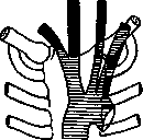
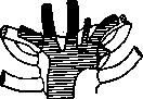
Aorta (Greek aorte, from aeiro - to lift, to suspend), the main trunk of the systemic circulation, carrying blood from the left ventricle of the heart to the chief parts of the trunk and giving rise to arterial trunks that go to all parts of the body. In its modern sense, the word A. first appears in Aristotle; in the Hippocratic collection, 'aortae' (plural) denote large bronchi, on which the lungs seem to be suspended. In the Aorta the following parts are distinguished: aorta ascendens (ascending A.) - the part of the A. rising from the left ventricle 4-6 cm upward; arcus aortae - the arch, the place where the A. changes its ventral position to a dorsal one; aorta descendens - the part of the A. extending from the third thoracic to the fourth lumbar vertebra, and the bifurcation - the place where the A. divides into two common iliac arteries and the median sacral artery. Corresponding to the position of the aorta descendens, in the chest and abdominal cavities, the thoracic aorta and abdominal aorta are distinguished. The diameter of the various parts of the A. averages from 32 mm (aorta ascendens) to 17 mm (aorta abdominalis); the thickness of the wall from 0.9 mm to 1.6 mm. - The aorta ascendens, from its beginning, directs itself obliquely from below upward, from left to right, and from back to front, and at the level of the manubrium of the sternum bends with a gentle arch to the left and backward, gradually passing into the arch. Its initial part is covered by the root of the pulmonary artery and is not visible in front; its further part lies between the superior vena cava on the right and the pulmonary artery on the left and in front; on the left and behind it is encircled by the right branch of the pulmonary artery and the left bronchus; in front lies the pericardium and the tissue of the anterior mediastinum. The ascending A. is covered by the epicardium (visceral layer of the pericardial sac), forming a common sheath for it and the pulmonary artery. The place where the visceral layer passes into the parietal layer lies at the level of the division of the pulmonary artery and corresponds to the boundary between the ascending part of the A. and its arch. In the initial part of the aorta ascendens there is a small, bulb-like dilatation called the bulbus. The three aortic valves and the place of exit of the coronary arteries are located in it. The valves of the A. (valvulae aortae) have a correct semilunar shape (valvulae semilunares); their outer convex edge is attached to the wall of the vessel, the free concave edge faces the lumen; it is bordered by a dense fibrous strip, which in the middle forms a small thickening - the nodule of Arantii. The outer edge is also denser, and only near the free edge of the valve is there a thin translucent semilunar place - the lunula of the semilunar valve. According to location, the right, left, and posterior valves are distinguished, located respectively on the right, left, and posterior walls of the aorta. Above each of them there is a small depression limited by the valve and the wall of the A. - the sinus of Valsalva. During systole of the ventricle, the valves lie parallel to the wall of the A. and give a free passage for blood; at the moment of diastole, under the pressure of the blood column, the valves move away from the wall, forming pockets filled with blood, their free edges approach each other and tightly close the lumen. In the right and left sinus of Valsalva are the openings from which the coronary arteries (a. coronaria cordis) originate, supplying: the left - mainly the anterior surface of the heart, the right - mainly the posterior surface of the heart. - The arch of the aorta is located at the level of the manubrium sterni and bends in an oblique direction backward and to the left, reaching the left lateral surface of the body of the third thoracic vertebra, where the arch passes into the aorta descendens. Further, the A. occupies an asymmetric, specifically left-sided position. In front of the arch lies the edge of the left lung, behind - the trachea and esophagus, above - the superior vena cava, and below - the place of division of the pulmonary artery and the left bronchus. Between the lower concave surface of the arch and the pulmonary artery runs a short ligament - the ligamentum arteriosum, representing the remnant of the artery that connected these vessels in the embryo (ductus arteriosus of Botalli). To the left surface, which is also the anterior surface, adhere the left vagus nerve, with its branch - the recurrent nerve - encircling it in a loop, the phrenic nerve, the pericardiaco-phrenic artery, and the lung; to the right (posterior) - mainly the trachea and the superior vena cava. From the arch of the A. three large vessels emerge: the brachiocephalic trunk, the left common carotid artery, and the left subclavian artery; from the concave surface of the arch begin several posterior bronchial arteries going to secondary bronchi. Corresponding to the places of exit of the large vessels, on the intima of the A. there are openings of a special form (ostia) and partitions formed by a duplication of the wall between them (rostra). The latter play the role of a kind of watershed. Lisitsyn drew attention to the fact that the location, shape, and size of these ostia, as well as the direction of the rostra, can vary, favoring in some cases and somewhat hindering in others the favorable distribution of the blood jets in the brachiocephalic trunk, left common carotid artery, and left subclavian artery. According to the research of Shevkunenko, the order of the vessels departing from the arch of the aorta is very inconstant. The extreme forms of the numerous variations are as follows: 1) brachiocephalic trunk - long, goes almost vertically, the places of exit of the carotid artery and left subclavian artery are close to each other, as if tending to merge (concentration); the A. has the shape of a hook and is located more caudally (see Fig. 1); 2) brachiocephalic trunk - short, goes more obliquely; the places of exit of the carotid artery and left subclavian artery are removed from each other by a significant distance (dispersion); the A. has the shape of a gentle arch and is located more cranially. The number of trunks in the latter case can increase due to the inferior thyroid arteries (30%), vertebral artery, as well as the separate exit of the carotid artery and right subclavian artery (see Fig. 2). The level of location of the arch in these extreme types fluctuates between the body of the second and fifth thoracic vertebra. The described extreme forms are a reflection of the two extreme degrees of concentration and dispersion, which have been perfectly studied in animals by Parsons, who drew attention to the fact that the order of location of the large arterial trunks is in a certain correspondence with the structure of the skeleton of the trunks of the aortic arch. The same is observed
and in humans: extreme forms of concentration and dispersion correspond to a special form of the superior thoracic aperture, compressed either from the sides or from front to back. In addition to these variations lying within the norm, the aortic arch sometimes presents anomalous deviations, which have their explanation in developmental irregularities. Among these, in particular, is the origin of the right subclavian artery from the descending aorta. The right and left common carotid arteries and the left subclavian artery arise from the aortic arch, while the right subclavian artery originates either from the concave edge of the arch or from the descending aorta and passes between the spine and the esophagus. Regarding the position of the aortic arch, it should also be noted that age exerts its influence. Due to the relaxation of the ligamentous apparatus and the elongation of the vessels, the level of the aortic arch in old age is found to be one vertebra lower than usual (Lisitsyn). The emergence of anomalous forms is explained by developmental irregularities; for example, a ring-shaped aorta develops from the remnants of its right and left roots, and the origin of the right subclavian artery from the descending aorta occurs in cases where the right aortic root is preserved, etc. The thoracic aorta, from the place of its origin—at the level of the third thoracic vertebra on the left—is directed toward the aortic hiatus of the diaphragm, describing a slight spiral on its path. Its cranial end lies, compared to the caudal, more laterally (to the left) and more posteriorly (dorsally). The esophagus, adhering to it from the right side, performs the same, but in the opposite direction, by virtue of which it transitions from a right-sided to a left-sided position in relation to the aorta. On its path, the aorta gives off the following branches: 1) bronchial arteries, 3-4 in number; 2) esophageal arteries, 5-6 in number; 3) mediastinal arteries, supplying blood to the posterior mediastinum, pleura, pericardium, and, with terminal branches, the diaphragm; 4) intercostal arteries (10 pairs), running in the intercostal spaces, starting from the 3rd. - Figure 2. Dispersion of the aortic arch trunks. The abdominal aorta extends from the level of the 12th thoracic vertebra to the middle of the height of the 4th lumbar (most often), where it divides into two common iliac arteries and the median sacral artery. The latter constitutes, as it were, a continuation of the aorta into the caudal region; in humans, it is rudimentary, but in animals that have a tail, it constitutes a powerful vessel. Having the inferior vena cava to its right, and the pancreas and the root of the mesentery in front, the abdominal aorta lies on the anterior surface of the vertebral bodies slightly to the left of the midline and is intertwined with a large number of plexuses of sympathetic, chromaffin, and lymphatic tissue. Its branches are divided into parietal and visceral. The former include the inferior phrenic arteries and 4 or 5 pairs of lumbar arteries. The visceral branches are as follows: celiac trunk, superior mesenteric artery, middle suprarenal arteries, renal arteries, internal spermatic arteries, and inferior mesenteric artery. Moskalenko, who studied the architecture of the abdominal aorta on a large amount of material, drew attention to the fact that the length of the vessel, as well as the order of origin of the large branches, are highly inconstant. He distinguishes a short aorta, with division at the 3rd lumbar vertebra, and a long one, with division at the 5th. The former is characterized by a crowding of the main branches, their early division, and, mainly, the multiplicity of renal arteries; for the latter form—a uniform origin of vessels, later division (main type—see Artery), and single renal arteries. The first type represents a legacy of the animal kingdom. The level of division of the abdominal aorta is determined, in addition to the type of structure, by age (Sozon-Yaroshevich). During life, a slow lowering of the aorta occurs along the spine, reaching one or two vertebrae by old age. Among the age-related changes is the formation of bay-like protrusions and zigzag curvatures,
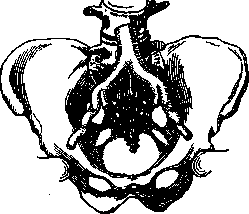
Figure 3. Age-related curvatures of vessels.
causing significant elongation of the vessel (see Figure 3). Usually, the aorta divides into two common iliac arteries and one median sacral artery. The length of the former reaches 8 cm (see Figure 4). But in a number of cases, the common iliac arteries are significantly shorter and, finally, are completely absent, in which case five trunks take their origin directly from the aorta: two external iliac arteries, two hypogastric arteries, and the median sacral artery (see Figure 5). This form, which repeats the architecture of vessels in some animals (Marsupialia and Monotremata), is called the dispersed type.
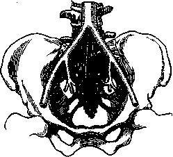
Figure 4. Main type of aortic bifurcation.
It is interesting that here, too, a specific type of architecture corresponds to a special form of the nearest part of the skeleton (pelvis): with the dispersed type, the pelvis is wide, flattened from front to back, while with the main type, it is narrow, compressed from the sides. Mention should be made of cases of asymmetry of the right and left iliac arteries, occurring in 80% (see Figure 6). Embryology. The formation of the aorta occurs simultaneously with the formation of the heart at very early stages of development. Corresponding to the paired primordium of the heart, two ascending aortas develop as its continuations, and on the dorsal side of the intestine, two descending aortas are laid down, giving off two vitelline arteries (omphalomesenteric arteries) and connecting with two
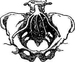
Figure 5. Dispersed type of aortic bifurcation.
umbilical arteries. The ascending aortas connect with the descending ones by means of arterial arches, six in number, passing through the thickness of the pharyngeal so-called gill arches, and the descending aortas fuse in the middle part, forming a single trunk. Such is the embryonic picture, from the individual parts of which the aorta and large vessels develop (see Figure 7). In mammals and humans, development proceeds as follows. When the paired primordia of the heart, having united, give rise to the unpaired heart, the anterior end
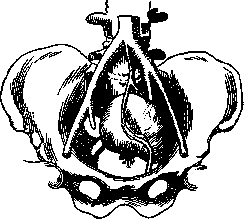
Figure 6. Asymmetrical form of bifurcation.
of it forms the truncus arteriosus, which connects with the ascending aortas, which also fuse in the initial part. The aortic trunk is formed from: 1) the truncus arteriosus, 2) the initial part of the ascending aortas, 3) the fourth pharyngeal arch on the left side, and 4) the left descending aorta, continuing into 5) the common descending trunk (all these parts are blackened in the figure). In view of the asymmetry of the development of the aorta, the development of large vessels also proceeds asymmetrically. To the right of the aortic arch, the innominate artery departs, branching into the right subclavian (4th right arch) and the common carotid; from the left of the arch, only the carotid artery departs, and the subclavian is laid down much further away as a separate vessel. All other parts of this complex system atrophy, as shown in Figure 8. All the numerous and diverse anomalies of large vessels are explained by the preservation and atrophy of various sections of the embryonic system. Embryology provides the key to understanding the comparative anatomy of the aorta. In fish, the embryonic picture is preserved throughout life; in amphibians and reptiles, two aortic arches remain, right and left; in birds, one arch, but, in contrast to mammals, it is right-sided (see Figure 8). Histology. The aorta belongs to the large-caliber arteries, the so-called elastic
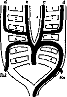
Figure 7. Scheme of the development of the vertebrate arterial system: v—ventral, d—dorsal longitudinal trunks; Bd and Bs—right and left roots of the aorta; 1-6 pharyngeal arches (according to Rathke).
type, and consists of the same three tunics: intima, media, adventitia, the boundaries between which authors do not always draw in the same way. The intima of the aorta is distinguished by its thickness; inside, it is lined with endothelium of a polygonal shape, followed by a thin-fibrous, striated layer with longitudinal networks of elastic fibers, including flat, branched Langhans cells. If the intima is silvered by running a silver nitrate stick over it and a flat preparation is made, the Langhans cells appear as light stellate spaces on a dark brown background. Outside this layer follows the internal elastic lamina, not
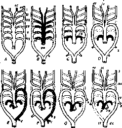
Figure 8. Modification of aortic arches in various vertebrates: A—primary scheme; B—lungfish; C—tailed amphibians; D—frog; E—snake; F—lizard; G—bird; H—mammal; c—intestinal artery; da—dorsal aorta; ab—ductus arteriosus; ec, ic—external and internal carotid arteries; p—pulmonary artery; s—subclavian artery; va—ventral aorta. Vessels carrying venous blood are blackened. Disappeared vessels are indicated by a dotted line (according to Boas).
in the form of a simple elastic membrane, as in the smaller arteries, and in the form of a plexus of elastic and adhesive fibers of considerable thickness; it can be split into two leaves, from which in the inner one longitudinal fibers predominate, in the outer one—transverse direction. The media is built from alternating layers of elastic and smooth muscle tissue, numbering 50-60; only at the very beginning of A. on some extent muscles are absent. Elastic tissue forms here the so-called annular membranes, arranged in the form of concentric tubes, the intervals between which are filled with fibrous tissue and networks of smooth muscle fibers, going mainly in the transverse direction; annular membranes are connected to each other by obliquely running bridges. The adventitia is developed relatively weakly, consists mainly of bundles of fibrous connective tissue interlacing in different directions, and, among them, networks of elastic fibers. Longitudinal smooth muscles in A. are found next to the media, so that some authors refer them to the media. The walls of A. have their own vessels—vasa vasorum in the adventitia and media and are abundantly supplied with nerves, partly motor, partly sensory, which form three plexuses (Ranvier): 1) in the adventitia, 2) at the periphery of the media, and 3) in the thickness of the media; sensory nerve endings in the form of plate-like branches are described in the intima under the endothelium. The semilunar valves are usually described as folds of the endocardium covered with endothelium, in the middle of which lie plates of dense fibrous tissue with a large amount of elastic fibers; there are especially many of them on the free edge and in the nodule. The layer of endocardium is thicker on the ventricular side. The semilunar valves of the blood vessels do not contain them. Pathological anatomy of diseases of A. Congenital malformations of A., for the most part, are associated with anomalies in the development of the heart and therefore are usually considered together with them (see Heart defects). Of independent significance are the narrowing of the isthmus of A. (stenosis of the isthmus of A.) and its congenital narrowness. The isthmus of A., to which the section of it between the left subclavian artery and the area of the opening of the ductus arteriosus (see) belongs, can become a place of narrowing of the lumen of A.; such narrowing is of two types: 1) tubular over the entire isthmus, encountered usually only in newborns and explained as a delay in the development of A. in this place; 2) funnel-shaped—in the area of the opening of the ductus arteriosus or directly below it, observed in children and adults, being a consequence of the spread of the obliteration process, i.e., the growth of connective tissue, from the ductus arteriosus to the inner coat of A. With a strong degree of narrowing, reaching sometimes complete closure (atresia) of A., circulation is carried out by collateral paths, through anastomoses between arteries departing from the subclavian arteries and the descending A. Congenital narrowness of the entire aorta (aorta ap-gusta) is such a state when A. in an adult turns out to be unusually narrow throughout (3 cm instead of 4-5 cm in circumference of the descending thoracic A.). This narrowness of A., accompanied by thinness of its wall, and sometimes combined with an abnormally small heart, is considered as a manifestation of congenital underdevelopment, hypoplasia of A. Some dispute the existence of congenital narrowness of A., expressing the thought that, with the elasticity of the wall, post-mortem contraction of it can simulate a narrow state of the aorta on a corpse. In A., infiltrative processes are frequent, of which the most common is atheromatosis and atherosclerosis (see Arteriosclerosis); in A. such processes usually begin at the places of departure of large vessels and intercostal arteries from it; there appear dense, whitish or yellowish plaques; with a strong degree, the entire inner surface may be affected. In some cases, sclerosis predominates, i.e., the formation of dense white plaques and 5 thickenings (see Figure 9), in others, a tendency to widespread atheromatosis (see), i.e., the development of atheromas and atheromatous ulcers; such ulcers, located in the places of departure of vessels, can become a place of formation of thrombi. Atherosclerosis serves as one of the causes of the development of aneurysms (see). Obesity of the inner coat of A. is often encountered, observed already from early childhood and expressed in the appearance of barely raised, light-yellow matte spots and stripes, located mainly along the length of the posterior wall of the descending A.; microscopically, the deposition of fat droplets in the cementing substance of fibers and in the cells of the superficial layers of the intima is found. These spots can decompose, giving rise to superficial ulcers, called fatty ulcers (ulcers). It is now known that this process of obesity can be the beginning of atherosclerosis. - Among disorders of circulation in A., thrombosis in it deserves mention. Thrombi in A. are formed on the ground of a violation of the integrity of its inner coat (for example, with atherosclerosis, inflammatory changes, small ruptures due to trauma and even with insignificant changes of the endothelium during infections and intoxications) and in the vast majority of cases relate to mural thrombi. Only in rare cases, specifically in the lower part of the abdominal aorta, is obstructive thrombosis encountered with the formation of collateral circulation between arteries departing from the iliac arteries and from the sections of the aorta lying above the place of obstruction. The role of collateral paths is taken mainly by the epigastric arteries (a. a. epigastricae superior et inferior), as well as the following arteries: iliolumbales, glutaeae, and a. circumflexa femoris. Thrombi of the aorta can serve as a source of embolism. Inflammation of the wall of A. is most often expressed in ulcerative thrombo-endaortitis (thrombo-endaortitis ulcerosa), in which on the inner surface of one or another part of A., an area of decomposition of the inner coat is noticeable with the imposition of thrombotic masses, usually loose and easily removable; sometimes thrombotic masses have a purulent character; the microscope opens in such places a combination of necrosis and purulent infiltration of tissue, with pus spreading also to the middle and outer coats. Ulcerative thrombo-endaortitis develops either as a result of the transition
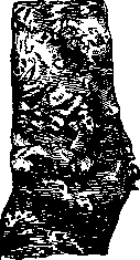
Figure 5. Atherosclerosis
of the thoracic aorta; large sclerotic plaques are visible. The process can involve the intima of the aortic valves during endocarditis, in which case the site of localization is the part of the ascending aorta adjacent to the valve ring, either due to infection of the intima from the bloodstream, or finally as a result of bacterial embolism in the vasa vasorum of the aortic wall; in the latter cases the process may begin in the outer or middle coat and then proceed to the intima. There are also cases of isolated non-suppurative inflammatory involvement of the middle coat, for example mesortitis caused by streptococcus, which sometimes bears a great resemblance to syphilitic mesortitis. In addition, suppurative inflammation of the aortic wall can be observed as a result of the process spreading to it from neighboring parts, for example from suppurated retroperitoneal lymph nodes, pararenal tissue, etc. On the basis of all the above inflammatory changes of the wall, a rupture of the aorta or the development of acute aneurysm of it may occur. Of specific inflammations, tuberculosis of the aorta is relatively rare; sometimes in millet tbc one can find tubercles on the intima of the aorta; in addition, the tuberculous process can spread to the aortic wall from neighboring lymph nodes, which usually leads to the formation of an aortic aneurysm. Syphilitic involvement of the aorta is much more common, which is usually observed in the gummatous period, although it can sometimes develop very soon (6-7 months) after infection with syphilis (see Figure 10). The formation of a true solitary gumma in the coats of the aortic wall, usually in its outer coat, is relatively rare; the more common form of syphilitic involvement of the aorta is the so-called syphilitic mesortitis (see Syphilitic Aortitis). Rupture of the aorta, apart from those ruptures that are the source of aneurysms (see), can occur with injuries and various inflammatory changes in the aortic wall. The majority of cases of aortic injury end in immediate fatal outcome. Nevertheless, there are cases (Regtes, 12 el.) when wounded persons live for many weeks and months. Sometimes spontaneous healing occurs (the vessel wound scars over), more often traumatic aneurysm develops, which, usually due to the complexity of topographic relations, also has a complex character. These are most often arteriovenous aneurysms. The obstacle to surgical treatment, which is theoretically very desirable, is the complexity of topography and the severity of the injury. Nevertheless, such attempts have been practiced, of course, on more accessible parts. In addition, spontaneous rupture of the aorta is sometimes observed without noticeable changes in the wall; such a rupture, as a rule, occurs in the ascending aorta directly above the valve apparatus and has the appearance of a transverse crack involving the intima and middle coat. It is explained by increased blood pressure in the ascending aorta with narrowing of the arch or isthmus of the aorta (sometimes rupture occurs during work in a bent position, during defecation); some have found insignificant degenerative or inflammatory changes in the aortic wall in this case. With rupture of the aorta, there is extensive, usually fatal, hemorrhage into surrounding parts; with rupture involving only the inner layers of the wall, a dissecting aneurysm of the aorta develops. Aneurysms of the aorta are most often observed in its thoracic part, occupying the ascending part or the arch of the aorta and relatively less often
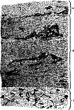
Figure 10. Syphilitic Aortitis, early stage (weak, enlargement): thickening of the intima of the aorta (o); foci of infiltration of the middle coat (o); infiltrates around vessels of the outer coat (c).
the descending part; in the abdominal part of the aorta aneurysms are more rare. In the ascending aorta aneurysms have a diffuse or sacculated type, and sometimes represent a combination of diffuse dilation with a sacculated aneurysm; in the region of the arch and in the descending aorta limited sacculated forms are more common. A rare form is the limited aneurysm of the Valsalva sinus of the aorta, located directly above its valves. Sometimes several aneurysms are observed along the aorta. Aneurysms of the aorta, by their pressure, cause displacement and ulceration of neighboring parts; the most frequent outcome of them is rupture (details see Aortic Aneurysm). Treatment of various diseases of the aorta - see the corresponding diseases lying at their basis (arteriosclerosis, syphilis, etc.). Among surgical measures, ligation of the abdominal aorta deserves attention - an operation developed by Pirogov and performed several dozen times; at present it has been abandoned, since the organism is usually unable to adapt quickly to new conditions of circulation. Relatively often these conditions are not fatal in independently developing thrombosis of the abdominal aorta. At present, ligation has been replaced by suture operation, and successful cases of suture, both parietal and circular, have been registered. One should also mention aortotomy, which was performed, in particular, in cases of embolism of the abdominal aorta in the region of its bifurcation. The operation consists in exposing the aorta, opening it, extracting the embolus and subsequent suture. It gave a number of successful outcomes. Accesses for exposing the aorta in those parts where surgeons venture to intervene have been developed quite well. There are dozens of methods, mostly osteoplastic, in which wide exposure of the aortic arch is achieved, for example by a scalpel-like incision with the base upward and including a significant part of the sternum in the flap (Shevkunenko and others). Accesses to the abdominal aorta can be both extraperitoneal (Pirogov's incision - sectio lumbo-ileo-inguinalis) and intraperitoneal (laparotomy along the midline). For an organ located extraperitoneally, Pirogov's incision should be considered normal, but a number of clinical circumstances (possibility of injury to neighboring organs) sometimes force one to resort to laparotomy. The conditions of access may, depending on the type of skeletal structure, turn out to be no worse in the second case than in the first.
Related articles
Mentioned in
Cite this page
“Aolan.” Soviet Medical Encyclopedia. English translation of Bolshaya Meditsinskaya Entsiklopediya, 1st ed. (Moscow, 1928–1936), ed. N. A. Semashko. https://sovietmedicalencyclopedia.pages.dev/article/aolan/