Botallus Duct
Historical document, translated for reference. It reflects medical knowledge of the 1920s–30s and is not medical advice.
Summary
This article from the 1928–1936 Soviet medical encyclopedia details the anatomy, embryology, and physiology of the Botallus duct, a vital vessel in fetal circulation that normally closes after birth.
Encyclopedia article (1928–1936)
Botallus Duct, ductus arteriosus Botalli (Leonardo Botallo, 16th century), represents a vascular trunk connecting the fetal aortic arch with the pulmonary artery (art. pulmonalis) and closing after birth. The development of the B. p. is connected with the metamorphosis of the branchial or pharyngeal arterial arches connecting the aortic root emerging from the heart with the two descending aortas (see); such arches are normally six in vertebrate embryos (counting from the front, see Figure 1). At early stages, the aortic root, starting from the heart, splits longitudinally into two trunks; of these, the posterior one, connecting with the 4th arch on the left side, gives rise to the aortic arch, while the anterior one enters into connection with the 6th arches and gives rise to the pulmonary artery with its two branches (the 5th arch disappears in mammals). In this process, only the proximal parts of the 6th arches go into the formation of the branches of the pulmonary artery; the distal part on the right side disappears in humans, while the one on the left is preserved and forms the B. p., connecting the pulmonary artery with the aortic arch. The B. p. is laid down in all vertebrates, starting from lungfish (Dipnoi) and ending with man, but later disappears, being preserved only in lungfish and caudate amphibians. In the human fetus, the B. p. is a wide and short trunk branching off from the pulmonary artery above the place where it divides into two branches; it runs in a slanting direction to the left and backward and opens into the lower side of the aortic arch, below the place of origin of the great vessels. Its diameter is slightly smaller than that of the pulmonary artery (equal to 5.6 mm in a 6-month-old fetus) and significantly exceeds the diameter of its branches, which is why during the intrauterine period the main mass of blood from the pulmonary artery is directed not to the lungs, which are in a collapsed state at this time, but to the aorta. This circumstance is usually linked to fetal circulation, in which the Botallus duct is assigned an important role. In the embryo, arterialized blood comes from the placenta through the umbilical vein (v. umbilicalis) and the ductus venosus Arantii into the inferior vena cava (v. cava inferior) and from there is directed to the right heart; from the right atrium, thanks to the existence of a special valve (valvula Eustachii) and the foramen ovale, it passes almost entirely into the left atrium and through the left ventricle into the aorta (see Figure 2). Thus, the aorta receives strongly arterialized blood. Venous blood flows into the right atrium from the superior vena cava, which, mixing with a small amount of arterial blood and remaining, for the most part, venous, goes further into the right ventricle and pulmonary artery, and from it through the B. p. into the aorta. As a result, the aortic arch and the a. anonyma, a. carotis, and a. subclavia sinistra branching off from it receive purer arterial blood than the descending aorta, in which the blood of the aorta mixes with the blood of the B. p. This causes faster growth of the head and forelimbs in the first half of pregnancy. By the end of pregnancy, conditions change due to the change in the place of entry of the inferior vena cava, as a result of which a larger amount of blood enters the right atrium, right ventricle, and B. p.: therefore, there is greater arterialization of the blood of the descending aorta and increased growth of the lower part of the body. This teaching, dating back to Haller and expounded in all manuals, is now contested in the sense that in the right atrium a mixture of arterial and venous blood occurs, in which case the aorta and B. p. receive blood of the same quality (Pohlmann's experiments on pig embryos); then the significance of the oval window and B. p. is reduced only to the outflow of blood into the aorta, bypassing the undeveloped lungs. This question, however, cannot be considered settled.
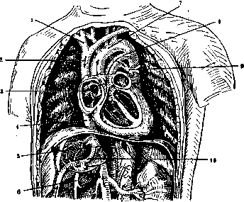
Figure 2. Scheme of fetal circulation: 1-a. anonyma dext.; 2-v. cava sup.; 3-for. ovale; 4-v. cava inf.; 5-vv. hepaticae; 6-v. portae; 7-arcus aortae; 8-ductus arteriosus (Botalli); 9-ram. sin. a. pulmonalis; 10-ductus venosus (Arantii). The aorta receives strongly arterialized blood. Venous blood flows into the right atrium from the superior vena cava, which, mixing with a small amount of arterial blood and remaining, for the most part, venous, goes further into the right ventricle and pulmonary artery, and from it through the B. p. into the aorta. As a result, the aortic arch and the a. anonyma, a. carotis, and a. subclavia sinistra branching off from it receive purer arterial blood than the descending aorta, in which the blood of the aorta mixes with the blood of the B. p. This causes faster growth of the head and forelimbs in the first half of pregnancy. By the end of pregnancy, conditions change due to the change in the place of entry of the inferior vena cava, as a result of which a larger amount of blood enters the right atrium, right ventricle, and B. p.: therefore, there is greater arterialization of the blood of the descending aorta and increased growth of the lower part of the body. This teaching, dating back to Haller and expounded in all manuals, is now contested in the sense that in the right atrium a mixture of arterial and venous blood occurs, in which case the aorta and B. p. receive blood of the same quality (Pohlmann's experiments on pig embryos); then the significance of the oval window and B. p. is reduced only to the outflow of blood into the aorta, bypassing the undeveloped lungs. This question, however, cannot be considered settled. After birth, the B. p. becomes empty and obliterates. With the first respiratory movements, the lungs expand, and a larger amount of blood begins to enter them through the branches of the pulmonary artery, as a result of which the pressure in the B. p. falls; at the same time, the mechanical conditions also change. Various hypotheses have been expressed in this regard. Previously, it was assumed that at the beginning of respiratory movements the B. p. twists. Schanz, in a special study, asserts that with the expansion of the lungs, the pulmonary artery moves forward and pulls the B. p. after it, which stretches in length. The pericardium, attached near the B. p., acts in the same direction; being fused with the diaphragm, it shifts downward with the onset of respiration and also stretches it. When stretched, the B. p., as an elastic tube, must constrict in the middle and its lumen close. According to Faber, stretching of the B. p. can be caused by the pressure of the left branch of the pulmonary artery. In any case, the collapse of the B. p. begins in the middle, from there it goes to the pulmonary artery and then to the aorta; obliteration is accompanied by thickening of the walls due to proliferation of the intima. On the 20th day after birth, the B. p. has already turned into an arterial ligament (ligamentum arteriosum), having a thickness of 2-3 mm and a length of 9 to 17 mm. As a result of obliteration, the descending aorta is deprived of admixture of venous blood. In rare cases, the B. p. remains open after birth, which is the basis of one of the types of congenital heart defects. In these cases, communication between the aorta and the pulmonary artery is preserved for life, with the blood usually flowing from the aorta into the pulmonary artery through the open B. p. (opposite to what occurs during the intrauterine period). Malformation in the form of a persisting B. p. (ductus Botalli persistens) is often combined with other developmental defects of the heart. An aneurysmatic expansion of the open B. p. may also be observed. X-ray examination can detect a persisting B. p. in the second left intercostal space as a sharply limited shadow contour. - Pathology and clinic - see Heart Defects.
Related articles
Mentioned in
Cite this page
“Botallus Duct.” Soviet Medical Encyclopedia. English translation of Bolshaya Meditsinskaya Entsiklopediya, 1st ed. (Moscow, 1928–1936), ed. N. A. Semashko. https://sovietmedicalencyclopedia.pages.dev/article/botallus-duct/