Hand
Historical document, translated for reference. It reflects medical knowledge of the 1920s–30s and is not medical advice.
Summary
The hand is the distal part of the upper limb beyond the forearm, functioning as a grasping organ capable of both coarse and fine movements. This article covers the embryology, anatomy, and development of the hand's bones and joints.
Encyclopedia article (1928–1936)
758 Hand (manus), the peripheral part of the upper limb, located distal to the forearm. Functionally, the hand represents a grasping organ, easily adapting to the volume of the object being grasped and performing with certain precision both the coarsest and the finest movements. The hand and fingers are the creative and protective force of man. Embryology. By the 6th week of embryonic development, the main parts of the limb can already be recognized: the hand resembles a flat fin, on the anterior edge of which indentations are already visible. Further indentations between the thumb and index finger and between the III and IV fingers on one side and the growth of these rudiments on the other reveal the radial outline of the latter (Hertwig). The future bony skeleton of the hand is outlined by the 2nd month. Cartilaginous epiphyses and cartilaginous outlines of the metacarpus appear only in the 1st year of extrauterine life and disappear with the cessation of growth in
Fig. 1.
Fig. g.
Fig. 1. Metacarpal bones of a 5-year-old child. Fig. 2. Carpal and metacarpal bones of a 7-year-old child. length of individual bones. On an X-ray of the newborn's hand, only the diaphyses of the finger phalanges and metacarpal bones are visible; by the 5th month - bone nuclei of os. naviculare and os. hamatum. In the second half of the 2nd year, a small flat, rapidly increasing bone nucleus forms on the lower epiphysis of the radius. In the second half of the 4th year, bone nuclei of all fingers are visible and at the end of the 4th year - os capitatum, hamatum and triquetrum; os multangulum majus et minus, naviculare, lunatum (semilunare) and pisiforme are absent (fig. 1). By the 7th year, all bones of the hand, with the exception of os pisiforme, are ossified or in the process of ossification (Wilms) (fig. 2). At the beginning of the 18th year, the epiphyseal line usually disappears below the end of the radius and ulna (Wilms). Significant deviations from these age changes in one direction or another are considered pathological, such as accelerated ossification in local tuberculous bone diseases (Rehn). - Philo- and ontogenesis - see Limbs.
Anatomy. The hand, starting from the forearm, is divided sequentially into three parts: wrist (carpus), metacarpus (metacarpus) and fingers (digiti). Wrist. The base of the wrist is formed by the following Fig. 3. Joints of the wrist: 1-art. interme-tac.; 2 and 4-lig. interos- }§. seum; a-os haraat.; л- oa tri(juetr.;6-os pisif.; 16. 7-os lunat.; S-disc. artic.;»-art.radio-uln.; 10-ulna; 11-art. car-17 po-metac; IS и 13-os is-multang. majus et min.; 14-art. intercarpea; IS-os capitat.; 16-os ia-navic; 17-art. radio-carp.; 18-Mn. epiphys.; 19-radius. (From Spalteholz.) groups of bones (from top to bottom): 1) the distal end of the radius and ulna; 2) the first row of carpal bones (counting from the radial edge): os naviculare (scaphoideum), lunatum, triquetrum, pisiforme (navicular, semilunar, triquetral, and pisiform); 3) the second row of carpal bones: multang. majus et minus, capitatum, hamatum (multangular - major and minor, capitate, and hamate); 4) the proximal ends of the five metacarpal bones. All these bones are connected to each other by three groups of joints: 1) radiocarpal, 2) intercarpal, and 3) carpometacarpal (articulatio radio-carpea, intercarpea et art. carpo-metaearpeae; figure 3). - The radiocarpal joint is located between the radius and the corresponding surfaces of os. navicularis, lunati et triquetri; the articular surface of the radius extends distally beyond the end of the ulna - a fact explaining the frequency of isolated fractures of the distal end of the radius in falls, when the palmar surface of the hand, becoming a point of support, plays a protective role (Gurlt). A consequence of such a relationship between the ends of the forearm bones is that the end of the ulna does not directly participate in the formation of the radiocarpal joint. This deficiency on the part of the ulna is compensated for by the so-called triangular cartilage (discus articularis), the base of which is connected to the notch of the radius. This triangular cartilage is of great importance for the mechanism of unhindered rotation of the radius around the ulna during pronation and supination (Fick). This cartilage does not belong to the ordinary interarticular cartilages, but represents a true continuation of the articular cartilage of the radius and participates in all its movements (Gerlach) (fig. 3). The joint capsule is distinguished by its spaciousness and, due to this, lacking sufficient strength, receives support from the strengthening radiocarpal and lateral ligaments. The fourth bone of the first row of carpal bones - os pisiforme - does not participate in the formation of this joint and is 'movably mounted' on os triquetrum, representing - a sesamoid bone of the ulnar flexor of the hand (Spalteholz). - Joint mechanism. The radiocarpal joint - a free joint (arthrodia). Movements occur in all directions, more in the dorsal-palmar direction (180°) than in the lateral (80°); at the same time, it is easier to bring the hand closer to the ulna than to the radius, which on the one hand is explained by the presence of the triangular cartilage (Rauber), and on the other hand by the fact that the styloid process of the ulna (proc. styloideus ulnae) ends proximally to the radial process.-Me JK O ей™ metacarpal joint. It connects the bones of the first row with the second. The center of the second row of carpal bones forms a joint head as a whole, with os capit. and part of os. hamati forming the largest convexity of this head (fig. 3). The distal row serves as a support for the bones of the first row and therefore cannot be as mobile as the first, which forms with the radius and ulna a very mobile joint.-The carpal bones are connected to each other by short ligaments located on the palmar and dorsal sides and between the lateral surfaces of the small carpal bones (lig. volaria, dorsalia, inter-ossea). - Carpometacarpal joint. Between the carpal bones and the bases of the metacarpal bones, there is a whole series of connections allowing minimal flexion and extension movements and at the same time simultaneous with the movement of the second row of carpal bones; both rows of joints represent as if one joint (Hyrtl). An exception is the carpometacarpal joint of the thumb, which has an independent connection with os multang. majus; in this joint, the most extensive movements of the hand are possible. On the palmar side, the boundary separating the forearm from the wrist is marked by two grooves, particularly prominently expressed in well-fed children. At this place, the skin is very thin and delicate, and the veins are clearly visible. In lean people, a series of long cords - the tendons of the flexors (flexor carpi radialis, ulnaris et m. palmaris longus) - are also visible here, disappearing in the thickness of thenaris and hypothenaris. At the radial edge, the protrusions of os. navicul. are clearly palpable, and at the ulnar - os. pisiformis.-The subcutaneous tissue here is quite firmly connected to the underlying layers, as a result of which inflammatory processes of the subcutaneous tissue of the palmar side of the forearm do not pass to the palm as quickly as processes in the area of the dorsal forearm to the dorsal side
Fig. 4. Lig. carpi volare-3; 1-thumb; 2-common flexor tendon of the fingers; 4-P. carpi rad.
of the hand.-The antebrachial fascia, when passing to the hand, forms the transverse ligament (ligamentum carpi volare), usually closely connected with m. palmar, longus (Rauber). Part of the forearm muscles ends here, and part passes over or under the transverse ligament to the hand. Over the ligament passes m. palm, long., under the ligament - all finger flexors. The friction created by the movement of all flexors under this ligament is reduced by the presence of a large bursa at this place, which extends distally to the point of origin of mm. lumbric. and proximally rises 4 cm above the transverse ligament (fig. 4). In the narrow depression between the tendons of m. palm, longi and fl. carpi rad. passes n. medianus, and on the radial side of the flexor, the pulsation of the rather superficially lying
Fig. 5.
Fig. 6.
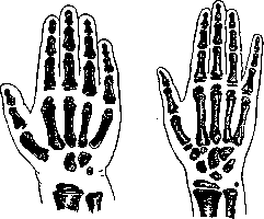
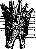
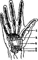
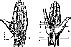
Fig. 5. Snuffbox; 1-m. extensor pollicis longus; 3-proc. styloideus; 4-m. extensor pollicis brevis et abductor pollicis. Fig. 6. Lines of the palm: 1-linea mediana; 2-linea cephalica; 3-linea fortunae; 4-hypothenar; 5-m. flexor carpi ulnaris; 6-tendom. palmaris; 7-linea vitalis; 8-thenar; 9-m. flexor carpi radialis, covered only by skin and a thin fascial sheet of the radial artery (a. radialis). N. ulnaris lies on the radial side of ossis pisiformis. The dorsal side of the wrist. Here the skin is very mobile and easily forms a fold. On this side the head of the ulna is particularly prominent, as well as the tendon of m. extensoris pollicis longi (fig. 6). With strong abduction of the thumb, on the dorsal side between the tendons mm. abductor pollicis longus et brevis and extensor pollicis longus, a triangular depression is formed (the so-called 'snuffbox'), at the bottom of which the pulsation of the radial artery can be felt, which here passes from the dorsal side of the Hand to the palm to participate in the formation of the deep arch (fig. 5). Through the skin of the dorsal wrist, a relatively large vein is visible-v. cephalica pollicis. As on the palmar side, the fascial sheet of the dorsum of the forearm thickens to form the dorsal ligament of the wrist (lig. carpi dorsale), forming sheaths for the extensors. The synovial membrane of each sheath, bulging out, envelops the tendon within it, forming a kind of 'mesenteriolum', in which the vessels supplying the tendon are located (Braus). Above these tendons, the formation of cystic tumors, known as 'synovial ganglion,' is often observed. Of the vessels of the dorsum of the wrist, the radial artery should be noted, which passes between the bases of the I and II metacarpal bones to the palm.
The metacarpus. The base of the metacarpus is formed by 5 independent tubular bones-metacarpal bones (ossa metacarpalia), connected with the second row of carpal bones and forming distally heads for articulation with the proximal phalanges of the fingers. The palmar surface, the palm (vola). On the palmar surface, the metacarpus is covered, in contrast to the dorsal surface, with a thick layer of soft tissues. The length of the palm is constant, its width is variable and depends on the position of the fingers during abduction and adduction. The palm forms a depression when bent and received its name from this shape (lad'ya - palm). The aforementioned concavity (depression), being limited on both sides by muscular ridges-thenar and hypothenar,-increases with adduction of the thumb and flexion of the others (fig. 6). The palm is abundantly supplied with nerves and vessels and therefore has a higher temperature and sensitivity than the dorsal side of the hand. The skin itself has a large number of sweat glands. The epidermis differs, as on the flexor surfaces of the limbs in general, in delicacy, especially in people not engaged in physical labor; conversely, in those engaged in physical work it is significantly denser. The Hand and its palmar surface, including the fingers, very often bear clear traces of profession, which deserves attention not only clinically but also in a forensic-medical respect (Lesgaft). On the skin of the palm, the presence of three lines (palmar creases) deserves attention, the origin of which, according to older authors (Girtl), must be explained by the flexion of the Hand in intrauterine life, since these lines are noted not only in newborns but also in the embryonic state. These lines can serve as a guide for determining underlying structures; thus, at the place where the second line intersects the III carpal bone, is the highest point of arcus volaris superficialis. These lines almost never disappear during inflammatory processes of the palm, with edema, etc. The name of these lines comes from those distant times when chiromancy still had the right of citizenship in medicine (fig. 6). The first is the line of life (linea vitalis)-begins at the wrist, between both muscular ridges of the palm, ascends upward along the border of thenaris and ends near the proximal phalanx of the index finger. The second is the cephalic line (linea cephalica)-usually begins at the point where the first line ends and in an oblique-transverse direction approaches the inner border of hypothenaris, not reaching the edge of the palm. The third is the line of 'reason' (linea mentalis)-begins at the base of the middle finger, runs transversely almost to the ulnar edge to the base of the little finger. The variable fourth line is the line of 'fortune' (linea fortunae)-runs almost parallel to the first.
The subcutaneous tissue of the palm is insignificant but very dense. In the middle of the palm, between fibrous bundles, fat lobules are embedded, which, with a vertical skin incision, seem to protrude from the wound. Subcutaneous hemorrhages of the palm are almost never observed. The dense sheet of aponeurosis represents a kind of protective organ for the vessels and nerves of the palm. Since the skin of the palm is fused with the aponeurosis, inflammatory processes of the subcutaneous tissue here are extremely painful. Subaponeurotic abscesses of the palm cannot break through the dense fascial sheet and seek an exit proximally on the forearm or even through the interosseous spaces onto the dorsum. Since the palmar aponeurosis sends to the skin and fibrous sheaths of four fingers processes, it plays a certain role in the flexion of the fingers. This becomes especially clear in Dupuytren's contracture. When the fingers are straightened, the mentioned processes are stretched, and the underlying fatty tissue diverges in the form of three tubercles between the proximal phalanges of four fingers.-Muscles of the palm are divided into 3 groups: middle and two lateral. The first is located in the depression between the palmar ridges and is represented by the tendons mm. flexores digitorum profundi et sublimis, flexoris pollicis longi, as well as mm. lumbricales (fig. 10). The interosseous muscle group (musculi interossei) stands apart.-Mucous sheaths. On the palm there are 2 mucous sheaths (fig. 4): one for flexor pollicis longus, another for flexor digitorum prof, et sublimis. The latter descends 3 cm distal to the transverse ligament and ends with a bulge for four fingers. The bulge for the little finger reaches the point of attachment of flexor prof., for the others-to the middle of the corresponding carpal bones. The sheath of the tendon of the thumb accompanies it to the point of attachment to the ungual phalanx. Separate sheaths for II, III and IV fingers extend from the ungual phalanges upward only to the heads of the metacarpal bones (fig. 4). These
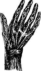
Fig. 7. Deep palmar arch, digital arteries and nerves of the palm.
relationships are illustrated by the well-known fact of transition of phlegmon of the thumb and little finger from the palm to the forearm, and due to the narrowness of the common sheath under the metacarpal ligament, the swelling takes the shape of an hourglass. 2 cm above the transverse ligament, above the flexor tendons and under the aponeurosis, is located arcus volaris superficialis. Above the bases of the bones lies arcus profundus.-The dorsal side of the Hand. Its skin, in contrast to the palmar side, is not so abundantly supplied with vessels and nerves, is very mobile and easily forms folds. The subcutaneous tissue is loose, quickly reacting to processes in the fingers with edema. Subcutaneous veins are clearly visible and when the Hand is lowered, they quickly fill. The fascial sheet of the dorsum is thinner than the palmar one, consisting of 2 sheets, of which the deep one covers the metacarpus and mm. interossei. The extensor tendons pass between the two mentioned sheets (fig. 5 and 8).-Fingers (digiti, or dactyli) represent the distal ends of the Hand, numbering 5, anatomically and functionally to a large extent independent of each other, each consisting of a system of bone levers connected by true joints allowing great and diverse mobility. The bony basis of the fingers consists of the so-called phalanges, connected by interphalangeal and metacarpophalangeal joints (fig. 9). The articular end of a phalanx is always thicker and wider than its middle part, consequently finger joints represent
Fig. 9. Arteries and nerves of the finger. 1-art. interphal.; 2-art. metacarpophal.; 3-art. interphal.; 4-art. carpometacarp.; 5-art. interphal.
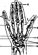
The s st L 6_tT1_ ГИ.KaK И ПЯСТНЫе KO- berosit. unguicul.; Г-art. СТИ,-трубчатые KO- carpo-metacarp. poll.; «- сти, но более корот- "* ^dkwan» 9~art-кие. Различают основную, среднюю и ногтевую костные фаланги (на большом пальце основную и ногтевую). Основная фаланга соединена c головкой пястной кости посредством шарнирного сустава. Последняя фаланга заканчивается так наз. tuberositas unguicularis-расширенным грибообразным костным наростом на ладонной поверхности. Длина пальцев на ладонной стороне кажется меньше, чем на тыльной, т. к. на ладонной они доходят только до борозды, отделяющей пальцы от ладони, a эта борозда соответствует только половине основной фаланги на тыльной стороне. Кожа на ладонной стороне пальцев имеет, так же как и на самой ладони, волнообразный рисунок, играющий, как известно, огромную роль в судебной медицине (см. Дактилоскопия). Наряду c этим на каждом пальце имеются три линии, соответствующие, кроме нижней из них, расположению фа-ланговых суставов. Кожа большого сустава несет на себе также три линии, из которых последние две лишены топографического значения.-Подкожная клетчатка пальцев состоит из плотного жирового слоя,
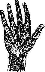
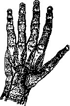
Figure 10. Superficial palmar arch and tendons of the fingers.
especially on the nail phalanx, where there is a dense network of vessels and nerves, which is connected with the extremely well-developed sensitivity of the nail phalanx, which is an organ of touch; this also explains the extremely severe pain of this phalanx during inflammatory processes, which is further increased due to the presence of dense connective tissue partitions. - Fibrous sheaths on the palmar side of the fingers replace the fibrous fasciae here. For the II, III and IV fingers, the sheaths begin above the heads of the metacarpal bones and end at the base of the nail phalanx. The sheaths of the little finger and thumb are much longer, as they begin on the forearm. On all four fingers, the aforementioned sheaths contain flex. prof, et sublim., while on the thumb only fl. long. The tendons do not lie freely in these sheaths, but are connected to them by synovial processes, which for the tendon play the role of the previously mentioned mesenteriolum (Braus); the vessels nourishing the tendon lie in them. In purulent tendovaginitis, the vessels of these mesenteriola can become blocked and thereby cause necrosis of the tendon.-On the dorsal side of the fingers, the skin is very mobile and, except for the nail phalanx, is often provided with hairs. Concentric folds (Fig. 11) are noted above the middle phalangeal joint, which in well-fed people take the form of dimples. The subcutaneous tissue is poor in fat; beneath it is the aponeurosis of the extensor tendon, which receives support from the tendons of mm.interossei et lumbric. The aponeurosis is divided into the middle - wide, attached to the second phalanx, and two lateral - narrow, attached to the third phalanx. Above the periosteum there is a thin layer of tissue, and below the periosteum - the bony mass.-The nail is a horny plate on the dorsal side of the distal phalanx of the fingers, which in humans has only a protective function (see Nail).-The thumb, in terms of its functional significance and structure, has many features compared to other fingers (Figs. 9 and 12). While all 5 fingers together have 18 short muscles and 18 tendons of long muscles, the thumb alone has 8 (Braus). It has the freest movements and the greatest strength of all fingers;

Figure 11.
the range of its movements is so great that it freely comes into contact with the palmar and partly with the dorsal sides of the other fingers. Such a range of movement is due to the presence in the base of the thumb between the metacarpal and carpal bones of a saddle joint (Fig. 13). The vascular system of the Hand differs by numerous anastomoses between the large arteries, which have the appearance of either arches, from which small vessels depart, or vascular networks located around the main joints, predominantly -carpal ones. Obviously, without such a structure with almost continuous action of the hand and fingers, circulatory disorders could occur. On the other hand, this abundance of anastomoses is the cause of difficult bleeding control. Art. Figure 12. Little finger: 1- i radius; 2- distal epiphysis; 3- art. radio-carp.; 4-os lunat.; 5- art. intercarp.; 6-os ca-pit.; 7-art. carpo-meta-carp.; 8-os. metacarp, V; 9- palmar side; 10- bone marrow; 11- epiphysis, cartilage; 12- distal epiphysis; 13- trochlea; 14- fatty fold; 15-art. metacar-io po-phal.; 16-lig.collat.; 17-lig. accessor, vol.; 18 and 19-art. interphal.; 20-lig. accessor, vol.; 21-tuberos. unguicul.; 22- epiphyseal cartilage. Figure 13. Thumb: 1-radius; 2-art. radio-carp.; 3-os navi-4-art. intercarp.; 5- os mul-tang. majus; 6-art. carpo-meta-carp.; 7- epiphysis; 8- os metacarp.; 9- bone marrow; 10- sesamoid Figure 14.
na bone; 11- main joint; 12- phalanx of the thumb of the great finger. The radial artery (a. radialis) at the wrist passes to the back of the hand below the attachment of m. supin. long., but before this it sends to the palm a superficial branch that goes over the small muscles of the thumb and directly under the fascia fusing with the terminal trunk of a. uln., forming the superficial palmar arch (arc. vol. superfic). The trunk of a. rad. passes to the back of the hand and lies between the tendons of m. extens. pol. long, et brev. and then passes again to the palm between the metacarpal bones I and II and, fusing with the deep branch of a. uln., forms the deep palmar arch (arc. vol. profundus). From the dorsal part of a. rad. three digital branches emerge: for both borders of the thumb and the radial border of the index finger. The continuation of the trunk of a. radialis on the palm is also called the deep palmar branch (ramus volaris profundus a. radialis). From it, before the formation of the arch, the first metacarpal artery (a. metacarpea volaris I) emerges; it sends palmar digital branches along the borders of the thumb and along the radial border of the index finger.-A. ulnaris- the ulnar artery sends to the hand one branch to the dorsal arterial network of the metacarpus (r. dorsalis a. ulnaris); from it a branch emerges to supply the ulnar border of the little finger. At the radial border of the pisiform bone, a. uln. divides into superficial and deep palmar branches. The first forms an arch with r. vol. superf. a. rad.; the second, penetrating into the depth of the palm, fuses in an arch with r. vol. prof. a. rad. (arc. vol. superficialis et profundus). The participation of a. uln. in the formation of arc. vol. superf. is much more significant than that of a. rad. The arch is often formed in a different way. The beginning of the palmar arch is covered, besides skin and fascia, by fibers of m. palm, brev., and the continuation- only by skin and fascia.- From the convex part of the arch emerge three common palmar arteries (aa. digitales volares communes). Each of them divides into two palmar digital arteries going along the palmar sides of two fingers. These arteries anastomose at the joints of the finger bones, forming an arch at the ends of the fingers.- Arc. vol. prof, is formed mainly by the deep branch of a. rad., which is thicker than the deep branch of a. uln. From this arch arise four palmar interosseous arteries, which fuse with the common digital arteries. Before this connection they give perforating branches (rr. perforantes), going backward to the dorsal aa. metacarpeae.- The dorsal carpal network (rete carpi dorsale) is formed from the dorsal branches of a. radialis et a. ulnaris. From this network emerge three dorsal interosseous arteries (aa. metacarpeae dorsales II-IV). These arteries divide into two branches to supply the opposite borders of two fingers.-Veins of the hand and fingers. The beginning of the venous system is formed by the digital venous plexuses: the vein of the thumb (v. cephalica), going along the dorsal side of the thumb and from there along the radial border of the forearm; the vein of the little finger (v. basilica), the beginning of which is near the little finger, from where it goes to the ulnar border of the forearm. On the dorsal side there is a fairly significant plexus of veins, connecting into a large venous trunk.- Lymphatic vessels are divided into superficial and deep. The first lie on the palmar and dorsal sides of the fingers and hand, converging mainly on the anterior surface of the forearm, where they form plexuses in the elbow bend. The deep ones also anastomose with the superficial ones, mainly in the elbow bend, where they pass through deeper lying glands. Nerves of the hand and fingers. N. medianus- the median nerve reaches the hand between the superficial and flexors along the midline of the forearm (hence its name median nerve); it supplies all flexors of the hand and fingers, except for m. flex. carp, ulnaris, as well as m. palm. longus and both pronators (teres et quadratus). R. palmaris n. mediani separates from the main trunk near the lower third of the forearm and ends in the skin of the hand (palm).-Nn. digitales volares com. I-IV (common palmar digital nerves). They constitute the end of the main trunk of the median nerve and supply with nerve branches all muscles of the thenar, as well as the skin on the radial palmar border of the thumb. The 2nd, 3rd, 4th palmar digital branches of n. med. supply on the palmar side both sides of the thumb, II, III and the radial border of IV finger. The nerves of both sides at the end of the finger connect and form terminal loops. For more details-see Medianus nervus.-N. ulnaris (ulnar nerve). Near the metacarpus n. uln. divides into dorsal and palmar branches. The first (r. dorsalis manus) passes to the back of the hand near the metacarpus under the tendon of m. flexor. carpi ulnaris, supplies the skin at the ulnar border of the dorsum and usually gives three branches: nn. digitales dorsales, which supply both sides of the little finger, IV and the ulnar side of the middle finger, the skin and tendon parts of the 1st and the upper part of the 2nd finger joints (phalanx prima).-R. volaris manus n. ulnaris (palmar nerve) divides near os pisif. into superficial and deep branches (rr. volares superficialis et profundus). The superficial branch sends nerves to supply the fingers not covered by the median nerve (little finger and ulnar border of IV finger) and further anastomoses with the 4th p. vol. n. mediani. The deep branch penetrates into the depth of the palm between the abductor and flexor muscles of the little finger, goes arch-like under all tendons flexing the fingers, supplying the muscles of the little finger, interossei, and ends in the muscle that abducts the thumb, i.e. supplies all those muscles that are not innervated by the median nerve. For more details-see Ulnaris nervus.-N. radialis (radial nerve). For the hand and fingers, the superficial branch of n. rad. is important, which passes below the middle of the forearm to the dorsal surface between the long supinator and the radial bone. Here it is called ramus dorsalis n. radialis and, after sending branches to the thumb, anastomoses with r. dors. n. ulnaris and with n. musculo-cutan. After this it sends r. digitalis dors, to the radial border of the thumb, r. digit, dors.- to the opposite borders of the thumb and index finger and r. digit, dors.- to the same borders of II and III fingers. For more details-see Radialis nervus.-On the main branches of the median and ulnar nerves on the palmar surface of the hand and on the fingers there are terminal nerve bodies, so-called Pacini bodies; on the dorsal side of the hand and fingers THEY are absent.
R. Gertsenberg. Pathology of the Hand. Congenital malformations. A comparatively rare malformation is the congenital giant growth of the entire hand or its individual parts. It affects either all elements of the hand (bony skeleton and soft tissues) or is expressed by excessive growth of one particular tissue. Most often it is a matter of excessive development of adipose tissue on the entire hand or in its individual parts. Sometimes it is accompanied by hyperplasia of the nervous and vascular apparatus. In this latter type of malformation, the excessive growth of soft tissues continues after birth according to the type of growth of neoplasms. In order to stop further growth, one can try to stop the process by bandaging the hand. But this rarely helps, and in those cases when the malformation interferes with the function of the hand, one should resort to surgical intervention, which should consist, depending on the case, of the hand
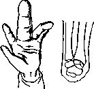
Figure 14.
Figure 15. In the removal of only excess tissue or in the amputation of fingers and even the entire Hand, especially in the tumor-like growth of blood and lymphatic vessels. With the giant growth of individual fingers (fig. 14), either their amputation or surgical removal is indicated, partly for cosmetic purposes, but mainly to improve the function of the Hand. Congenital deviations in the skeletal structure of the Hand can be divided into the following groups: excess number of individual rays of the Hand, defects in the skeleton, and congenital malpositions of the fingers. The first group is characterized by an excess of metacarpal bones and fingers (see Polydactyly). Malformations with defects in the skeletal structure of the Hand are relatively common and can in turn be divided into several subgroups: 1) the mildest degree of impairment-shortening of one or more fingers with a normal number of their bony elements; 2) shortening of fingers due to the absence of one or another phalanx; 3) absence of one or more fingers with the presence of metacarpal bones; 4) absence of fingers together with the corresponding metacarpal bones; 5) complete absence of the Hand. In addition, various combinations of the above forms are encountered. With complete absence of the Hand, which is very rare, the condition amounts to the complete absence of fingers and metacarpal bones. The wrist, although in a rudimentary state, is usually preserved (fig. 15). The absence of several fingers together with a defect in the corresponding metacarpal bones often forms the so-called forked cleft Hand, compared to a crab's claw. In this malformation, only parts of the thumb and little finger are present, while the middle fingers are absent (fig. 16). Sometimes the ring finger on the ulnar side is preserved, usually in such cases intimately fused with the little finger. Much more frequently than the above malformations, syndactylies are encountered (see).--The causes of Hand malformations remain unclear to this day. The role of scar constrictions by strands in the amnion should be considered very modest. Traces of such constrictions are noted in only a small percentage of malformations and are easily noticeable as scars on the stumps in cases of finger defects and in some cases of syndactyly. The occurrence of most other, often complex malformations cannot be attributed to amniotic constrictions, as for example in cases of absence of the proximal or middle phalanges in two-phalanx fingers. The occurrence of malformations should be attributed to the earliest periods of embryonic development. Fere and a number of other authors attribute great importance to syphilis and alcoholism of parents in the origin of malformations. -Treatment of the above-mentioned Hand malformations is limited to the removal of excess and giant fingers and the correction of syndactyly (fig. 17). Injuries to the Hand are very diverse and complex and usually involve a complex of individual elements (bones, muscles, nerves, etc.). When planning treatment, it is necessary to consider the injury as a whole, taking into account the characteristics of injuries to individual parts and their interrelationship.--Injuries to the wrist bones are rarely isolated. Due to the small size of individual bones, their dense connection by ligamentous apparatus into one common mass, and the accompanying injury to soft tissues, it is often difficult to make a local diagnosis without X-ray examination. Isolated fractures most often affect the os pisiform and os naviculare (scaphoideum). These are usually indirect fractures from falling on the Hand in the position of dorsal flexion. With direct violence, fractures are observed simultaneously in the os capitatum, hamatum et lunatum. Fracture of os. navicul. due to its proximity to the radius bone often combines with a fracture of the latter. In order to preserve joint function in fractures of the wrist bones, it is recommended to begin massage and movements early in combination with measures that promote resorption processes (hot baths). Fractures of the metacarpal bones are observed somewhat more frequently. Like most traumatic injuries, fractures of these bones occur less frequently in women than in men. Fractures most often occur as a result of direct violence, from impact, push, gunshot wound, or from falling on the back of the Hand, when it is possible for the metacarpal bones to bend toward the palm. Fractures from indirect violence are observed when falling on the Hand clenched into a fist, when the impact acts along the axis of the Hand against the metacarpophalangeal joint. Statistics (Le Dentu) indicate that fractures from direct violence are more common. In most cases, fractures are oblique with indirect force and transverse with direct violence (impact). Displacement of fragments is noted infrequently, and when it occurs, the fragments are usually at an angle with the apex directed toward the dorsal surface of the Hand. Lateral displacements are less common. In treating closed fractures with displacement, it is first necessary to apply traction on the corresponding finger and pressure on the fracture site to bring the fragments into correct position and fix them with an immovable bandage for 2-3 weeks to achieve contact between the fragments. Subsequent treatment consists of massage and the application of heat. Absorption of the callus and restoration of Hand function are observed only after a long period (up to 6 months).
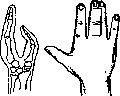
Figure 17. The mechanism of fractures of the phalanges is the same as that of the metacarpal bones. Phalanges also break much more often from direct violence than from indirect. The method of treatment is the same. In open fractures (e.g., gunshot wounds), the matter is mainly reduced to preventing infection or combating infection complicating the injury; therefore, the period of healing of the fracture itself is significantly longer. In complicated fractures, the matter often ends with insufficient consolidation (fibrous) or its complete absence. If it is a matter of one metacarpal bone (except for the first metacarpal bone), the resulting pseudarthrosis does not very strongly disrupt the function of the Hand. In pseudarthrosis of all metacarpal bones or the first metacarpal bone, there is a significant disruption of function, and it is necessary to resort to surgical intervention to achieve consolidation of the ununited fractures. The severity of open fractures also consists in that defects in consolidation are joined by the consequences of infection and damage to soft tissues. As a result, defects occur not only in the bony skeleton but also in tendons, muscles, and scar changes in the corresponding areas of the skin. Often the Hand is so disfigured that it becomes completely non-functional. Fractures near joint surfaces often lead to ankylosis. After a long period of time, when infection can be considered eliminated, surgical treatment can be undertaken to restore the bony skeleton, but treatment is complicated by damage to soft tissues. The inability to compensate for losses in muscle tissue, fill defects in nerve branches and tendons often makes the task of restoring Hand function insoluble. Dislocations of individual carpal bones are rare and are even less frequently recognized. More often than others, dislocations of os. pisiform and os. navicul are observed. Dorsal dislocations occur from forced flexion or from falling on a flexed Hand. Palmar dislocations occur from violence in the opposite direction. In fresh cases, reduction is possible. For this, with palmar displacement, it is necessary to produce maximum extension, with dorsal displacement - strong palmar flexion. Simultaneously, by pressure on the displaced bone, it must be made to return to its normal position. But due to the fact that dislocations of carpal bones are recognized late, bloodless reduction does not always succeed, and it is necessary to resort to surgical intervention. Dislocations of metacarpal bones are also rare due to their strong connection with the carpal bones. More often than others, dislocation of the first metacarpal bone is observed. The characteristic violence in the occurrence of these dislocations is excessive palmar flexion with simultaneous adduction. Dislocations in the metacarpophalangeal and interphalangeal joints occur more often (6.7% of all dislocations according to Kronlein), and usually dorsal displacement is observed. Rarely is there displacement in the palmar direction. In diagnosis, differentiation should be made with fracture of the head of the metacarpal bone or base of the phalanx. Reduction is accomplished without great difficulty by means of hyperextension in dorsal displacement and hyperflexion - in palmar displacement. Of the dislocations of the fingers, the dorsal dislocation of the thumb is most often observed. It occurs with excessive extension of the finger and begins with rupture of the joint capsule on the palmar side. The head of the metacarpal bone emerges through the resulting gap onto the palmar surface between the bundles of the short flexor. The width of this gap decreases due to the contraction of the short flexor. The degree of displacement of the sesamoid bones characterizes the severity of the injury itself according to Farabeuf. In incomplete dislocation, they lie on the articular surface of the metacarpal bone; in complete dislocation, they are displaced to the dorsum; in complex dislocation, they lie between the articular surfaces. At the same time, the tendon of the long flexor usually slips onto the inner surface of the head of the metacarpal bone and may become caught at the place where the head transitions to the neck. The position of the thumb in dorsal dislocation is perpendicular to the metacarpal bone in the initial period and parallel to the metacarpal bone if flexion of the finger subsequently occurred. On palpation, a spherical head of the metacarpal bone is determined on the palmar surface, and on the dorsal side - the base of the phalanx. Obstacles to reduction may be the falling between the articular surfaces of the ruptured capsule with the sesamoid bones and the catching of the head of the metacarpal bone by the tendon of the long flexor. Reduction is begun by pulling the thumb forward in the direction which the finger took after dislocation. Then strong extension is applied to it to bring the dorsal edge of the phalanx close to the dorsum of the metacarpal bone while simultaneously flexing the nail phalanx to relax the long flexor. In cases of failure of bloodless reduction, surgical intervention is recommended, which consists in eliminating the obstacles to reduction (often it is necessary to remove the sesamoid bone). In old cases, reduction often succeeds only after resection of the head of the metacarpal bone. After reduction by one method or another, movements in the joint should be begun in the next few days. Dislocations of the thumb toward the palm are very rare. In addition to injuries to individual systems of the Hand, complex injuries are often observed with disruption of the integrity of bones, tendons, vessels, and nerves. Such injuries are very varied and range from single incised wounds of the superficial layers of soft tissues to severe crushes with avulsion of parts of the Hand. Often in the first hours after injuries in severe cases, it is difficult to recognize the degree of loss of tissue viability and to draw the line between dead tissue and living tissue. Wounds of the Hand are easily infected; infection enters both from the skin of the hands and from objects causing injury. In addition to eliminating the dangers of infection to the whole body, the treatment of Hand injuries should be planned in such a way as to preserve maximum Hand function. Not only gross injuries but also milder ones, limited only to the skin and subcutaneous tissue, affect the function of the hand. Simple scars on the ends of the fingers, especially on their palmar surface, reduce finger function due to loss of sensitivity and tactile ability and due to pain in these scars. Furthermore, they disrupt finger function open and closed injuries to tendons. In closed injuries, avulsions of tendons (more often extensors) usually occur at their place of attachment and arise with maximum flexion of the terminal phalanx. Open injuries can be observed in any location with incised and puncture wounds. In order to preserve finger movement, the integrity of the tendons should be restored by suturing according to tendon plasticity methods as soon as possible after injury. In avulsions of fingers with bone exposure, their stumps should be urgently processed in such a way as to preserve maximum Hand function. Special care should be taken with the thumb to preserve grasping function. In amputations of fingers, it should be borne in mind that the stump after disarticulation in the interphalangeal joints, as it atrophies, turns into a protruding pointed process, easily traumatized. Therefore, it is more rational to perform amputations than disarticulation, preserving the mobility of the basal part of the phalanx. Severe consequences of Hand injury should include contracture and limited mobility or immobility of the fingers. Such a Hand results from destruction of tendons, their fusion in scars, bony and fibrous ankyloses in the finger joints (see Contracture). Extensor contracture may not involve all fingers, but only some. In the latter case, these protruding fingers, with normal function of the others, extremely hinder the work of the Hand, and their removal usually improves Hand function. Flexion contracture to a lesser degree reduces the work of the Hand, since a flexed Hand can carry small loads. Contractures often form after prolonged immobilization of the Hand. Therefore, during the treatment period for preventive purposes, passive and active movements of the fingers should be begun as soon as possible. Ankyloses in the radio-carpal joint disrupt Hand function to a greater or lesser degree depending on the position of the Hand relative to the forearm. The most favorable position is extension of the Hand, and the least favorable is ankylosis in the flexion position. A tendency to this must be combated during the treatment period, as patients usually hold the affected Hand in the flexion position. Diseases of the Hand. The skin of the Hand is the site of development of many diverse diseases; here are observed furuncles, anthracic carbuncles, eczema, Pudend ulcer, lupus, burns, chilblains, warts, calluses, and many others. More often than other diseases on the Hand, purulent inflammatory processes are observed, which can be localized in individual elements of the Hand or can simultaneously spread to many systems (skin, subcutaneous tissue, tendon sheaths, nerves, bones). Purulent inflammations are most often observed in the fingers (see Panaritia).
Paronychia often serves as a source for the development of purulent processes in other parts of the hand. Phlegmons of the hand should be divided into 2 groups: superficial and deep. The first develop superficial to the palmar aponeurosis, the second beneath it. Superficial phlegmons can arise from superficial skin damage or primarily in the subcutaneous tissue without damage. The clinical symptoms consist of pain and elevated temperature. Due to the thickness of the skin and its low compliance, the inflammatory swelling does not quickly reach significant size. The abscess approaches the superficial layers of skin most often in the area of the interdigital fold and from this area can pass to the dorsal surface, where in such case a significant inflammatory edema appears very quickly. Soon there appears swelling of the regional lymph glands in the elbow bend and in the axillary region. The entire hand becomes edematous and very painful. The fingers take a flexed position. Unlike deep phlegmons (beneath the aponeurosis), movements of the fingers remain possible and painless. Treatment consists in the opening of purulent accumulations. Phlegmons developing beneath the palmar aponeurosis are a severe disease. The usual route for their development besides deep wounds is purulent inflammation of the tendon sheaths. Pain due to the low compliance of the palmar aponeurosis and the abundance of nerve endings is often very severe. Movement of the fingers is impossible. In the purulent process from the very beginning, the tendon sheaths participate, through which pus can penetrate to the forearm, causing there the formation of a deep phlegmon. In those cases when evacuation of the abscess is not done in a timely manner, purulent melting of the interosseous muscles occurs and penetration of pus to the back of the hand. Under the same conditions, involvement of the hand joints in the purulent process is possible. For the purpose of localization of the process it is necessary to provide timely drainage for the pus. In view of the fact that the palmar aponeurosis does not provide the necessary space for the wound canal, it is rational in most cases to make a counter-opening on the back of the hand and to conduct treatment with through drainage. In the postoperative period, one should keep in mind the possibility of subsequent bleeding from the arteries of the hand, the walls of which may also be ulcerated by the septic process. A consequence of deep phlegmon is impairment of hand function, caused by wrinkling of the palmar aponeurosis, adhesions of tendons and their sheaths, and subsequent neuritis caused by infection and fibrous contraction of the surrounding connective tissue. For prevention it is necessary during the treatment period to early apply movement of the hand and its fingers simultaneously with the prescription of heat treatment. Isolated purulent inflammation of tendon sheaths is characterized by elevated temperature and painful swelling along their course. Treatment should consist in opening the tendon sheaths with evacuation of pus. In cases of purulent melting when the process has reached the stage of necrosis, it is necessary to remove the dead areas of the tendons. The inflammatory process in tendon sheaths can have a chronic course, mainly in specific infections (tuberculosis, syphilis), sometimes also after injuries. In tuberculous lesions various pathological-anatomical forms can be present: either in the form of serous effusion, often with the presence of rice bodies (see), or in the form of fungous growths. In serous forms, the elongated swelling extends to the lig. carp, transversum, approaches beneath it and rises above. At the level of lig. carp, transv. a constriction is formed between two sac-like fluctuating swellings, and the fluctuation is transmitted from the upper accumulation to the lower and back. In the non-tense sacs, rice bodies can sometimes be palpated, moving freely or hanging on a stalk. In the fungous form, the disease presents as a limited focus of dough-like consistency. Benign serous forms sometimes pass into severe fungous forms. In serous forms, good results are obtained from treatment with immobilization and injection of iodoform emulsion. In fungous forms, however, one has to resort to injection of a stronger irritant in the form of camphor-naphthalene emulsion (1:5). Simultaneous or subsequent treatment with sunlight or mercury-quartz lamp leads in the final result to satisfactory hand function. Syphilitic lesions are usually observed in the form of chronic serous forms, do not reach large volumes and do not impair hand function. Differential diagnosis with tuberculosis is facilitated by the presence of other manifestations of syphilis. Treatment is specific. A characteristic lesion of tendon sheaths is the so-called tendovaginitis crepitans, usually observed in the sheaths of finger extensor tendons and expressing itself as swelling, pain, and characteristic crepitation noise during movement of the tendons. The pathogenesis of this disease is associated with overexertion of the fingers, mainly in people unaccustomed to a particular form of labor. This disease is often encountered among laundresses, washerwomen, carpenters, etc. It is generally believed that in tendovaginitis crepitans, fibrin is deposited on the inner surface of the tendon sheaths, and from the friction of which during movement the crepitation results. Through the research of Kiittner and Frisch, the pathological-anatomical picture of this disease has been thoroughly elucidated, and it has been established that crepitation is observed not in those areas where the tendon is enveloped by the synovial sheath, but where it, devoid of the sheath, passes among loose connective tissue. This tissue is divided into two layers with delicate connective tissue bridges between them. Mayer named this tissue paratenon. With excessive labor, an exudative process occurs and fibrin is deposited in the spaces between the layers of the paratenon. Therefore, Hauck proposes to replace the old name tendovaginitis crepitans with another-paratendinitis crepitans. Treatment in the acute stage of this disease should consist of rest; subsequently-treatment with heat and massage. Prevention of recurrences consists in moderate, not too strenuous labor. The bones and joints of the hand are subject to the same diseases that affect other areas of the body. The joints of the hand are often affected in rheumatism; in severe cases (deforming polyarthritis) the hand takes on characteristic outlines. The interphalangeal joints are disfigured by nodular thickenings; in the metacarpophalangeal and wrist joints, the hand deviates to the ulnar side partly due to subluxation in these joints, partly due to displacement in the ulnar direction of the extensor tendons and shortening of the corresponding muscles. Due to this displacement, with the simultaneously developing muscle weakness, the disfigured hand becomes non-functional. The disease is more often encountered in middle and old age (see Rheumatism). Severe disfigurements of the hand are difficult to reverse and are often irreparable. Gouty lesions of the hand are quite common. Chronic forms are observed in the form of recurrent pains in the joints, often with focal deposits of salts in the joints or near them. In far-advanced cases, the hand is disfigured by persistent thickening of the soft tissues with deviation of the fingers in the metacarpophalangeal joints to the ulnar side. In treatment, anti-gout therapy should be conducted (see Gout). Exacerbations are expressed by the appearance of significant pain, swelling, and redness of the joints without involvement of the regional lymph pathways. Relatively rarely, osteoarthropathies in locomotor ataxia and syringomyelia are observed in the hand. Among other trophic lesions, diabetic and spontaneous gangrene, causalgia, acromegaly should be mentioned. Often encountered and to this day remains unexplained is the trophic disorder characteristic of the nail phalanges in the form of their club-like swelling, the so-called drumstick fingers (see). Among infectious lesions, characteristic features are presented by tuberculosis of the metacarpal and phalangeal bones in the form of the so-called spina ventosa. The disease is encountered mainly in childhood and youth and is expressed as painless swelling of one or
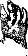
Figure 15
of another phalanx (Fig. 18). The soft tissues surrounding the affected bone may be edematous with areas of redness. The swelling is usually of elastic consistency, sometimes with areas of softening. The pathological-anatomical picture is expressed in the lesion of either the diaphysis or the epiphyses of the bone and consists in the gradual disappearance of the spongy part of the bone and its replacement by granulation tissue impregnated with pus. The corticalis together with the periosteum is pushed apart by the granulation tissue growing inside the bone. The foci of distribution are distributed depending on the topographic features of the blood supply to the bones and are usually found where the bone arteries have the character of terminal arteries. Therefore, in the metacarpal bones, foci of tuberculosis are more often found in the peripheral ends of the diaphyses, while in the phalanges they are found in the central parts. In rare cases, the process begins in the periosteum with the occlusion of a tuberculous embolus in a periosteal vessel, and from here the process can secondarily spread to the bone. With prolonged progression of the disease, deformities of the affected bone may occur, expressed in its curvature and shortening. (According to the latest research by Kozlovsky, spina ventosa is caused by staphylococcal osteomyelitis, to which tuberculous infection can subsequently be added hematogenously.) Treatment is carried out according to the methods of conservative treatment of bone tuberculosis in general (care for nutrition, hygienic housing, sun, quartz lamp, etc.). Surgical intervention should be as conservative as possible, sparing the growing young bone (removal of small sequestra and curettage of the bone cavity). Impairment of function of the Hand can be caused by the formation of contractures after injuries and diseases. Elimination of contractures often presents great difficulties. Therefore, when treating the affected Hand, preventive measures against their formation should always be taken. Contracture of the Hand and its fingers often forms due to inelastic skin scars after injuries, superficial inflammations, burns, etc. Prevention of such contractures consists in early initiation of movements. When there is a threat of skin contractures forming on the palmar side, the Hand must be kept in the position of extension and vice versa. In dense skin scars, good results are observed from treatment with heat, especially diathermy, mud packs, as well as iontophoresis with 1-2% KJ, and ionization through mud, especially in combination with mechanotherapy. In case of prolonged failure of conservative treatment, surgical intervention should be attempted, which can be varied depending on the peculiarities of the individual case. In general, the operation should consist in the excision of scars and replacement of the resulting defect with skin plasty in the form of transplantation of skin flaps on a pedicle (Italian plastic surgery or according to Filatov) or in the form of free transplantation according to Thiersch. Contractures caused by disruption of the integrity of tendons can be very diverse. The tendon may be restricted in its movement by abnormal adhesion to the underlying tissues, shortened by contraction after suppuration, or destroyed over various lengths by trauma. In such cases, it is necessary to surgically restore either the possibility of tendon movement or its continuity. In cases where the ends of the tendon have diverged, to fill the defect, one can use plastic surgery with the formation of flaps on a pedicle (Fig. 19) or transplant a piece of tendon or fasciae latae, which is attached to the divergent ends with sutures. Regarding contracture caused by contraction of the palmar aponeurosis, see Dupuytren's contracture.-A severe disease is the so-called ischemic contracture of Volkmann. This contracture is believed to be a consequence of insufficient blood circulation when the limb is held in tightly applied immovable bandages. If the arterial blood supply is significantly reduced due to bandage pressure, but not to the extent of causing gangrene, then the contractile substance of muscle fibers, poorly supplied with blood, clots, and the result is their scarring. Usually, the flexor muscles suffer more than others. The sclerosing process affects the muscles not continuously, but in the form of separate nests scattered among healthy muscle areas. The symptoms of ischemic contracture appear soon after the application of the bandage and consist of severe pain, edema in areas free of the bandage, and cyanosis. The Hand, freed from the bandage, assumes a characteristic position with the fingers strongly flexed except for the thumb, which usually remains in the extension position. At the same time, a decrease in skin sensitivity is observed with preservation of muscle excitability by electricity. Treatment begins with removal of the bandage and consists of measures aimed at increasing the blood supply to the affected limb: heat, massage, movement on special devices for finger extension. Changes in the Hand in lesions of the central and peripheral nervous system-see Medianus nervus, Radialis nervus, Ulnaris nervus. The working capacity is significantly reduced in the so-called professional cramps of the hand. These cramps are observed in persons whose work consists in prolonged
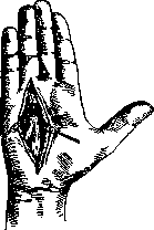
Figure 19.
with uniform small movements of the Hand: among scribes, typists, tailors, violinists, pianists, etc. In this disease, only one specific type of movement related to the profession is impaired. All other types of movements are performed by patients freely. The spasms manifest as either tonic or clonic spasms of the muscles. As a result, in the spasms of scribes, the patient exhibits excessive movements in the form of trembling, twitching; the letters come out irregular and distorted. Sometimes instead of spasms, only rapid fatigue and pains in the hand are noted. The pain occurs soon after the start of work and is so severe that the patient is unable to make the slightest movement. Sometimes pain or spasms appear even when attempting to fold the fingers as needed for holding a pen, bow, etc. The cause of professional spasms should be considered as overexertion of the neuromuscular apparatus functioning during this particular work. It should be noted that such patients usually exhibit symptoms of neurasthenia. It is possible that professional spasms more easily occur in individuals with reduced stability of the nervous system in general. The disease often drags on for a long time, and after periods of improvement, relapses are often observed. Treatment consists first of all in prolonged rest from the work that causes spasms. Next, a general strengthening regimen is necessary to eliminate neurasthenia. From local measures, massage of the hand from the wrist to the brachial plexus and galvanization are recommended. In the spasms of scribes, various apparatuses are used that hold the fingers during the act of writing. New growths. On the hand and fingers, all kinds of new growths are encountered, which are also observed in other areas of the body. For certain types of new growths, the Hand is a favorite site of origin. Such new growths include ganglia, enchondromas, and traumatic epithelial cysts. Enchondromas much more frequently affect the phalanges than the metacarpal bones. Often these tumors are multiple and reach very large sizes (as large as a child's head). In the initial stages of development, the phalanx appears swollen and in shape resembles spina ventosa. With further growth, destruction of the cortical layer of the bone occurs, and the enchondroma continues to grow in the soft tissues. The consistency of the tumor is usually hard, only in late stages, especially in the stage of malignant transformation into chondrosarcoma, the tumor becomes softer. In this same stage, the enchondroma acquires an infiltrating nature of growth. Enchondroma most often develops in young age and grows very slowly, over many years. Treatment is surgical. When there is certainty of absence of malignant transformation, during the operation it is possible to limit oneself to removing only the tumor; with incomplete removal, relapses are possible. If there is suspicion of transformation, the entire affected bone is removed. On the palmar surface of the Hand and on the flexor side of the fingers in adults, mainly in men, small round tumors resembling atheromas are encountered. The skin over such tumors is mobile and usually bears a scar mark from a previous injury. The wall of the tumor consists of connective tissue and is lined inside with stratified epithelium. The origin of such cysts is associated with the introduction of pieces of skin inward during injuries. Treatment is surgical removal. On the hand, angiomas, fibromas, neuromas, atheromas (exclusively on the dorsal surface), sarcomas of all kinds, and cancers are also encountered. The latter are most often observed on the dorsal surface of the Hand and originate either from the covering epithelium or from the epithelium of the skin glands. Osteomas and cartilaginous exostoses are encountered on the Hand not very often and present clinical interest only when, due to their size, they interfere with the function of the Hand or when, due to compression of nerve branches, they cause painful sensations. Treatment for these tumors consists in their surgical removal. Operations on the Hand. Ligation of the superficial volar arch is done through an incision in the middle of the palmar hollow, crossing the middle palmar crease. After incision of the skin and subcutaneous tissue, in the same direction the superficial aponeurosis is incised along a grooved probe, under which lies the superficial volar arch. - Disarticulation at the wrist joint with a palmar flap according to Farabeuf. The palmar flap is designed so that its width is equal to the width of the palm, and the greatest length is located along the axis of the third finger, at the level of the bases of the metacarpal bones (see Fig. 20). The incision is made in a wide arc from the radial styloid process to the ulnar. On the dorsal surface of the Hand, the incision is made 1 cm below the joint line. The skin flap is raised, the tendons on the dorsal surface of the joint are incised, then the joint itself is opened; when the latter is opened and the volar part of the capsule is cut, the flexor tendons are cut. The ulnar, radial, and interosseous arteries are ligated. The median, ulnar, and radial nerves are resected. To prevent muscle atrophy, the flexor tendons are sutured to the extensor tendons. - Disarticulation of the thumb together with the metacarpal bone. The incision begins 2 cm below the styloid process, proceeds along the dorsal surface of the first metacarpal bone to its middle.
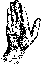
Figure 20. Incisions for disarticulation of the hand: aaa - formation of the palmar flap; bbb - formation of the ulnar flap.
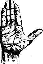
From here the incision curves in an arc toward the ulnar side and at the base of the first phalanx turns to the palmar surface, then goes along the palmar-digital crease and from the radial edge of the latter along the dorsal surface of the metacarpal bone reaches the starting point of the lateral incision. The incision is deepened, flaps are raised, including in the palmar flap all the thenar muscles. After exposing the joint area, the finger is disarticulated. - Simultaneous disarticulation of the second, third, fourth, and fifth fingers. Two incisions are made: on the dorsal surface the incision goes along the heads of the metacarpal bones, on the palm - along the palmar-digital crease. At the lateral edges of the palm the incisions meet. The palmar flap is raised to the level of the heads of the metacarpal bones, the dorsal flap is raised upward, and the fingers are disarticulated one after another. The finger being disarticulated is bent toward the palmar side, the dorsal part of the capsule is cut, the finger is deviated to the right, the lateral ligament on the left is cut, then the palmar part of the capsule, finally the right lateral. Then they proceed to the next finger and so on (Fig. 21). - For disarticulation of individual fingers, an incision in the form of a racquet is used; the incision is started slightly above the head of the metacarpal bone (en raquette), goes straight down through the head, after which it is deflected to one side and the other toward the palmar surface. Disarticulation is performed as described above. - Disarticulation in the interphalangeal joints. The line of the joint is determined by the prominence formed by the head of the proximal phalanx and noticeable when the finger is bent. It is advantageous to perform disarticulation with formation of a palmar flap, which in disarticulation at the first interphalangeal joint ends slightly above the head of the middle phalanx; in disarticulation at the second interphalangeal joint, the flap should extend to the end of the nail phalanx. Through a dorsal incision, slightly arched with convexity toward the tip of the finger, entry is made into the joint, the lateral ligaments are cut, and disarticulation is performed. In the wound, the digital arteries are ligated. - Amputations of metacarpal and phalangeal bones are performed through the same incisions as in disarticulations; only these incisions are made not along the line of the joints, but somewhat below the level of the proposed
amputation.
*
Related articles
Cite this page
“Hand.” Soviet Medical Encyclopedia. English translation of Bolshaya Meditsinskaya Entsiklopediya, 1st ed. (Moscow, 1928–1936), ed. N. A. Semashko. https://sovietmedicalencyclopedia.pages.dev/article/hand/