Foot
Historical document, translated for reference. It reflects medical knowledge of the 1920s–30s and is not medical advice.
Summary
The foot is the distal part of the lower limb, functioning as a support and spring apparatus during various body positions and movements. This article details the embryological development, skeletal formation, and anatomical structure of the foot.
Encyclopedia article (1928–1936)
FOOT (pes), the distal part of the lower limb. Functionally, the foot is an organ that works as a support and spring apparatus in various positions and movements of the body, for example, when standing, walking, running, jumping. Development. The paddle-shaped primordia, in the form of which the lower limbs are laid down, correspond mainly to the foot in their subsequent development. The division of these primordia into three main parts - thigh, lower leg, foot - and the initial formation of the foot occurs in the 2nd month of embryonic development. At the beginning of the 3rd month, the toes of the foot appear as tubercles. When laid down, the plantar surface of the foot is turned toward the trunk. The general character of the process of foot formation is connected with its gradual pronation (with the exception of the calcaneus and talus bones). This same process of pronation continues after birth and in cases of more pronounced course can lead to the appearance of flattening of the foot or even to flatfoot (see). The skeleton of the foot passes in its development through membranous, cartilaginous, and bony stages. Most of the tarsal bones have one ossification center. The calcaneus bone begins to ossify earlier than other tarsal bones, specifically in the 6th month of intrauterine life, somewhat later - the talus. Ossification of the cuboid bone occurs at the time of birth, of the third cuneiform - at 1-2 years of life, of the first and second cuneiform - at 2-4 years, and of the navicular - at 5 years of life. Ossification nuclei in the diaphyses of the metatarsal bones appear in the 8-10th week of intrauterine life, i.e., earlier than in other parts of the foot. The times of synostosis (ossification of epiphyseal cartilages) of the metatarsal bones of the foot and its phalanges are seen from the following table: Cartilage In men In women Phalanx I........ 18-22 years 17-22 years 16-20 years 16-18 years 15-20 years 15-18 years 14-17 years 13-16 years Thus, in women the foot is finally formed 2-3 years earlier than in men, but a difference of 4-5 years is also possible. To this should be added that the sesamoid bones of the foot appear in girls at 8-10 years of age, and in boys at 11-13 years (Hasselwander). The skeleton of the newborn's foot consists mostly of cartilage. It clearly bears the traces of the intrauterine position of the fetus. Certain features of the newborn's foot can be noted, connected with the type of fetal presentation. In breech presentation, the foot is usually in a more extended position and is pressed more strongly to the lower leg with its dorsal surface than in head presentation. If in the newborn the foot is placed in the 'plantar' flexion position, then its longitudinal axis becomes approximately at a right angle to the longitudinal axis of the lower leg. Conversely, with 'dorsal' extension, the dorsal surface of the foot and the anterior surface of the lower leg almost touch. If, however, extension is produced in the knee joint in the newborn, the end of the foot will drop due to 'passive insufficiency' (insufficient length) of the muscles (mm. gastrocnemii lat. et med.). For this reason, small children beginning to stand upright usually prefer to stand on the anterior part of the foot, i.e., with slight 'plantar' flexion. At the same time, in contrast to the adult, in the child, and especially in the newborn, movement in the ankle joint is possible more in the dorsal direction and less in the plantar direction. The arched shape of the newborn's foot can always be determined (Spitz), but the fatty layer on the sole smoothes out this arching, and the foot appears flat. The shape of the child's foot changes gradually. A strong factor influencing its formation is the load on the foot when teaching the child to stand and walk. When walking in small children, the step is made not only on the heel but also on the lateral edge of the foot, which is connected with the supinated position of the foot. This same, as well as the desire to have a larger support area, to some extent explains the usually observed in children beginning to walk slightly splayed position of the legs. Anatomy. Like the hand, the skeleton of the foot consists of three parts: 1) tarsus, 2) metatarsus, and 3) toes, consisting of phalanges. The tarsus includes the following bones: 1) talus, having a body, head, neck, trochlea, and articular surfaces for articulation with the bones of the lower leg, as well as with the calcaneus and navicular bones. On the lower side of the talus there is a deep groove (sulcus tali). 2) The calcaneus has a body, tuberosity, articular surfaces for articulation with the talus and cuboid bones, and sustentaculum tali - a process supporting the talus. 3) The cuboid bone has on the plantar side the sulcus m. peronaei longi, behind which lies the tuberosity (tuberositas ossis cuboidei). On the anterior and posterior sides, the cuboid bone has articular surfaces for articulation with the fourth and fifth metatarsal bones and the calcaneus. 4) The navicular bone (os naviculare s. centrale pedis) articulates posteriorly with the talus and anteriorly with the three cuneiform bones. At the medial edge of the navicular bone there is a tuberosity (tuberositas ossis navicularis). 5) The first, second, third cuneiform bones (ossa cuneiformia primum, secundum, tertium). The metatarsus consists of five tubular bones, of which the first metatarsal bone is thicker and shorter than the others, and the second metatarsal bone is the longest compared to the other metatarsal bones. The fifth metatarsal bone has a large tuberosity (tuberositas ossis metatarsalis quinti). The heads of the metatarsal bones are compressed from the sides, have articular surfaces for articulation with the proximal phalanges. The phalanges of the toes of the foot (phalanges digitorum pedis) in the great toe (hallux) are two in number, proximal and ungual, while in the others there are three. Often the middle and ungual phalanges of the fifth toe fuse with each other. The constant sesamoid bones of the foot are located on the plantar side of the metatarsophalangeal joint of the great toe, while the inconstant ones are found on the plantar side of the interphalangeal joint of the great toe and on the same side of the metatarsophalangeal joints of the second and fifth toes (ossification of inconstant sesamoid bones in women occurs at 15, in men at 20 years of age).
Figure 1. Joints of the right foot. View from the lateral side: 1 - lig. malleoli lateralis ant.; 2 - lig. talofibulare ant.; 3-lig. talo-naviculare (dorsale); 4-pars calcaneo-cuboidea and pars calcaneo-navicularis ligamenti bifurcati; 6-lig. cuboideo-naviculare; 7-lig. naviculari-cuneiformia dorsalla; 7-lig. cuneo-cuboideum dorsale; 8-lig. cuneo-cuboideum interosseum; 9-lig. calcaneo-cuboideum dorsale; 10-lig. talo-calcaneum laterale; 11-lig. talo-calcaneum interosseum; 12-lig. talo-calcaneum ant.; 13-retinaculum mm. peronaeorum inferius; 14-lig. calcaneo-fibulare; 16-lig. talo-fibulare post.; 16-lig. malleoli lateralis post.
Figure 2. Joints of the right foot. Dorsal side: 1-lig. basium (ossium metatarsalium) dorsalia; 2-lig. cuneo-cuboideum dorsale; 3-tendo m. peronaei brevis (cut off); 4-lig. cuboideo-naviculare dorsale; 5-pars calcaneo-cuboidea lig. bifurcati; 6-pars calcaneo-navicularis, lig. bifurcati; 7-lig. calcaneo-cuboideum dorsale; 8-lig. talo-calcaneum laterale; 9-retinaculum mm. peronaeorum inferius; 10-lig. calcaneo-fibulare (cut off); 11-lig. talo-calcaneum post.; 12-lig. talo-naviculare dorsale; 13-lig. naviculari-cuneiformia dorsalia; 14-lig. intercuneiformia dorsalia; 15-lig. tarso-metatarsalia dorsalia.
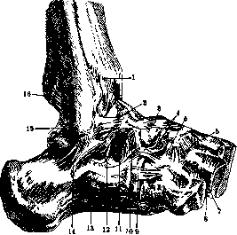
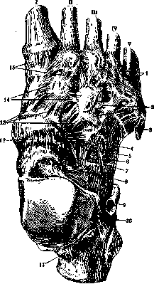
Pfitzner also described os peronaeum, a sesamoid bone in the tendon of m. peronaei longi. All the bones of the F. are connected to each other by joints (figs. 1-3), which can be divided into five groups: 1) the talocrural joint (see) (articulatio talo-cruralis), 2) the joints between the tarsal bones (art. intertarseae), 3) the tarsometatarsal joints (art. tarso-metatarseae), 4) the metatarsophalangeal joints (art. metatarso-phalangeae) and 5) the interphalangeal joints (art. interphalangeae). To art. intertarseae belong several joints, namely: 1) art. talo-calcanea, 2) art. talo-calcaneo-navicularis, 3) art. calcaneo-cuboidea, 4) art. cuneo-cubideo-navicularis. Practically, the joints between the talus and navicular bones on one side and between the calcaneus and cuboid on the other are combined into a special joint called art. tarsi transversa, s. Choparti. The shape of the joints of the F. is varied: art. talo-calcanea has a cylindrical shape, art. talo-naviculare has a ball-and-socket shape, art. calcaneo-cuboidea has a flat-saddle shape, and the remaining joints are flat. Among the numerous ligaments that participate in strengthening the tarsal joints, mention should be made of lig. talo-calcaneum interosseum, which fills the so-called sinus tarsi, of lig. plantare longum, which runs from the inferior surface of the calcaneus forward to the cuboid bone and the bases of the II-V metatarsal bones, as well as lig. calcaneo-cuboideum plantare. In the strengthening of the Chopart joint, lig. bifurcatum participates, connecting the calcaneus with the navicular and cuboid bones. In addition to the ligaments located between the metatarsal bones on both its dorsal and plantar sides, interosseous ligaments are also located between the cuneiform bones. The joints between the tarsal and metatarsal bones are combined under the name art. Lisfranci. They have a flat shape. The metatarsophalangeal and interphalangeal joints are similar in shape and in the structure of their ligamentous apparatus to the corresponding joints of the hand. The former are ball-shaped, the latter are block-shaped. The mechanisms of the foot, the axes of rotation of its individual joints, and the mobility in them, the structure of the arches of the F.-see Leg. In the strengthening of the F. and in its movements, muscles (figs. 4-7) that go to the F. from the leg (see Leg), as well as muscles proper to the F., participate. Flexion is produced by: m. tibialis post., m. flex. digit. long., m. flex. halluc. long., m. triceps surae, m. peronaeus long, and brevis. Extension: m. tibialis ant., m. extens. halluc. long., m. extens. digit, long., m. peronaeus tertius (an inconstant muscle). In the adduction of the F., m. tibialis ant. and m. tibial. post. participate to the greatest extent, which, working together, adduct the F. according to the law of the parallelogram of forces. In addition, in
Figure 3. Joints of the right foot. View from the medial side: 1-lig. deltoideum; 2-lig. talo-tibiale post.; 3-lig. calcaneo-tibiale; 4-lig. talo-calcaneum post.; 5-loop holding the tendon of m. flexor hallucis longus in sulcus m. flexoris hallucis longi; 6-lig. talo-calcaneum mediale; 7-lig. plantare longum; 8-lig. calcaneo-naviculare plantare; 9-lig. naviculocuneiforme plantare; 10-lig. naviculari-cuneiformia dorsalia; 11-lig. talo-naviculare (dorsale); 12-lig. tibio-naviculare; 13-lig. talo-tibiale ant.
this movement, the long flexors of the toes and to some extent m. triceps surae (Biesalski) participate. Abduction of the foot occurs with the combined work of the long extensors of the toes and the peroneal muscles. The F. is pronated mainly by the peroneal muscles and to some extent by the long flexor of the great toe and the posterior tibial muscle, and it is supinated by the anterior tibial muscle and the long extensor of the great toe (fig. 8). Alternating contraction of all the named muscles-extensors, adductors, flexors, and abductors-in the indicated or reverse order produces circumduction of the F. The muscles proper to the F. are located on its dorsal, extensor side and on the plantar, flexor side, and, as on the hand, the latter group of muscles is developed significantly stronger than the former. On the dorsum of the F., in addition to the tendons of the aforementioned long muscles of the leg, are located the short extensors of the toes, namely m. extensor hallucis brevis and m. extensor digitorum brevis. On the sole, the short muscles of the F. are grouped on the side of the great toe (m. abductor, m. flexor hallucis brevis and m. adductor) and on the side of the little toe (m. abductor digiti quinti brevis and m. flexor digiti quinti brevis). These two groups form elevations (eminentiae plantares med. and later.), between which is located the middle group of muscles, forming eminentia plantaris media. This group includes: m. flexor digitorum brevis, m. quadratus plantae, mm. lumbricales in the amount of four, and mm. interossei plantares (3) and dorsales (4). For details concerning these muscles, see the article Muscles of the human body. The fascia of the leg, passing to the F., forms a series of thickenings, namely: lig. laciniatum, retinaculum peronaeorum, lig. transversum, lig. cruciatum (see also Leg). Fascia dorsalis pedis covers the tendons of the long extensors, firmly attaching to the bones at the edges of the F., and becomes thinner toward the toes. The second fascia of the dorsum of the F., fascia interossea dorsalis pedis, passes between the extensors and the interosseous muscles, transitioning to the periosteum of the dorsal side of the metatarsal bones. On the plantar side of the foot is strongly developed aponeurosis plantaris, which stretches from tuber calcanei forward, dividing into bundles corresponding to the five toes. The lateral parts of this aponeurosis, covering eminentia plantaris medialis et lateralis, are developed significantly weaker than its middle part. Septa extending from this aponeurosis go between the middle and lateral elevations of the sole and pass into fascia interossea plantaris, covering the interosseous muscles from below. All these fascias participate in the formation of three connective tissue sheaths, in which are located the three main groups of muscles of the sole.
Synovial sheaths of the tendons of muscles going to the foot are located 1) in front under lig. cruciatum for the extensors, 2) inside under lig. laciniatum for the deep flexors and 3) outside under retinaculum peronaeorum for the peroneal muscles. To the first group belong independent synovial sheaths 1) m. tibialis ant., 2) m. extens. hallucis longi and 3) m. extens. digitorum longi. To the second group belong independent sheaths of the tendons 1) m. tibial. post., 2) m. flexoris digit. long, and 3) m. flex. hallucis longi. Communications between the synovial sheaths of the last two muscles are not uncommon. Under retinaculum peronaeorum is vagina tendinum mm. peronaeorum communis for the tendons of both peroneal muscles. In addition, on the sole there is a special sheath along the course of the tendon
Figure 5. Muscles of the right sole: 1-lig. vaginale; 2-bundles of aponeurosis plantaris to lig. vaginalia; 3-tendon of m. flexoris hallucis longi; 4-m. flexor hallucis brevis; 5-m. flexor digitorum brevis; 6-m. abductor hallucis; 7-tuber calcanei; 8-m. abductor digiti V; 9-strengthening bundle of aponeurosis plantaris; 10-tuberositas oss. metatarsalis V; 11-m. flexor digiti V brevis; 12-mm. lumbricales; 13-lig. cruciatum; 14-lig. plantare.
of the long peroneal muscle (vagina plantaris tendinis m. peronaei longi).-In the area of the plantar side of the toes are located fibro-osseous canals, in the formation of which the phalanges of the toes and the thickened fascia participate. These canals are lined with a synovial membrane, forming vaginae tendinum digitales pedis. These canals do not communicate with the synovial sheaths located under lig. laciniatum. For a comparative characteristic of the foot and hand, see Limbs.-The skin of the sole is coarse, thick, and has no hair. From the plantar aponeurosis to it go short but dense fibrous cords, between which are formed cells containing fat lobules. When cut, these lobules protrude in the form of large fat grains. The areas of greatest sensitivity of the skin of the sole are located on the tactile pads (toruli tactiles), the degree
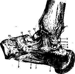
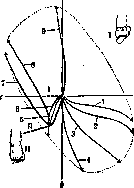
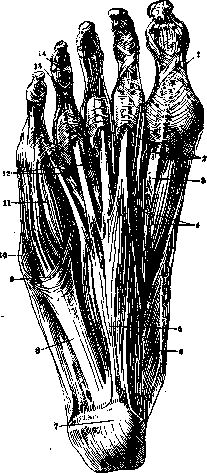
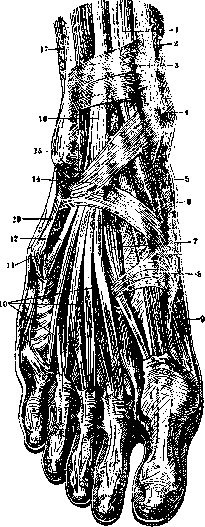
Figure 6. Muscles of the dorsum of the right foot: 1-tibialis ant. muscle; 2-tibia; 3-transverse ligament of the leg; 4-medial malleolus; 5-extensor hallucis longus muscle; 6-skin; 7-extensor hallucis brevis muscle; 8-reinforcing bundle; 9-abductor hallucis muscle; 10-extensor digitorum brevis muscle; 11-tuberosity of the fifth metatarsal bone; 12-peroneus tertius muscle; 13-peroneus brevis muscle; 14-cruciate ligament of the leg; 15-lateral malleolus; 16-extensor digitorum longus muscle; 17-peroneus longus muscle.
the intensity of which decreases with age. The arrangement of skin folds on the sole is very diverse. See also Callus, Perspiration, Blisters. The blood supply to the foot comes from branches of the dorsalis pedis artery, which is a continuation of the anterior tibial artery, and from branches of the medial and plantar arteries, which are the terminal branches of the posterior tibial artery (see the table of arteries in the article Blood vessels; in the same table-see the tarsal arteries medialis, lateralis, arcuata, ramus).
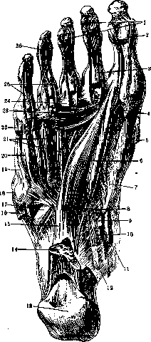
Figure 7. Muscles of the right sole: 1-tendons of the flexor digitorum longus muscle (cut); 2-tendon of the flexor hallucis longus muscle (cut); 3-tendinous chiasma; 4-laminae of the plantar aponeurosis to the ligamenta vaginalia; 5-abductor hallucis muscle (cut); 6-adductor hallucis muscle (oblique head); 7-flexor hallucis brevis muscle; 8-lamina to the flexor digitorum longus muscle; 9-tendon of the flexor hallucis longus muscle; 10-tendon of the flexor digitorum longus muscle; 11-origin of the abductor hallucis muscle; 12-deep layer of the ligamentum laciniatum; 13-tubercle of the calcaneus; 14-quadratus plantae muscle (cut); 15-long plantar ligament; 16-abductor digiti V muscle (cut); 17-tendon of the peroneus longus muscle; 18-tuberosity of the fifth metatarsal bone; 19-opponens digiti V muscle; 20-flexor digiti V brevis muscle; 21-interossei muscles; 22-abductor digiti V muscle (cut); 23-adductor hallucis muscle (transverse head); 24-transverse ligament of the heads; 25-tendons of the flexor digitorum brevis muscle; 26-tendons of the flexor digitorum longus perforans, metatarsal, dorsal, plantar, dorsal digital, plantar digital). The superficial veins of the foot are the great saphenous vein, running along the inner edge of the foot, and the small saphenous vein, running along its outer edge. Between these veins on the dorsum of the foot is an anastomosis in the form of the dorsal venous arch of the foot (see Blood vessels, table of veins, in particular the dorsal metatarsal veins, first dorsal metatarsal vein, fourth dorsal metatarsal vein, plantar metatarsal veins). The deep veins accompany the arteries. Lymphatic vessels of the foot see Lymphatic system.
The foot is innervated by terminal branches of the longest nerves of the lumbar and sacral plexuses. In the innervation of the dorsum of the foot, the saphenous nerve (medially), sural nerve (laterally), and deep and superficial peroneal nerves (intermediate segment) participate. On the plantar side of the foot, both the skin and muscles are innervated by the medial and lateral plantar nerves, which are terminal branches of the tibial nerve. See Nerves of the human body and figure
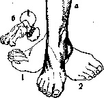
Figure 8. Movements of the foot: a-the diagram shows two extreme positions of the foot: 1-during its pronation, combined with abduction; 2-during supination, accompanied by adduction of the foot. In the middle position, the foot is shown with bent; b-axis of rotation, around which in the talo-calcaneo-navicular joint pronation and supination of the foot occur. The head of the talus is depicted as completed to a sphere. The given scheme is taken from Braus. It demonstrates the movement which German authors call "Maulschellen-bewegung des Fusses" ("ear-slapping movement of the foot"). The method of measuring the foot. At present, Yu. P. Zybyin has designed and widely applies for measuring the foot the "universal foot-measurer", which allows determining the position of each reference point of the foot in its three projections. See also Flatfoot and Shoe. Regarding changes in the shape of the foot in connection with function-see Leg.
M. Ivinitsky. Pathology. The basis of developmental defects of the foot lies in processes of excessive or insufficient growth, which may affect the entire foot as a whole or individual tissues of it. General gigantism is rare, more often there are changes in the peripheral segment of the lower extremity. If there is hypertrophy of soft tissues, particularly fatty tissue, diffuse or localized, then such developmental defects are classified as false gigantism. In gigantism of the foot, it is often combined with changes in the vascular system with the formation of naevi vasculosi and telangiectasia, as well as with disturbances of innervation leading to the formation of trophic ulcers. Subsequently, in most cases, disproportionate growth in the direction of its intensification is noted in the defective part of the extremity. One type of partial gigantism of the foot is megalodactyly, which is limited primarily to excessive growth of the big toe, less often the second toe, with the entire ray segment, i.e., metatarsus and phalanges, being hypertrophied. Often gigantism of the foot (macropodia) is combined with polydactyly and syndactyly, although the latter occur relatively rarely on the foot compared to the upper extremity. In cases of partially retarded embryonic development of the foot, brachymicro- and ectrodactyly are observed. Treatment of developmental defects of the foot consists of amputation of the entire foot or its defective part if there are significant disturbances of statics. In false gigantism of the foot, all changed tissue should be radically removed. Anomalies of development of the bony skeleton of the foot include the formation of accessory bones, so-called tarsalia, whose presence causes certain functional disturbances, which is why their diagnosis has clinical significance. The most common, approximately in 10%, is the os trigonum, located between the calcaneus and talus bones and being an excessively developed processus posterior-tali. The presence of os trigonum can lead to limitation of plantar flexion of the foot. Further, accessory bones are found: on the medial side of the foot at tuber os. navicularis-os tibiale externum, at the cuboid bone-os peroneum, at the base of the V metatarsus-so-called os Vesalii, between the bases of the IV and V metatarsals-os intermetatarsale. In the presence of trauma, small accessory bones can mislead the physician, as they can simulate a fracture. In the differential radiodiagnosis of accessory bones, it is necessary to take an X-ray of the other side, and attention should be paid to the typical position, symmetry of the anomaly, sharp demarcation of contours, and absence of the corresponding defect in the bone near which the accessory bone is located. The foot, being a support point of the body, often undergoes traumatic injuries, and both direct and indirect action of the injuring force may occur. In the latter, the position of the foot in terms of pronation, supination and flexion with simultaneous fixation of the ankle joint plays a major role at the moment of injury. Traumatic injuries of the proximal part of the foot, i.e., bones of the tarsus, are mostly observed with indirect action of force, fractures of the metatarsal bones occur predominantly under the influence of direct violence. Diagnosis of injury to the bony skeleton of the foot cannot always be established on the basis of clinical picture and in many cases is decided only by radiological data, and as a rule, radiographs in two projections are needed. Dislocations of the bones of the foot are observed relatively rarely, if we exclude dislocations and subluxations of the talus bone in the ankle joint, which are often combined with malleolar fractures (see Ankle joint). A complete dislocation of the talus with disruption of articulation with all surrounding bones, so-called double dislocation according to Malgaigne and Bouvier, occurs extremely rarely and arises under the action of a crude injuring force, which often leads to simultaneous damage to soft tissues-rupture of muscles, vessels and nerves. Lesser injuries are found in so-called luxatio pedis sub talo, where the dislocation occurs in the talo-tarsal joint at the weakest point of articulation of the talus bone, and at the moment of dislocation, the head of the talus presses against one or the other edge of the navicular bone. According to the direction of displacement of the head, 4 types of dislocations are distinguished: medially, laterally, backward and downward, and forward and backward. According to American statistics covering 178 years, medial dislocations were observed in 50%. Very close in clinical picture to luxatio pedis sub talo, dislocations in the Chopart joint occur even more rarely and are described in the literature as individual observations (Fuhr, Müller). From the first, they differ in the unchanged position of the calcaneus bone in relation to the talus bone. Dislocation of the foot in the Lisfranc joint can be partial and complete. In the first, there is a violation of the connection of individual metatarsal bones with one of the three cuneiform or cuboid bones. Displacement can occur in the dorsal, plantar, medial and lateral directions. Dorsal-plantar dislocations are more common. Of 58 cases collected by Lenormant, only in 19 cases was there a lateral dislocation. Lateral dislocations are almost always accompanied by simultaneous fractures of bones. In dislocations of the big toe, there is usually dorsal displacement of the base of the phalanx. Treatment of dislocations of the foot consists of possible early reduction with the application of extension along the longitudinal axis and with direct pressure on the dislocated area. However, open reduction is not always successful due to the difficulty of applying extension force and the power of the ligamentous apparatus. Thus, according to Lenormant's data, reduction of dislocations in the Lisfranc joint is successful only in 1/3 of cases. In open reduction, it is often necessary to resort to partial resection of bones; in cases of irreducible dislocations of the talus bone, good results in many cases are achieved by its complete extirpation. Fractures of individual bones of the foot are significantly more common than dislocations, with fractures of the calcaneus bone taking first place, which according to literature data (Ehret, Golebiewski) occur from 2.33% to 3.8% (in relation to all traumatic cases). If not recognized and improperly treated, they can lead to severe disturbances of the supporting function of the foot, with atrophy of the calf muscles and the formation of pes valgus. The most severe form is the compression fracture of the calcaneus, so-called fracture par ecrasement by French authors, occurring when falling on feet in a vertical position. The direction of the fracture line can be transverse, mostly passing through sinus tarsi, and longitudinal-in the horizontal plane, closer to the lower surface of the calcaneus. Clinical symptoms consist of expansion of the calcaneal area, valgus position of the posterior part of the foot, pain on pressure mainly from below and from the sides, relatively free flexion and extension, with sharply painful supination and pronation. In fractures of the calcaneal tuberosity, there is usually a more or less vertical direction of the fracture line. According to Gussenbauer, in the mechanism of occurrence of this type of fracture, two moments play a role: the action of the injuring force and the active muscular traction. Dislocation of fragments upward due to the traction of the Achilles tendon is moderated by the influence of the plantar aponeurosis and plantar short muscles. In pure forms of avulsion of the calcaneal tuberosity (so-called Rissfracturen), there is separation of a part of the corticalis from its upper surface. Isolated fractures of the talus bone occur less often and are often combined with dislocations. Compression of the talus bone occurs due to its being crushed between the articular end of the tibia and the calcaneus. In fractures of the body, a T-shaped fracture line is mostly observed, more often there is a fracture of the neck. In the absence of displacement, a fracture of the talus bone may remain undiagnosed without radiological data. In favor of it, there is usually significant hemorrhage and sharp pain on dorsal flexion. With large displacement, it is necessary to resort to open reduction, with Binney recommending preliminary tenotomy of the Achilles tendon, or partial or complete astragalectomy. Pathological fractures of the talus and calcaneus bones are observed in the initial forms of tabes. Fractures of small bones of the tarsus are rare and usually there is no displacement of fragments. In some cases, there is an avulsion of the tubercle of the navicular bone due to strong contraction of m. tibialis post. Fractures of metatarsal bones occur with direct action of force, mostly when heavy objects fall on the foot. The direction of the fracture line can be different, and no large displacement of fragments is observed. With strong contraction of m. peronaei brev., there can be an avulsion of tuberositas metatarsi V, which should not be confused with the independent ossification center encountered (Wenzel-Gruber's nucleus). To traumatic injuries of the foot belongs a specific disease, so-called foot tumors, first described by Breithaupt and observed in military personnel, usually in the first years of service, after long marches and with significant load.
On the dorsum of the foot, a swelling of the soft tissues forms and there is significant pain on pressure and walking, localized in the area of the metatarsal bones. A severe trauma in the anamnesis is usually not noted. According to the research of Schultze, Kirschner, and Matti, the cause of the disease is in most cases a fracture or periostitis of one of the metatarsal bones, in the overwhelming majority of cases (90%) of the second or third. In the examined cases, a fracture was found in 43.4%, periostitis in 13.2%, and the absence of bone changes in 43.4% of cases. For these latter cases, one of the possible causes is indicated as an inflammatory process of the tendon sheaths and interarticular ligaments of the foot. A factor contributing to damage to the foot, according to Momburg, is fatigue of the muscles, due to which the foot becomes poorly adapted to uneven ground at the moment of push-off during walking, when the main support points are the heads of the II and III metatarsal bones. According to the research of Turner, the decisive role in the development of this disease belongs to the involvement of the branches of the peroneal nerve in the pathological process, which is the result of trauma. The frequency of appearance of this symptom complex in injuries to the II and III metatarsal bones is explained by him by the particular richness of the terminal branches of the n. peronaei in the area of the interosseous spaces of the three middle metatarsal bones. Treatment is conservative—rest with support of the arch of the foot with a supinator. Very close to the 'tumor of the foot' is the disease described by Deuchlander—thickening of the metatarsal bone, which he attributed to inflammatory osteoperiostitis. Miller and Janssen adhere to the mechanotraumatic view. The thickening of the metatarsal bone is the result of a great demand on the function of this bone due to a change in the entire statics of the foot. Keller classifies Deuchlander's disease among osteochondropathies. Usually at the age of 25-35 years, without visible trauma, limited pain appears in the area of the II, less often III, metatarsal bone. After 8-9 weeks from the onset of the disease, deposits of a regular spindle-shaped shape are found on the X-ray at the border of the middle and distal parts of the bone from the side of the periosteum. In the center there is a transparent zone with a regular transverse direction, which sometimes extends to the periosteum (Looser's zone). After 3-4 months, bone remodeling occurs. Treatment is only rest.—To this same group belongs Morton's disease. This disease was described in the pre-X-ray era, and therefore from the modern point of view it appears as not quite a clear nosological unit. After prolonged work, transitions, and loads, pains develop at the outer edge of the foot, most often in the area of the metatarsophalangeal joint of the IV toe, which Morton interpreted as neuralgia of the n. plantaris lat. (Morton's metatarsalgia). Also belonging to such indeterminate diseases of the foot is the so-called talalgia (heel neuralgia) (Blum), called by Duplay 'pternalgia'. These terms denote a clinical syndrome caused by the most diverse reasons, connected either with trauma or with rheumatism, gonorrhea, gout, etc. The disease is expressed in sharp pains in the area of the heel, caused by the most diverse causes—exostoses, bursitis, neuritis, etc. At present, with the development of X-ray diagnostics, this term, as too indeterminate, is losing its significance. ' To traumatic injuries of soft tissues without violation of the integrity of the skin cover belong subcutaneous ruptures with hematoma, which are most often observed in athletes in the area of the Achilles tendon. With a complete rupture of the tendon, there is a divergence of the ends, its contours are not palpable, and a greater than normal range of motion in the dorsum is noted. In old, untreated cases, pes calcaneus develops. As an exceptional rarity, a rupture of the tendon of the m. tibialis ant. is described. The active action of muscles with simultaneous rupture of the ligamentous apparatus can lead to the so-called dislocation of tendons, which is observed almost exclusively in the peroneal group, and the tendons are palpable anterior to the lateral malleolus. Maydl collected only 20 cases of dislocations of the tendons of the n. peronaei. Cases of congenital dislocations of tendons are described, which apparently depend on the congenital absence of retinaculi peronaeorum. A predisposing moment is an anatomic variant of development of the ankle with flattening of its bony prominences. Treatment is either conservative or operative, based on the method of Lannelongue—strengthening of the ligamentous apparatus with a periosteal flap.—Traumatic injuries of the foot, complicated by injuries of soft tissues with violation of the integrity of the skin cover, are often encountered in fractures and dislocations and arise either due to damage to the skin from within by bone fragments or due to the direct action of a coarse traumatic force. Such traumatic injuries are the most dangerous from the point of view of subsequent development of gangrene or severe suppuration, sometimes lightning-fast septic infection, which is favored by the existing anatomical conditions. Gunshot wounds of the foot, in frequency, occupy the fourth place, and according to the data of the 1914 war, in most cases, namely 80%, they are wounds from artillery shells. The mortality rate for this type of wound is about 5.7%. Landois divides all wounds of the foot into injuries of the upper joint, i.e., the ankle joint, and other parts of the foot. In closed injury of the ankle joint, the main symptom is hemorrhage into its cavity. In wounds complicated by infection, empyema of the joint and panarthritis develop. The latter is already a serious complication of the wound, leading to the spread of infection along the tendon sheaths and to the involvement of the bones of the foot in the osteomyelitic process. In fresh wounds, after properly performed primary treatment of the wound by thorough excision of its edges and bottom, primary suture can be applied, and in some cases it is recommended to suture only the capsule. Tappeiner, with conservative treatment with prolonged application of congestive hyperemia according to Vir, had good results. In severe cases, primary resection or amputation is indicated. Fragments located along the line of the joint should be removed in order to avoid violation of its function. Immobilization of the foot is performed either by applying a circular plaster cast with a window or various splints are used—Folsman's, Kramer's, Bruns', etc. Erlacher, in 239 wounds of the foot, had in 60.8% cases the formation of complete ankylosis of the ankle joint. Bearing in mind the frequency of such an outcome, it is necessary to fix the foot in the correct position, i.e., to set it at a right angle to the axis of the leg and in a neutral position between supination and pronation. Acute inflammation of the soft tissues is encountered on the foot less frequently and has less significance than on the hand. Subcutaneous suppurations on the sole are often observed, which should be opened as early as possible, since the thick skin prevents the opening of the abscess to the outside and pus can easily spread under the aponeurosis. Deep phlegmons usually give sharp pains, especially on the plantar side of the foot. The presence of a strong plantar aponeurosis masks the typical symptoms of the formed abscess; swelling and redness spread mainly to the dorsum of the foot, and sometimes careful examination of the foot is required to determine the localization of the abscess. When opening the abscess, the location of the incision should be accurately taken into account in order to avoid in the future the formation of scars on the sole, which impair the supporting function of the foot. Chronic inflammations of the soft tissues of the foot mainly belong to the group of keratoses.—Among the diseases of the nails, the one of greatest practical importance is the frequently encountered suffering of unguis incarnatus (ingrown nail), the causes of which lie in the variant of the shape of the nail, the size of the toe, and the wearing of irrational footwear. The formation of an ulcer due to pressure from the altered edge of the nail is observed more often on the outer side of the toe. The most radical surgical method is the removal of the nail with part of the nail bed.—Acute purulent processes on the foot are often complicated by purulent tendovaginitis. Chronic tendovaginitis are observed in most cases in the area of the tendons of the peroneal muscles and extensors and are mainly of tuberculous origin.—Of the mucous bursae, the most frequently affected is the anterior bursa of the Achilles tendon (bursa retrocalcanea), which due to its position is easily subjected to injuries, both more significant single ones and barely noticeable multiple ones. Many cases of achillodynia have as their cause inflammation of the mucous bursa. Acute bursitis with symmetric affection are observed, according to Noble, in gonorrhea. In acute infectious diseases, the inflammatory process is localized mainly in the ankle joint (see Ankle joint). In purulent infection spreading from the surrounding soft tissues (due to trauma, wound, etc.), in addition to the joints and tendon sheaths, the spongy tissue of the small bones of the foot is involved.
Due to unfavorable conditions for the outflow of pus and the rapid sequestration of bones, it is often necessary to resort to partial resections of the Foot. In severe suppurations, one should not fear transverse incisions, since in most such cases the active function of the Foot is lost. The primary hematogenous osteomyelitic process most often affects the heel bone among the bones of the foot, with the rapid formation of fistulas localized under the malleoli. Osteomyelitis of the metatarsal bones and phalanges with their partial or complete sequestration leads to the subsequent development of persistent contractures and deformities of the toes, creating vicious conditions for the statics of the foot. In such cases, it is more rational to amputate or exarticulate the deformed toe, making an exception for the big toe, which plays a special role in the supporting function of the Foot. Among chronic infections, tuberculous lesions of the bones of the Foot are often encountered, occupying, according to Billroth, sixth place, and according to the material of the Göttingen clinic, constituting 14% of all bone and joint tuberculous diseases. Primary bone lesions significantly predominate over synovial ones, the process proceeds as a fungoid and fungoid-destructive form. The primary focus is most often localized in the body of the talus bone, less frequently in the heel bone, somewhat posterior to the sinus tarsi. In the heel bone, delimited tuberculous foci are also encountered (according to Garre in 0.8%), in which the process is strictly localized and the ankle joint remains free. Tuberculous disease of the metatarsal bones and phalanges in children proceeds in the form of spina ventosa, in adults there is more often involvement of the articular ends of the bones. Conservative treatment of tuberculous bones of the Foot according to the material of Rollier and Kisch gives good results. In addition to therapeutic measures lying at the basis of the principles of conservative treatment of tuberculosis in general, in tuberculous lesions of the Foot with effect, exposure to a mercury-quartz lamp and radiotherapy are applied. The latter, according to the observations of Iselin and Wilms, does not act bactericidally, but promotes the replacement of tuberculous granulations with connective tissue. Surgical treatment according to the material of König, covering 274 cases, leads to healing of the process in 66.4%. In delimited foci, the complete removal of all affected bone tissue is undoubtedly indicated. Luetic lesions of the Foot are encountered less frequently than in the knee and elbow joints. With localization of the specific process in the toes, there is a form of the disease dactylitis syphilitica, extremely resembling spina ventosa. A very typical lesion for arthritis urica is the involvement of the metatarsophalangeal and interphalangeal joints of the big toe. Gouty lesions-see Gout.-To chronic diseases with a definite localization should be included the so-called heel spurs, the pathogenesis of which has not yet been fully elucidated; most authors see the cause of the disease in an excessive proliferative reaction of the periosteum to various irritants. The occurrence of the pain syndrome apparently depends not on the presence of osteophytes, but on inflammatory changes in the surrounding tissues. Often in individual bones of the Foot, changes are observed, grouped together under the name of osteochondropathies, in the basis of which lie phenomena of aseptic necrosis with subsequent complex reparative processes (see Osteochondritis). The most common in this group is Köhler's disease II, in which the head of the second metatarsal bone is affected, less frequently the third (about 10%) (see Köhler's disease). In Köhler's disease I, the process is localized in the navicular bone, which significantly decreases in size and takes the shape of a lentil or sickle.-Osteochondropathy of the heel tuberosity, studied by Schinz, is rarely observed, mostly in the age group from 7 to 14 years. The clinical symptoms are: usually sudden onset of the disease, pain on walking and especially on pressure on the heel tuberosity from behind and swelling in this area. A characteristic radiological sign is a sequestra-like picture of the apophyseal shadow, sometimes with displacement of individual fragments to the side. Among the rare osteochondropathies of the Foot is also Rendu-Müller's disease, in which bone necrosis is localized in the sesamoid bone of the first metatarsophalangeal joint, more frequently the medial one. Pain appears when standing and walking and especially with hyperextension of the big toe. Since regenerative phenomena in this disease are expressed insignificantly, surgical treatment-removal of the affected bone-is recommended.-Among vascular diseases, aneurysms on the Foot are rarely observed, mostly they belong to the a. dorsalis pedis and depend on traumatic damage to the vascular wall. The most common disease associated with lesions of the vascular system is spontaneous gangrene, which is divided into a number of separate forms (gangraena senilis, praesenilis et juvenilis), diabetic, toxic, symmetrical gangrene of Raynaud, etc. (Clinical symptoms and treatment-see Gangrene.) Among neuropathic lesions of the Foot, trophic ulcer-mal perforant du pied Nelaton's, which forms at the main support points of the Foot, occupies a prominent place. The cause of the disease lies in changes in the central or peripheral nervous system. Tabetic lesions of the bones of the Foot have great diagnostic significance, as they are often observed in the initial forms of the disease and precede the stage of ataxia and pronounced symptoms from the nervous system (Ludloff, Levy). A picture of severe deforming arthritis develops, affecting both the ankle joint and the small bones of the Foot; sometimes complete disappearance of entire phalanges without suppuration and sequestration is observed; the slightest trauma leads to pathological fractures with separation of bone fragments, the subsequent formation of pseudoarthroses, despite significant periosteal proliferations. Often there are bone proliferations in the area of the Achilles tendon (Wilms). Tabetic lesions lead to severe deformities of the Foot, most often to the formation of pes valgus.-Among benign tumors of the Foot, fibromas, neuromas, angiomas and more often than others chondromas with localization in the area of phalanges and metatarsal bones and often with simultaneous involvement of the distal parts of the upper extremities are encountered. Among malignant tumors, sarcomas and squamous cell carcinomas are not often observed. The Foot plays an outstanding role in the conditions of statics and dynamics of our body, which is why the doctrine of deformities constitutes one of the most important chapters in the pathology of the Foot. All deformities are divided into two groups-congenital and acquired. The latter, depending on the cause of their occurrence, break down into traumatic, inflammatory, static and paralytic deformities. According to the nature of the deformity, five basic types are distinguished: pes varus, valgus, equinus, calcaneus and excavatus, which can occur both in pure form, and more often in combination with each other, with each form being one of the components in the general picture of the faulty position of the Foot (for example pes equino-varo-exca-vatus, pes valgo-calcaneus, etc.). The most common deformity is clubfoot, pes varus, pes equino-varus (pathogenesis, clinic and treatment-see Clubfoot). No less common is the deformity opposite to clubfoot-pes planus, pes plano-valgus, pes valgus, especially its static forms, grouped together under the name of flatfoot (see Flatfoot). Horse's foot (pes equinus) is characterized by the position of fixed plantar flexion without lateral deviation of the Foot; thus the contracture develops strictly in the ankle joint. In severe forms of pes equinus, dislocation of the talus bone and secondary changes in the position of other bones of the tarsus are observed; often there is the formation of contractures of the toes, dorsal or plantar, depending on the position of the Foot during walking (support on the plantar or dorsal side of the toes). The calf muscle is sharply shortened, with the taut, like a string, Achilles tendon protruding, the plantar muscles are tense, which together with secondary changes in the bones of the skeleton contributes to an increase in the arch of the Foot, and to the equinus deformity a component of excavation is added. Pure forms of pes equinus are rarely congenital, since the flexed Foot of the fetus is easily subjected to mechanical influences leading to its deviation to the side, more often inward, i.e. to the formation of pes equino-varus. Horse's foot of traumatic and inflammatory origin is encountered much more frequently due to damage to the peripheral nerve, formation of scar contracture or on the basis of an inflammatory process, when with insufficiently careful fixation of the Foot in the correct median position, stiffness or ankylosis of the ankle joint develops in a faulty equinus position. The most frequent etiological factor is a disease of the nervous system and in particular infantile paralysis (see Heine-Medina disease).
Pes equinus in this disease is observed both with partial damage to the extensor muscle group and with complete paralysis of the limb, when among the mechanical-antagonistic causes of the development of paralytic deformation, the exceptional role belongs to mechanical influences, namely the force of gravity. When deciding on the surgical treatment of pes equinus, the existing shortening of the limb should be taken into account, since in some cases the deformation should not be eliminated at all, and the patient is only provided with orthopedic shoes with a corresponding compensatory wedge. In mild cases of deformation, one can be limited to staged redressation of the foot. If the Achilles tendon is an obstacle to eliminating the faulty position of the foot, it should be lengthened in a Z-shape by open method, which makes it possible to accurately determine which part of the tendon should be lengthened, while its continuity is not disrupted. In many cases of paralytic pes equinus with incomplete paralysis of the extensors, such a simple surgical intervention gives an excellent functional result, since eliminating the element of overstretching of the muscles puts the latter in normal physiological conditions (Stoffel). With pronounced changes in the bony skeleton, where plantar flexion is hindered by a dislocated and deformed talus bone, a wedge with a dorsal base is excised from it or an astragalectomy is performed. The heel foot (pes calcaneus) is formed either due to an increase in the volume of dorsal flexion of the foot or due to a change in the shape of the calcaneus bone depending on the improper distribution of muscle traction. Accordingly, Nicoladoni distinguishes two forms of the heel foot: pes calcaneus sursum flexus and pes calcaneus sensu strictiori. The first form can be congenital or acquired, the second is only a paralytic deformation. The pathogenesis of congenital pes calcaneus apparently consists of the shortening of the dorsal muscle group, the tension of which serves as an obstacle to plantar flexion in a normal volume. This deformation develops due to improper intrauterine pressure and often combines with abduction and pronation of the foot (pes valgo-calcaneus). Acquired pes calcaneus can be of scar origin, but more often of paralytic origin, with the posterior muscle group being paralyzed. The foot is in the position of dorsal flexion, but due to the force of gravity, the anterior part of the foot often sags, which contributes to an increase in its arch. With paralysis of the muscles from the flexor group attached to the calcaneal tuberosity and simultaneous preservation of the function of the peroneal group and plantar musculature, a change in the direction of the axis of the calcaneal tuberosity occurs due to muscle traction dyssynergy, which occupies a plantar position instead of a posterior one (pes calcaneus sensu strictiori). In many such cases, the foot has an ugly shape, and the patient's gait is significantly impaired. The most common surgical intervention is the shortening of the Achilles tendon with the addition of muscle plasty - transplantation, most often of the tendon of the m. peronaei longi. Shortening can be performed either by simple suture of the tendon or by excision of a part of it according to Vill. A strong scar is obtained by longitudinal splitting and transverse section of the tendon with subsequent cross-shaped suture of its four ends. In pes calcaneus sensu strictiori, tendon surgery is often insufficient, and to it should be added either an oblique osteotomy of the calcaneal tuberosity according to Hoffa or a wedge-shaped resection according to Gocht, with the base of the wedge directed backward and upward. In many cases of severe deformation, preference should be given to arthrodesis of the ankle joint with mandatory correction of the faulty position of the foot. For the hollow foot (pes excavatus, pes cavus, pes arcuatus), the characteristic sign is a pathological increase in the arch of the foot. In its pure form, pes excavatus is not often encountered, while the component of excavation enters as a secondary deformation into a number of other types of foot curvature, such as pes varus, equinus, calcaneus. The etiology and pathology of pure forms of pes excavatus are not yet fully clear. Riedel, Lackner, and Kuslik consider poliomyelitis the most common cause. According to some authors (Henneberg, Leriche), a significant role in the formation of pes excavatus belongs to myelodysplasia. Schulthess considers the cause of the formation of a hollow foot to be dyssynergy of muscle traction, attaching particular importance to the function of the following muscles: triceps surae, short muscles of the sole, m. peronaeus longus, possibly extensors of the foot, tibialis post, and flexor hallucis longus. Very often, pes excavatus is accompanied by contracture of the toes, which take on a claw-like shape. In the bloodless treatment of pes excavatus, manual redressation usually gives little effect; when using a Schultze redressator (according to the data of the clinic of Payer and Besalsky), it is possible to eliminate the most severe forms of deformation. The purpose of surgical intervention in pes excavatus is to correct the existing deformation by operations on the bony skeleton of the foot (excision of a wedge, crescent-shaped osteotomy according to Kuslik, etc.) and to eliminate dyssynergy of muscle traction by means of tendon transfers. Among the curvatures of the toes, the most common and having the greatest practical significance is the deformation of the big toe with its abduction at the metatarsophalangeal joint - hallux valgus (see). The opposite deformation - hallux varus, i.e., the position of adduction of the big toe toward the midline of the body, is rare and is mostly a congenital disorder. Among the lateral curvatures of other toes, the deformation of the little finger is most often observed, which is in the position of adduction and lies on the IV toe. This deformation usually develops due to wearing narrow, irrational shoes. Among the rarely occurring lateral curvatures of the terminal phalanges, the deviation of the nail phalanx of the big toe outward should be mentioned, the so-called phalanx valga. The curvatures of the toes relative to the frontal axis reduce to extension and flexion contractures of them, both in the metatarsophalangeal and interphalangeal joints, and are often concomitant changes in paralytic deformations of the foot. From this type of deformation, the most typical are the so-called hammer and claw toes. The former are characterized by flexion contracture in the metatarsophalangeal and extension in the interphalangeal joints. In the pathogenesis of development, besides paralysis of the tibialis and myelodysplasia, static changes also play a role, in particular the presence of flatfoot (Nicoladoni). According to Hoffmann, such a position of the toes is caused by the compensatory adaptability of the patient, who seeks to unload the area of the metatarsophalangeal joints. In the presence of hallux valgus, hammer curvature is most pronounced on the II toe. In claw deformation of the toes, the opposite relationships are observed: the proximal phalanges are in the extension position, the middle and nail ones in plantar flexion. This deformation is often encountered in pes excavatus. Duncker points to a connection between its formation and spina bifida occulta. The treatment of curvatures of the toes, when they accompany other deformations of the foot, is of secondary importance and can be limited to conservative methods: wearing special splints, the use of passive gymnastics with redressation techniques, etc. In severe cases, tenotomy or lengthening of tendons is indicated; with subluxation and the presence of significant bone changes, resection of the joint is possible. In many cases of curvatures of individual toes, the most rational surgical intervention is amputation or disarticulation of the toe. In the doctrine of operations on the foot, the greatest importance is attached to the question of surgical approaches and methods of surgical intervention on the ankle joint (see Ankle joint, Arthrodesis, Arthroplasty, Amputation). Operations on the foot itself have a much more limited application. It should be taken into account that in amputations distal to the Lisfranc joint, one should strive for conservatism in terms of preserving the maximum length of the foot. In amputations behind the line of the joint of the navicular bone with the cuneiform, the length of the stump should not be considered, but the most anatomically-mechanically favorable ratios that create the best static conditions should be taken into account. Most of the proposed bone-plastic amputations are variants of the idea of Pirogov (see Pirogov amputation) and are used for special indications. In lesions of the coverings of the heel and sole, as well as in complete destruction of the calcaneus bone, Levschin and Spasokukotsky proposed an amputation with the formation of a dorsal flap containing wedge-shaped parts of the navicular bone and the base of the IV metatarsal bone. The flap is placed with the sawn surface of the tarsal bones against the sawn surface of the tibia and is fixed with sutures.
Localization of the lesion in the area of the talus and calcaneus bones serves as an indication for the application of the Vladimir-Mikulich operation, which consists in the removal of the talus and calcaneus bones; the anterior part of the Foot is connected with the bones of the lower leg, and the Foot is given a sharply equinus position, so that the heads of the metatarsal bones serve as support when walking, and the fingers are set at a right angle to the sole. In diseases or injuries of the Foot, in which the talus is preserved, it is possible to perform disarticulation of the Foot under the calcaneus - exarticulatio sub talo. The stump in most cases turns out to be sufficiently durable, and mobility is preserved in the ankle joint. Depending on the degree and extent of skin lesions, various methods of flap incision are used (Malgaigne, Kocher, König). Disarticulation of the Foot in the Chopart joint is performed with the formation of a flap cut from the sole so that its edge does not reach 4 cm to the plantar-digital fold. The identifying points from which the incision begins are the tuberculum naviculare on the medial edge of the foot and a point located 1.5 cm posterior to the tuber metatarsi V on the lateral side. After the Chopart operation, the statics of the Foot significantly suffer, as the point of support is transferred to the top of the arch of the Foot, and often later due to the predominance of flexors, an equinus deformity of the stump develops. A number of authors suggest performing removal of the talus bone and inserting the calcaneus bone into the fork of the ankle joint - exarticulatio intercruro-calcanea (Ricard-Samfiresky operation). The indication for disarticulation according to Lisfranc is mainly injuries and frostbite of the anterior part of the foot. The Lisfranc joint, consisting of the connection of five metatarsal and four tarsal bones, has a complex broken line and a strong ligamentous apparatus; one of the ligaments appears especially strong, namely between the first cuneiform bone and the second metatarsal, called the Lisfranc key; only its crossing achieves complete opening of the joint during disarticulation. The identifying points are the tuber metat. V and the small tubercle located at the base of the first metatarsal bone, which corresponds to the middle of the medial edge of the Foot. Disarticulation is performed with the formation of a plantar flap, as in the Chopart operation. To avoid the formation of pes equino-valgus in the subsequent period, it is recommended to suture the tendons of the extensors and flexors. Amputation of the Foot along the metatarsal bones - amputatio metatarsea according to Sharpe - is the oldest and usually gives good functional results, as part of the muscles retains its normal function. One of the conditions for its application is completely healthy skin on the sole, as a powerful plantar flap is needed to close the long stump. Disarticulation of all fingers is usually performed by a two-flap method: the incision on the sole is made along the plantar-digital fold, but in the area of the big toe it should pass to the plantar surface of the toe in order to have enough tissue to cover the voluminous head of the first metatarsal bone. Disarticulation of individual fingers with the metatarsal bone is best done through an en guette incision. Removal of the first metatarsal bone causes significant disturbances in statics, especially if there is a valgus position of the Foot. The technique of disarticulation of individual fingers is the same as on the upper extremities (see Hand). When disarticulating individual phalanges, dorsal contracture of the fingers often develops. Of the operations for removing individual bones of the Foot, the greatest significance is astragalectomy, which in the modification according to Whitman is now widely used in the treatment of deformities of the Foot (see Clubfoot). Operations on soft tissues and in particular on tendons - their transplantation - thanks to the work of a number of authors constitute an extensive chapter in the doctrine of the surgical treatment of paralytic deformities of the foot and are described in the corresponding articles (see Heine-Medina disease, Clubfoot, Flatfoot, Transplantation).
Related articles
Cite this page
“Foot.” Soviet Medical Encyclopedia. English translation of Bolshaya Meditsinskaya Entsiklopediya, 1st ed. (Moscow, 1928–1936), ed. N. A. Semashko. https://sovietmedicalencyclopedia.pages.dev/article/foot/