Varus
Historical document, translated for reference. It reflects medical knowledge of the 1920s–30s and is not medical advice.
Summary
Varus is a medical term referring to the inward deviation of a limb toward the midline of the body, creating an angle that opens toward the center. This article describes various forms of varus deformities affecting different joints and bones, including cubitus varus, coxa vara, genu varum, crus varum, and pes varus.
Encyclopedia article (1928–1936)
VARUS (Latin), a term opposite to valgus and used to denote the inward curvature of a limb, i.e., the bending of the limb goes toward the midline of the body and the angle opens in that direction. This term is most often used to describe curvature that occurs in the bones or near joints or in the joints themselves, which is why the inward curvature of the limbs is usually denoted by the word varus and the name of the joint around which the curvature occurred. This form of curvature on the limbs is very common, especially in the form of congenital deformities, but can also develop for other reasons. - Cubitus varus - curvature in the elbow joint, consisting of abnormal adduction of the forearm toward the ulnar side. It occurs rarely, usually after improperly healed fractures. - Coxa vara - curvature of the femoral neck, consisting in the neck being as if bent toward the shaft of the femur and forming with it an angle less than 120°, which in this deformity sometimes reaches a right angle (see Figure 1). This relationship of the neck and femur creates a position of constant adduction of the thigh. - Genu varum - curvature in which the lower leg is abnormally brought toward the midline, due to which an angle open inward is formed between the thigh and lower leg (see Figure 2). With such bilateral curvature, O-shaped legs occur, i.e., when standing with ankles tightly together, the knees do not come close together; it occurs less frequently than genu valgum (see Knee joint). - Crus varum develops due to curvature of the tibial bones inward, so that the angle opens toward the midline. With bilateral curvature, O-shaped legs also occur; it develops more often on the basis of rickets. - Pes varus - deformation of the foot, consisting in the deviation and curvature of the foot inward (supination) (see Figure 3). The deformation is more often congenital; pes varus occurs very frequently (see also Clubfoot). T. Zatsepin.

Figure 1. Coxa vara.
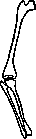
more often called curvature that occurs in the bones or near joints or in the joints themselves, which is why the inward curvature of the limbs is usually denoted by the word varus and the name of the joint around which the curvature occurred. Such a form of curvature on the limbs is very common, especially in the form of congenital deformities, but can also develop for other reasons. - Cubitus varus - curvature in the elbow joint, consisting of abnormal adduction of the forearm toward the ulnar side. It occurs rarely, usually after improperly healed fractures. - Coxa vara - curvature of the femoral neck, consisting in the neck being as if bent toward the shaft of the femur and forming with it an angle less than 120°, which in this deformity sometimes reaches a right angle (see Figure 1). This relationship of the neck and femur creates a position of constant adduction of the thigh. - Genu varum - curvature in which the lower leg is abnormally brought toward the midline, due to which an angle open inward is formed between the thigh and lower leg (see Figure 2). With such bilateral curvature, O-shaped legs occur, i.e., when standing with ankles tightly together, the knees do not come close together; it occurs less frequently than genu valgum (see Knee joint). - Crus varum develops due to curvature of the tibial bones inward, so that the angle opens toward the midline. With bilateral curvature, O-shaped legs also occur; it develops more often on the basis of rickets. - Pes varus - deformation of the foot, consisting in the deviation and curvature of the foot inward (supination) (see Figure 3). The deformation is more often congenital; pes varus occurs very frequently (see also Clubfoot).
Figure 2. Genu varum.
VASILYEV'S METHOD of preparing transparent anatomical specimens is, in essence, a modification of the well-known original method of Spalteholz, consisting in that if an anatomical specimen is immersed in a mixture of ether oils having the same coefficient of refraction as the tissues of the specimen, then the specimen becomes transparent, since the rays pass through without refracting through the ether oil and the specimen. According to this method, the specimen is fixed with formalin or alcohol, sublimate. If the specimen contains bones, decalcification in a 1-5% HCl solution for 1-4 weeks or more is necessary, depending on the size of the object. After washing in distilled water, it is dehydrated in absolute alcohol. Then it is passed through benzene (change 2-3 times) and placed in a clarifying mixture, which includes ether oils with the following coefficients of refraction: Oleum Gaultheriae (Wintergrunol) - 1.538, Benzylbenzoate - 1.570 and Isosafrol - 1.577. Within these limits of the coefficients of refraction of oils are all human tissues and some animal tissues. Knowing the coefficient of refraction of the tissue of a given object, it is easy to prepare a suitable clarifying mixture for the specimen. For the convenience of the worker, Spalteholz provides a ready-made scale of mixtures for all kinds of anatomical specimens (see Spalteholz's method).
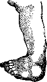
Figure 3. Pes varus.
VASILYEV'S METHOD of preparing transparent anatomical specimens is, in essence, a modification of the well-known original method of Spalteholz, consisting in that if an anatomical specimen is immersed in a mixture of ether oils having the same coefficient of refraction as the tissues of the specimen, then the specimen becomes transparent, since the rays pass through without refracting through the ether oil and the specimen. According to this method, the specimen is fixed with formalin or alcohol, sublimate. If the specimen contains bones, decalcification in a 1-5% HCl solution for 1-4 weeks or more is necessary, depending on the size of the object. After washing in distilled water, it is dehydrated in absolute alcohol. Then it is passed through benzene (change 2-3 times) and placed in a clarifying mixture, which includes ether oils with the following coefficients of refraction: Oleum Gaultheriae (Wintergrunol) - 1.538, Benzylbenzoate - 1.570 and Isosafrol - 1.577. Within these limits of the coefficients of refraction of oils are all human tissues and some animal tissues. Knowing the coefficient of refraction of the tissue of a given object, it is easy to prepare a suitable clarifying mixture for the specimen. For the convenience of the worker, Spalteholz provides a ready-made scale of mixtures for all kinds of anatomical specimens (see Spalteholz's method).
Related articles
Mentioned in
Cite this page
“Varus.” Soviet Medical Encyclopedia. English translation of Bolshaya Meditsinskaya Entsiklopediya, 1st ed. (Moscow, 1928–1936), ed. N. A. Semashko. https://sovietmedicalencyclopedia.pages.dev/article/varus/