Varolius' Bridge
Historical document, translated for reference. It reflects medical knowledge of the 1920s–30s and is not medical advice.
Summary
Varolius' bridge (pons Varoli) is part of the brainstem, appearing as a broad white ridge located between the medulla oblongata and cerebral peduncles. It connects to the cerebellum via cerebellar peduncles and forms the floor of the fourth ventricle.
Encyclopedia article (1928–1936)
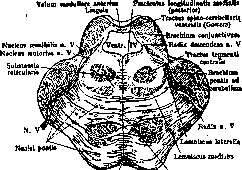
VAROLIUS' BRIDGE (pons Varoli), belongs to the trunk part of the brain; in the form of a broad white ridge, located transversely, lies between the medulla oblongata and cerebral peduncles, from which it is sharply separated (see figure 1); on the sides this ridge narrows and passes into the cerebellar peduncles (pedunculi cerebelli ad pontem Varoli, or brachia pontis), which end in the cerebellar cortex. The boundary between the Varolius' bridge and cerebellar peduncles serves as the place of exit of the roots of the trigeminal nerve. The anterior surface of the Varolius' bridge, turned not only forward but also downward, lies on the clivus Blumenbachi of the base of the skull; between them are the inferior petrosal sinus and basilar plexus. On the anterior surface of the Varolius' bridge there is a groove - sulcus basilaris, in which lies the basilar artery. The posterior surface of the bridge forms the floor of the fourth ventricle, its upper triangle (see Ventriculi eerebri). Along the middle line of the floor of the fourth ventricle, or rhomboid fossa, runs the fissura fossae rhomboideae (fissura longitudinalis posterior), on each side there is an elevation - eminentia teres, which in the lower parts of the Varolius' bridge consists of two parts - the outer one, corresponding to the nucleus of the abducens nerve, and the inner one - the nucleus of the funiculus teres. At the base of the triangle pass the striae medullares, or acusticae, in the outer corner lies the area acustica, slightly above a depression - fovea superior, corresponding to the motor nucleus of the trigeminal nerve, still higher the locus coeruleus, colored in a dark grayish-blue color, depending on the presence of a large number of pigmented cells. The Varolius' bridge, together with the cerebellum and medulla oblongata, develops from the hindbrain - rhombencephalon r the cavity of which is transformed into the fourth ventricle (development see Brain). According to histological structure the Varolius' bridge can be divided into two parts: the anterior, or ventral part of the bridge forms the basis pontis (base), while the posterior, or dorsal part is called tegmentum pontis (covering).-The basis pontis throughout the height of the Varolius' bridge has the same structure; here lie the pyramidal, temporal-frontal-bridge fibers; they are surrounded by cells of nuclei pontis, in which end collaterals of pyramidal fibers and other cortico-bridge systems and where begin fibrae propriae pontis (own fibers of the Varolius' bridge); they are arranged in a transverse direction forward, backward and between the bundles of pyramidal fibers, forming three layers - stratum superficiale, profundum and complexum; the fibers pass to the opposite side, crossing with fibers of the opposite side; at the outer edge of the Varolius' bridge the fibers all come together, forming the pedunculus cerebelli ad pontem Varoli, and end in the cerebellar cortex.-The tegmentum is not identical in structure throughout the height of the Varolius' bridge; it can be divided into three parts. In the lower part (see figure 2), at the boundary of basis and tegmentum, lies the lemniscus medialis (main loop) transversely; it is intersected by fibers of the corpus trapezoidei, going transversely; these fibers begin in the nucl. ventralis nervi cochleae, pass to the opposite side and end in the oliva superior (superior olive), lying outward from the lemniscus med.; in the olive begins the lemniscus lateralis (lateral loop); even more outward lies the bundle of Gowers. Behind these formations is the substantia reticularis, in it behind and outward from the oliva superior are seen two nuclei of the facial nerve (for the upper and lower facial musculature); fibers from this nucleus first go backward, surround the nucleus of the abducens nerve, lying at the bottom of the fourth ventricle, form the knee of the facial nerve, and then, piercing the entire thickness of the Varolius' bridge, exit on its anterior-lateral surface. Near the nuclei of the facial nerve lie nuclei of the parasympathetic system (nucl. salivator. super, and nucleus for the lacrimal gland). At the bottom of the fourth ventricle is the nucleus of the abducens nerve, inward from it the fasciculus longitudinalis posterior, outward the nucleus of Deiters. In the lateral parts of the substantia reticularis lies the central bundle of the tegmentum (tractus thalamo-olivaris) and nuclei reticularis tegmenti.-The middle part of the tegmentum is characterized by the presence in the tegmentum of nuclei of the trigeminal nerve and the place of exit of its roots (see figure 3); the motor nucleus is located inward from the root, and the sensory nuclei-outward. Into the motor root also enter fibers from the mesencephalic nucleus, lying near the aquaeductus Sylvii. The nuclei of the VI and VII nerves have disappeared, the substantia reticularis has become larger, in it are seen tractus thalamo-olivaris (central bundle of the tegmentum) and nucleus reticularis tegmenti. At the bottom of the fourth ventricle lies the fasciculus longit. post., nucleus of the funiculus teres, under them pass fibers of the central path of the V nerve, more outward-locus coeruleus and nucleus of Bekhterev. On both sides of the fourth ventricle are the pedunculi cerebelli ad corpora quadrigemina s. brachia conjunctiva, connected between themselves by means of the velum medullare anterius, in which cross the cerebellar fibers of the bundle.
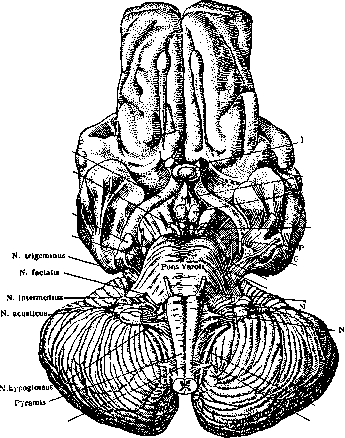
Figure 2. Cross-section through the Varolius' bridge
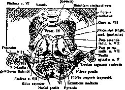
in its lower part. N. abducens and n. facialis (according to Mliller-Spatz).
Stratum superficialis pontis
Ругатш»
Figure 3. Cross-section through the Varolius' bridge in the middle part. N. trigeminua (according to Muller-Spatz).
Gowers' bundle (see Gowers' bundle). At the boundary between basis and tegmentum lie the medial, lateral lemnisci and the sensory part of Gowers' bundle (fasciculus spino-thalamicus).-In the upper part of the tegmentum the fourth ventricle narrows, due to the displacement of the brachia conjunctiva in the ventral and inward direction (see figure 4); in the velum medullare anterius, besides the fibers of Gowers' bundle, cross the fibers of the root of the IV nerve; on the sides of the fourth ventricle, in its gray matter, passes the mesencephalic motor root of the V nerve. Then follows the locus coeruleus and fasciculus longitudinalis posterior; part of the tegmentum is occupied by brachia conjunctiva, directed toward the middle line, nucleus ventralis and dorsales tegmenti, in which ends the tractus mamillo-tegmentalis; the lemniscus medialis occupies a more outward position, and the lemniscus lateralis is placed perpendicularly to it; in the posterior part is the sensory part of Gowers' bundle.
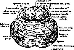
Figure 4. Cross-section through the Varolius' bridge
in the upper part. Decussatio brachiorum conjugtivorum (no Muller-Spatz'y). The vessels supplying the V. bridge depart from art. vertebralis, art. basilaris or from their branches (art. cerebelli anter., med., posterior); they belong to the terminal arteries, i.e. they do not anastomose with neighboring arteries. They are divided into arteriae medianae, or nucleares, and arteriae radiculares; the first penetrate through the midline to the nuclei, the others enter together with the roots and also reach the nuclei. Art. medianae and radiculares for the V. m. take their origin from art. basilaris and only for the nuclei of nerves VI and VII sometimes depart from a. vertebralis or a. cerebelli media. The veins follow the course of the arteries. Despite their relatively small size, the V. m. is an extremely important organ in functional terms, as indicated by the complexity of its structure. The V. m. is a conductor of fibers going from the brain to the periphery and back; in the base of the V. m. passes the pyramidal tract, going from the anterior central gyrus to the anterior horns of the spinal cord; from this tract collaterals depart, ending in nuclei pontis, in addition, part of the cortico-nucleus fibers (going from the cortex to the nuclei of the pons) branches off from the common pyramidal tract, crosses to the opposite side and ends at the corresponding nuclei; an exception exists for the nucleus of nerve VII, innervating the upper half of the facial muscles and receiving fibers not only from the pyramidal tract of the opposite side but also its own (this explains the preservation of movement in the upper half of the face in lesions of the central pyramidal neuron). Lemniscus medialis is the second sensory neuron carrying deep sensitivity and the sense of touch for the opposite half of the body; among the loop layer go fibers in a descending direction, belonging to the pyramidal system and ending in the nuclei of some cranial nerves (nerves VI, XI)-librae cortico-nucleares. To it are added conductors of sensitivity from the nuclei of V, IX, X cranial nerves. Superficial sensitivity is conducted in the lower parts of the V. bridge by the bundle of Govers, and then these fibers join the lemniscus lateralis, which is the conductor of auditory sensations. In the V. m. is the second auditory neuron-corresponding to trapezoideum of the vestibular system nuclei (nuclei Deiters'a and Bekhtereva). The extrapyramidal system is represented by fasciculus rubro-spinalis, tecto-spinalis, thalamo-olivaris, etc. Along the entire length of the V. bridge, along the bottom of the IV ventricle, lies fasc. longitudinalis posterior, in charge of combined deviation of the eyes in head movements. Through the V. m. pass brachia conjunctiva. Finally, in the V. m. occurs a connection between the cerebral cortex and the cerebellum-in nucl. pontis end fibers going from the cerebral cortex (collaterals of the pyramidal paths, fronto-temporo-occipito-pontine systems) and begin librae propriae pontis, giving rise to pedunc. cerebelli ad pontem. Due to the accumulation of nerve cells in the V. m., it plays not only the role of a conductor but also a central apparatus. In the V. m. are located the nuclei of cranial nerves: n. trigemini-motor (innervating the masticatory muscles) and sensory (innervating the skin and mucous membranes of the corresponding half of the face), n. abducentis (innervating muse, rectus externus), n. facialis (innervating the facial musculature) and nuclei of n. acust. (auditory and vestibular). The motor nuclei of these nerves are simple motor centers-centers of muscles. Besides them, in the V. m. are: 1) centers belonging to the autonomic nervous system (see): centers of lacrimation, salivation (nucl. salivatorius super.), the main sweating center; 2) centers in charge of more complex acts requiring the work of two or several nuclei of cranial nerves in a certain sequence and proportion. Anatomically such centers are formed by separate parts of the nuclei of the corresponding nerves; for their joint work it is necessary to assume the existence of coordinating mechanisms, i.e. a group of cells that connect the simple centers with each other, without being themselves connected to the muscles. Attempts have been made to find such centers, but their localization is still unknown. Some authors generally deny the existence of such centers, and assume a connection between simple centers through associative neurons, found in large numbers in substantia reticularis of the V. m. Such complex centers include centers related to the acts of swallowing (in which participate nerves V, VII in the V. m., nerves IX, X, XII in the medulla oblongata), voice formation (nerves VII, X, XII), blinking (nerves V, VII), chewing (nerves V, XII). Some of the above centers work not only under the influence of volitional impulses, but also reflexively and automatically, others only reflexively, for example, the centers of the autonomic system. The general symptomatology of diseases of the V. m. Relatively often focal lesions are observed in the V. m. Their symptomatology depends on the size and localization of the process; with small foci the process can develop without symptoms. Foci involving one half give alternating syndromes (see). Bilateral lesions of the V. m. can give a picture of bulbar paralysis (see) due to the interruption of fibers going from the cerebral cortex to the nuclei of the medulla oblongata. Sometimes with bilateral lesions, disorders of sphincters are observed. By some authors the center of convulsions is localized in the V. m., irritation of which leads to convulsions similar to epileptic; others connect this center with the vasomotor center, the lesion of which causes convulsions of a reflex nature, without the participation of the cortex. Of the pathological processes, softenings are most often found in the V. m., on the basis of occlusion of art. basilaris; hemorrhages are less frequent; among neoplasms are found gliomas or tubercles. Inflammatory diffuse processes in the V. m. are also observed in general infections, but these processes rarely involve the V. m. in isolation, more often simultaneously with other parts of the nervous system. Lit.: Blumenau L. V., The Human Brain, L.-Moscow, 1925; L? i 1 1 i g e r E., The Brain and Spinal Cord, Moscow, 1914; Rauber F., Handbook of Human Anatomy, SPB, 1911; Danilevsky V. Ya., Textbook of Human Physiology, Kharkov, 1923; Zarchenko M. L., Vascular Diseases of the Brain Stem, M., 1911; Handbuch der Neurologie, hrsg. von M. Lewandowsky, B. V, 1914; Oppenheim H., Lehrbuch der Nervenkrankheiten, B. II, B., 1923.
Related articles
Mentioned in
Cite this page
“Varolius' Bridge.” Soviet Medical Encyclopedia. English translation of Bolshaya Meditsinskaya Entsiklopediya, 1st ed. (Moscow, 1928–1936), ed. N. A. Semashko. https://sovietmedicalencyclopedia.pages.dev/article/varolius-bridge/