Fasciculi
Historical document, translated for reference. It reflects medical knowledge of the 1920s–30s and is not medical advice.
Summary
Fasciculi are collections of nerve fibers of the same function located adjacent to each other, originating from similar cells and ending near cells with identical functions. They can be associative, commissural, or projection fibers, serving various pathways in the nervous system.
Encyclopedia article (1928–1936)
FASCICULI, s. tractus (syn. bundles, conducting pathways), a collection of nerve fibers of the same function, located directly next to each other, over a greater or lesser distance; all fibers of the same bundle originate from similar cells and end near cells having the same function. In their course, F. send collaterals to cells or nuclei, past which they pass; some of these, approaching their termination, gradually decrease due to fibers leaving them; others, conversely, increase due to the addition of new neurons to them. Thinner bundles are known under the name fibrae, filae (fibers). The length of bundles is very diverse, from very long F., which extend throughout the entire spinal cord (fasc. radiculo-bulbaris or fasc. Golli), to very short ones, not extending beyond one segment. - From a physiological point of view, the following are distinguished: 1) associative bundles, connecting different parts of the cortex or segments of the spinal cord; 2) commissural bundles, connecting similarly named places of different halves of any part; 3) projection bundles, connecting various parts located lower or higher, and in the final stage with the periphery; these F. may be centrifugal, carrying impulses from centers to the periphery, and centripetal-from the periphery to centers. Sometimes an impulse passes successively through several F., until it reaches its destination. In the table (pp. 555-580) all possible F., tractUS and fibrae are given as completely as possible. E. Kononova. FASCICULUS LONGITUDINALIS POSTERIOR, s. dorsalis, posterior longitudinal bundle, paired, represents a complex system of direct and crossed, descending and ascending fibers of varying lengths, originating from various cellular groups of the brain stem (reticular formation, nuclei of the oculomotor, lateral and abducens nerves, Deiters' nucleus, etc.); it begins near the opening of the Sylvian aqueduct into the third ventricle, passes along the entire stem and descends into the anterior column of the spinal cord (see separate table, figure 3), where it ends, with some fibers reaching the lumbo-sacral segment. In the spinal cord, F. contains only descending fibers, is located next to the anterior pyramidal bundle and is not distinguished in a normal preparation (see fig.). In the medulla oblongata, the bundle is located dorsally to the medial lemniscus, on both sides of the raphe; when transitioning into the pons Varolii, it is located near the floor of the fourth ventricle, and then the Sylvian aqueduct, clearly standing out on a frontal section as a comma between the central gray matter surrounding the Sylvian aqueduct and the reticular formation of the tegmentum; it passes directly under the nucleus of the trochlear nerve (see separate table, figures 1 and 2) and then along both sides of the oculomotor nerve, traced to the posterior commissure, where it receives fibers from the nucleus of Darkshevich, and according to some authors from
^
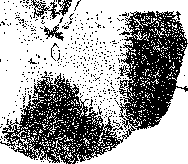
Cross section of the cervical part of the spinal cord of a dog; descending degeneration: 1- fasciculus longitudinalis posterior; 2-fibrae reticulo-spinales; 3-fasciculus vestibulospinalis Lowenthal; 4-scattered fibers in the area of the lateral pyramidal bundle. "interstitial nucleus", nucl. interstitialis (Cajal), representing a collection of large cells located below and more laterally than the nucleus of Darkshevich, between F. and the red nucleus. Fasciculus longitudinalis posterior-phylogenetically ancient bundle and topographically very constant, starting from the lamprey and ending in man. Connecting the centers of eye muscles and the end apparatus of the vestibular nerve and transmitting impulses to the anterior horns of the spinal cord, F. 1. p. conducts impulses for combined movements of the eyeballs, coordination of head and eye movements and is one of the efferent pathways of labyrinthine reflexes and other reflexes of position and movement, localized in the trunk.-With slight unilateral damage to F, 1. p., nystagmus in the same direction is observed; with complete interruption. F. the ability to combine the rotation of the eyeballs toward the side of the lesion (combined gaze paralysis) is lost; when pressure is applied to F. or its irritation (e.g., by a tumor), forced abduction of both eyeballs toward the focus may occur-the patient looks at the focus.
o. Blankov.
Related articles
Mentioned in
Cite this page
“Fasciculi.” Soviet Medical Encyclopedia. English translation of Bolshaya Meditsinskaya Entsiklopediya, 1st ed. (Moscow, 1928–1936), ed. N. A. Semashko. https://sovietmedicalencyclopedia.pages.dev/article/fasciculi/