Auditory Pathways
Historical document, translated for reference. It reflects medical knowledge of the 1920s–30s and is not medical advice.
Summary
The article describes the neural pathways of hearing, from the cochlear nerve to the auditory cortex, detailing the nuclei and tracts involved in auditory processing. It also discusses the pathology of auditory pathways, including symptoms like hearing loss, tinnitus, and various auditory distortions.
Encyclopedia article (1928–1936)
AUDITORY PATHWAYS, CENTERS. The auditory pathways consist of several neurons. The first neuron—the cochlear nerve (the root of the acoustic nerve)—like any sensory nerve, originates in the peripheral ganglion, the spiral ganglion of Corti, located in the labyrinthine cochlea. The peripheral processes of the ganglion cells terminate near the cells of Corti's organ (see), while the central processes, as the root of the VIII nerve, enter the brainstem at the pontocerebellar angle above the retroolivary fissure; passing lateral to the restiform body, they terminate in two nuclei of the medulla oblongata (see figure): in the anterior nucleus (nucleus ventralis n. cochlearis), located on the anterior surface of the restiform body between it and the cerebellar flocculus, and in the posterior nucleus (nucleus posterior, s. tuberculum acusticum—the acoustic tubercle), lying on the floor of the fourth ventricle at the level of the lateral recess on the lateral-posterior surface of the restiform body. From these two nuclei originates the second neuron of the auditory pathway. Part of the fibers emerging from the ventral nucleus terminate in the pons Varolii in the superior olive, while the largest part first ascends and then toward the midline
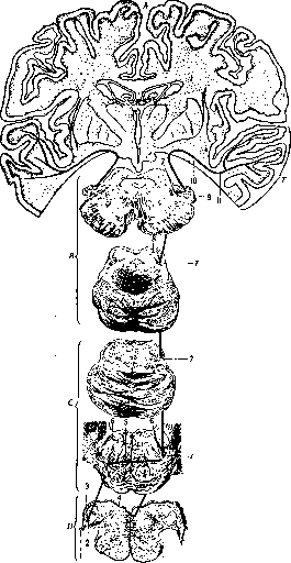
Auditory pathways and centers: A—cerebral hemisphere, B—cerebral peduncle, C—pons Varolii, D—medulla oblongata, E—temporal lobe. 1—cochlear nerve; 2—ventral nucleus of cochlear nerve; 3—acoustic tubercle; 4—medullary striae; 5—trapezoid body; 6—superior olive; 7—lateral lemniscus; 8—inferior colliculus; 9—medial geniculate body; 10—sublenticular part of internal capsule; 11—central auditory pathway and its termination in the temporal lobe.
and forms the system of the trapezoid body (corpus trapezoides), which passes through the layer of the medial lemniscus and is penetrated by scattered nuclei—nuclei corporis trapezoidi. The corpus trapezoides includes fibers from the ventral nucleus as well as fibers from the superior olive and the trapezoid body nucleus of the same side. Crossing the midline in the ventrolateral part of the pons Varolii, they form a bundle—the lateral lemniscus. Another part terminates in the superior olive and the trapezoid body nucleus of the opposite side; from these structures originate fibers that join the lateral lemniscus. Fibers of the second neuron originating in the posterior nucleus (in the acoustic tubercle) take a different path: part of the fibers from the acoustic tubercle along the floor of the fourth ventricle go toward the midline in the form of white striae—striae medullares, s. acusticae. In the median sulcus of the rhomboid fossa, they descend deeply and run for some time along the raphe in the ventral direction, then cross the midline and reach the superior olive of the opposite side to join the lateral lemniscus; but not all fibers run superficially; some (Held's bundle) upon leaving the acoustic tubercle immediately go deeply, then are directed to the opposite side, forming a decussation—Held's decussation, and reaching the superior olive of the opposite side, enter the composition of the lateral lemniscus. This entire system of fibers ascends as part of the lateral lemniscus; on this path, fibers from the nucleus of the lemniscus itself (nucleus lemnisci lateralis) join them; the fibers are directed to the primary auditory centers—corpus quadrigeminum posterius et corpus geniculatum mediale (see Corpora quadrigemina and Corpora geniculata). Some fibers pass by the posterior colliculus nucleus to the anterior. In the medial geniculate body originates the last central neuron of the auditory pathway, which through the sublenticular part of the internal capsule (see Capsula interna) are directed to the cortex of the temporal gyrus (see Temporal Lobe, Brain). Among the auditory fibers going to the cortex of the auditory area, fibers run in the reverse direction—from the cortex to the primary auditory centers. In the cortex of the auditory area, a large number of fields are distinguished cytoarchitecturally (see Architectonics of the cerebral cortex). The following features are characteristic of the cortex of the temporal gyri: 1) the cortex is very wide; 2) the transverse and longitudinal striations are very clearly expressed in it; 3) the molecular layer is very wide; 4) the II and IV layers (outer and inner granular) are narrow and poor in cells; 5) the III layer is narrow, poor in cells, but the cells are of large size; 6) the V layer is wide and the cells in it are large; 7) the VI layer is wide, the cells are large and very numerous. In some fields of the temporal lobe this structure is very clear, but there is no complete unity. The first temporal gyrus somewhat deviates from this type; in some of its fields, the pyramidal cells of the III layer are of very large size, both horizontal and vertical striations are very sharply expressed. Regarding the exact place of termination of the auditory pathways, opinions vary greatly. Some authors (Monakho, Déjerine, etc.) assume that the main place of termination of the auditory pathways is the superior temporal gyrus, its middle and posterior parts, but generally the entire temporal lobe participates in auditory perceptions; according to others, the Heschl gyri are related to the auditory sphere (Flechsig, Nissl, Mayendorf, Pfeiffer). There is also a compromise opinion according to which both the superior temporal gyrus and the Heschl gyri are related to auditory sensations. The most widespread is the first opinion regarding the relation of hearing to the superior temporal gyrus. Through the system described above, auditory sensations are transmitted from the periphery to the centers, with the exception of the fibers going to the inferior colliculus. Regarding the significance of this connection, views have changed, and the former doctrine of the inferior colliculus as one of the main centers of the auditory pathway is currently not considered correct; the fibers going to the colliculi serve for conducting reflexes, for implementing reflex movements in the form of ear movements, vocal cord movements; these reflexes are carried out by fibers going from the colliculi to the medulla oblongata, to the spinal cord. On the basis of some experiments on dogs (Munk), special centers for sounds of different heights were distinguished in the auditory cortex of the cortex—the posterior parts serve for perception of low, and the anterior—high tones; recently some scientists (Pfeiffer and others) support the view that in humans high and low tones are perceived by different parts of the auditory area: high tones by the inner part of the Heschl gyrus, and low tones by its outer part. However, many dispute the existence of such tone centers (Goldstein and others). Pathology. According to most observations, unilateral destruction of the auditory area leads to changes in hearing on both sides, but on the opposite side to a greater degree than on the corresponding side. This is explained by the incomplete decussation of auditory fibers (a smaller part of the fibers remains on their side). One of the first symptoms in diseases of the auditory pathways or centers, especially the peripheral auditory neuron, is tinnitus of various characters and intensities. This tinnitus can be a source of hallucinations. Along with tinnitus there is a decrease in hearing, which can extend to the entire range of sounds or affect only certain low or certain high sounds. The acuity of hearing is tested by various tuning forks and voice (whispering or loudly). A decrease in hearing of central (i.e., nervous) origin must be distinguished in differential diagnosis from peripheral deafness; experiments by Weber, Rinne, Schwabach (see the respective words) are of great help here. Additionally, in disorders of central origin, a number of accompanying phenomena are observed—vomiting, dizziness, headaches, nystagmus and other symptoms depending on the localization of the lesion—disorder of movement, sensitivity, vision, etc. As a disorder of hearing, besides decrease, the following are observed: 1) hyperacusia—all sounds seem sharper, which causes distressing sensations; 2) paracousia, or distortion of sounds—the patient perceives one sound as another, hears better among noise (paracousie de Willis), excessively increased perception of oscillations of the range (paracousie de Weber); in spatial paracousia sounds are perceived as coming from the opposite side; 3) colored hearing (audition colorée); 4) word deafness (see) and 5) mind deafness (see). In some diseases of the nervous system, disorders of hearing of various intensity are observed: hydrocephalus is accompanied by a decrease in hearing, reaching complete deafness. Vascular diseases of the brain (hemorrhage, thrombosis, sclerosis) are often accompanied by tinnitus. Brain tumors, especially in the posterior cranial fossa, cause almost complete deafness and tinnitus; meningitis, pachymeningitis—the same phenomena. In tabes, disseminated sclerosis, in chorea, epilepsy, migraine and finally in hysteria, neurasthenia and other diseases, a disorder of hearing can be observed. Finally, a disorder of hearing is a frequent phenomenon in trigeminal neuralgia, in facial nerve paralysis.
Related articles
Mentioned in
Cite this page
“Auditory Pathways.” Soviet Medical Encyclopedia. English translation of Bolshaya Meditsinskaya Entsiklopediya, 1st ed. (Moscow, 1928–1936), ed. N. A. Semashko. https://sovietmedicalencyclopedia.pages.dev/article/auditory-pathways/