ARCHITECTONICS OF THE CEREBRAL CORTEX
Historical document, translated for reference. It reflects medical knowledge of the 1920s–30s and is not medical advice.
Summary
The doctrine of the morphological structure of the cerebral cortex, established through the study of local peculiarities of its structural elements. This doctrine evolved from viewing the cortex as a uniform organ to recognizing it as a complex organ with distinct areas having specialized functions.
Encyclopedia article (1928–1936)
ARCHITECTONICS OF THE CEREBRAL CORTEX, the doctrine of the morphological structure of the cortex, based on the study of local peculiarities of its structural elements. The essence of this doctrine is as follows. To earlier researchers, the cerebral cortex appeared to be built uniformly, therefore they viewed it as a single organ. But through the efforts of histologists, partly experimental physiologists and pathologists during the second half of the 19th and in the 20th century, this view was destroyed. The cerebral cortex is a complex organ; besides being divided into layers, it further breaks down on its surface into separate territories, or fields, each of which individually has the significance of a more elementary organ in structural and functional relation. The term "architectonics" also has a narrower meaning - it also signifies the microscopic structure of the cortex on a section perpendicular to the surface1. If on such a section the nerve cells are stained, cytoarchitectonics is obtained, if myelin fibers - myeloarchitectonics. In the same sense, fibro-, glio- and vasoarchitectonics are spoken of. History of the question. The basis of the architectonic doctrine lies in the fact of the unequal structure of different parts of the cerebral cortex on the surface and in depth. The data pertaining to this had been accumulating since the time of Gennari (1782). But Meynert (1868) was the first to describe five types of cortical structure and divided it into regions. He taught that pyramidal cells carry motor function, spindle-shaped cells carry associative function, and granular cells are sensory elements. As for the individual regions of the cortex, he believed that since they are built differently, each must have its own special function. Thus, Meynert arrived at the idea of "organology" of the cerebral cortex. His successor, the Kiev anatomist Betz, discovered (1874) in the anterior central gyrus special giant cells. He found the same cells in animals, and established the identity of the area of their distribution with the electrically excitable zone of Fritsch-Hitzig. Betz also indicated the cellular peculiarities of other parts of the brain. His assertion that the brain surface should be divided not on the basis of furrows but on the basis of microscopic structure also proved correct. Hammarberg (1893) expressed the same viewpoint, giving descriptions of individual parts of the cortex and pointing to their changes in idiocy. Campbell performed cyto- and myeloarchitectonic studies of the entire cortex, divided it into fields-areas, and gave the first areal map of the brain surface (of 20 fields). Elliot Smith (1907) proposed an areal map (50 fields) based on structural features of the fresh brain. Since 1903, a series of very valuable architectonic works by Brodmann, Cecile and Oscar Vogt appeared. Brodmann, on the basis of cytoarchitectonic studies of the cerebral cortex, compiled his map of the brain surface (52 fields). He deepened the problem of architectonics himself, using embryological and comparative-anatomical methods. S. and O. Vogts studied the areal structure of the cortex, using mainly myeloarchitectonics, on the basis of which O. Vogt compiled maps of the brain surface of more than 200 fields. In the work of Economo and Koskinas (1925), there are cytoarchitectonic areal maps of 107 fields. Architectonic works continuously continue. Among the most recent can be mentioned Rose's research on cortical genetics. Parallel to the study of the cerebral cortex of humans, its study in various animals (Mauss, Flores, Zunino, Preda, Meyer and others) proceeded. In the process of development of the doctrine of the structure of the cerebral cortex, the doctrine of localization of individual functions in the cortex was closely interwoven with it. Some localization ideas (Broca), even the fantastic doctrine of Gall about localization of mental faculties in the brain, influenced its development. Flechsig's doctrine of cortical centers, based on the myelogenic principle, was also of great importance. Methods of architectonic research. The usual methods for staining cells (Toluidinblau, Kresylviolett) or myelin fibers on celloidin or paraffin sections are used. Economo and Koskinas, in order to obtain strictly perpendicular to the surface sections, cut small pieces of cortex for research. When serial preparations are already available, the researcher's task is to identify on those places that differ in any structural peculiarities, and to determine their boundaries. Attention is paid to the width of the cortex, and mainly the general layered structure is studied, as well as each layer of the cerebral cortex individually. The main type of cortex, with which others can be compared, is the six-layered according to Brodmann or seven-layered according to Vogt (see figure 1).
ARCHITECTONICS OF THE CEREBRAL CORTEX
348 width of the cortex, and mainly the general layered structure is studied, as well as each layer of the cerebral cortex individually. The main type of cortex, with which others can be compared, is the six-layered according to Brodmann or seven-layered according to Vogt (see figure 1).
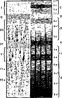
Figure 1. Left - six basic cellular layers of the cortex according to Brodmann: I-lamina zonalis, molecular or tangential layer; II-l. granularis externa, outer granular layer; III-l. pyramidalis, pyramidal layer; IV-l. granularis interna, inner granular layer; V-l. ganglionaris, ganglionic layer, or layer of deep pyramids; VI a and b-lamina multiformis, polymorphic layer, divided into layer of triangular cells (VIa) and layer of spindle-shaped cells (VIb). Right - myeloarchitectonic picture according to Vogt: 1-l. tangentialis with subdivision into sublamina superficialis (i°), subl. intermedia with two parts-pars externa (1a) and pars interna (1b) and subl. profunda (1c); 2-l. dysfibrosa; 3-l. suprastriata with three sublaminae: subl. superficialis (3a1), or stria Kaes-Bechterewi, subl. intermedia (3a2) and subl. profunda (3b); 4-stria Baillargeri externa, or outer transverse stripe; 5-l. interstriata (5a) and stria Baillargeri interna (5b), or inner transverse stripe; 6-l. substriata (6a1, 6b1), l. limitans interna (6b2) and album gyrorum (6b3).
and stria Baillargeri interna (5b), or inner
transverse stripe; 6-l. substriata (6a1, 6b1), l. limitans interna (6b2) and album gyrorum (6b3). In some parts of the cortex the six-layered cellular type is not expressed, in others it seems underdeveloped and in third it undergoes various modifications. Each layer individually may turn out to be wider or narrower; the nerve cells of it larger or smaller; their layer denser or sparser; besides, they are also arranged differently. Sometimes the main layer
is divided into sublaminae. The main layers can also completely disappear. Thus, in area gigantopyramidalis lam. granularis interna disappears; in area striata it splits into three layers. Figure 1 shows that myeloarchitectonics gives an even more detailed division of the cortex into layers than cytoarchitectonics. The difference in the structure of myeloarchitectonic pictures can be expressed as follows: radial fibers can be of different thickness, and their bundles of different density; their length can also vary; transverse fibers differ in thickness or in the formation of various laminae. Embryological foundations. The biological significance of the six-layered type of cortex and its variations has been clarified by embryological studies of Brodmann and supplemented by recent studies of Rose. By the end of the third week of development of the human embryo, the anterior part of the neural tube divides into five vesicles, of which the outermost - the telencephalon - gives the hemispheres. The initial epithelial structure of the hemisphere wall first transforms into a myelospongium, consisting of spongioblasts and neuroblasts (see figure 2) - the embryonic plate. Then it is stratified into matrix and marginal layer (middle section of fig. 2), and from matrix the pyramidal or cortical layer (Py, lower section of fig. 2) differentiates. The pyramidal layer grows and thickens, and at the 5th month of intrauterine life its neuroblasts begin to group in layers. Initially, at the 6th month of intrauterine life, layers V and VI are isolated. Then, during the period from the 6th to the 8th month, the other layers are also isolated from the general cell mass. Thus, asynchronously - in one place of the cortex earlier, in
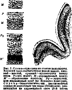
intrauterine life).
another somewhat later - the textogenetic six-layered cortex appears. But it does not form everywhere: there are areas of the cerebral cortex that do not pass through this stage of development, i.e., in them six layers either do not form or they are underdeveloped. The outer surface
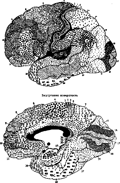
Figure 3. Brodmann's areal map. Field names: 1-a. postcentralis intermedia; 2-a. postcentralis caudalis; 3-a. postcentralis oralis; 4-a. prae-centralis gigantopyramydalis; 5-a. praeparietalis; 6-a. frontalis agranularis; 7-a. parietalis superior; 8-a. trontalis intermedia; 9-a. frontalis granuiaris; 10-a. frontopolaris; 11-a. praefrontalis; 17-a. striata; J*-a. occipitalis; 19-a. praeoccipitalis; 20-a. temp oralis infer.; 21-a. tempor. media; 22-a. temper. sup.; 23-a. limbica post, ventralis; 24-a. limb. ant. ventr.; 26-a. subge-nualis; 26-a. ectosplenialis; 27-a. praesubicularis; 28-a. entorhinalis ventralis; 29-a. retrolimbica granuiaris; 30-a. retrolimbica agranularis; 31-a. limbica post, dorsalis; 32-a. limb. ant. dors.; 33-a. praegranularis; 34-a. entorhinalis dors.; 35-a. perirhinalis; 36-a. ectorhinalis; 37-a. occipito-temporalis; 38-a. temporopolaris; 39-a. pariet. inf. post. s. angularis; 40-a. pariet. inf. ant. s. supragranularis; 41-a. tempor. transversa ant.; 42-a. temp, transv. post.; 43 -a. subcentralis; 44 - a. opercularis; 45 - a. triangularis; 46 - a. frontalis media; 4 7-a. orbitalis. (heterogenetic formations). Such is the phylogenetically oldest cortex of the large brain (see below). In the new cortex, the six-layered stage of development is mandatory for all its parts (homogenetic formations); however, in significance, the six layers here are not identical to the six-layered cortex of the mature brain. This is only the primary six-layered cellular foundation, subject to further differentiation. The differentiation of it does not occur all at once, but in separate areas of the cortex. In some of them, the six-layered type is preserved later on (homotypic formations), in others the number of main layers either decreases or multiplies (heterotypic formations). Other structural features also appear. Thus, gradually separate fields-areas-are formed with those structural characteristics that are determined in the mature brain during architectural studies. Comparative-anatomical parallels. Comparative anatomy teaches that at different levels of phylogenetic development, derivatives of the forebrain, in particular the hemispheres, are by no means equivalent. Their structure and function in fish and amphibians are closely connected with the olfactory area. But already in amphibians, in this oldest cortex (archipallium), two departments are distinguished: palaeocortex, connected with bulbus olfactorius, and archicortex-the future Ammon's horn. Further, at an even higher stage of development-in reptiles-the new cortex (neopallium, neocortex) begins to develop, reaching enormous development in higher vertebrates. As stated above, in humans archipallium, i.e., the old olfactory cortex, and in the process of ontogenetic development, has its own special type of development: it does not pass through the stage of six-layered cortex. For neopallium (neocortex), this stage is mandatory and characteristic. The old and new cortex in the mature brain also have significant structural features. O. Vogt established that radial fibers in the new cortex reach either to the upper border of 5Ъ (type infraradiatus) or to the boundary between 3Ъ and 3c (type intradiatus). According to his terminology, this is isocortex-the part of the cortex corresponding to neocortex. In the old cortex, radial fibers reach the upper part of the third layer (type supraradiatus). He calls it allocortex. Recently, O. Vogt, based on the research of Rose and Jan, has classified infraradiatus as allocortex. Laminar structure and function. When studying the layered, or laminar, structure of the cortex, it is necessary to distinguish, properly, the structure of the layers, their number, the composition of each layer, etc. The corresponding data concerning individual areas are so diverse and extensive that they can only be presented when illustrative material is available. It is only necessary to note that the myeloarchitectonic method, as seen in Fig. 1, allows dividing the cortex in laminar relation even more detailed than the cytoarchitectonic method. Thanks to this, a greater areal differentiation of the cortex is obtained by this method, and hence also a greater number of fields (Vogt). In addition to the composition of the layers, the connection between them (synaptology) is also studied (best by the Golgi method). Much has been done in this respect by Ramon y Cajal (S. Ramon y Cajal). But the connection between the layers and adjacent fields is also studied experimentally, by cuts made in different directions, insertion of insulating plates, electrical stimulation, etc. As for laminar function, there is no complete unanimity regarding it. According to Kappers, in the old cortex (palaeocortex) there are two mutually connected main layers: the superficial-granular layer and the deep-pyramidal layer. The cells of the first have a receptor function, of the second-projection-commissural and associative. In the new cortex (neocortex), in turn, two departments or zones can be distinguished, only more developed: the outer, which includes the outer layers starting from the fourth, and the inner, consisting of the fifth and sixth layers. The same author establishes that the outer zone is genetically and morphologically equivalent to the granular, and the deep zone to the pyramidal layer of the old cortex. And in functional relation between them there are analogies. A series of experiments has proven that the deep zone of neocortex is relatively independent of the superficial one. Corticospinal, corticobulbar, corticopontine, corticothalamic fibers have their source in cells of the deep infragranular zone. From there also originate the fibers of the corpus callosum. Conversely, afferent fibers go to the supragranular zone. There are especially many of them in sensory centers, where the layer of internal grains is also strongly developed. In the outer zone, associative fibers also begin, connecting with each other both intragriseally and extragriseally individual parts of the cortex. Thus, the inner zone is, predominantly, the starting station for the transfer of neurodynamic energy; the outer cell layers, mainly, perform receptor and associative functions. Areal structure of the cortex. The genetic principle of differentiation of the cortex not as a whole, but in separate areas, concerns both the old and new cortex. As a result, everywhere in the mature cortex we find fields-areas developed according to the general laws of organogenesis, i.e., composed of different structural elements. Very characteristic in their construction is that the morphological boundary of each field has a linear form. It only coincides in places with the brain sulci, but can also run along the convex surface of the gyri. Areas are also present in the cortex of various mammals. Comparing with each other the areal structure of the cerebral cortex in various mammals, the following can be established: 1) the general arrangement of fields has a segmental character, 2) there are fields that retain the same layered structure and occupy the same places in the cortex in all these animals, 3) in all mammals, complexes of fields similar in structure and location are revealed, the so-called areas. In the lowest of these vertebrates, there are fewer fields and they are less differentiated. The highest complexity of areal structure is reached in humans. At the same time, the difference in the fine areal structure between the human brain and higher anthropoid apes turns out to be much greater than between anthropoid and lower apes [contrary to Huxley's law]. However, there are areas in the human cortex that are less developed in humans than in other animals. Division of the cortex into areas. On the basis of some general morphological features, part of the fields can be distinguished into one common complex-area, and the entire cortex breaks down into several such areas (regiones). Areas are found in all mammals, which indicates some constant and common function for each of them. Brodmann distinguishes 11 areas: 1) regio postcentralis (includes fields of the posterior central gyrus 1, 2, 3, 43); 2) regio praecentralis (includes fields of the anterior central gyrus 4, 6); 3) regio frontalis-all fields of the frontal lobe in front of regio praecentralis and cingularis; 4) regio insularis-area of the insula; 5) regio parietalis-fields 5, 7, 39, 40; 6) regio temporalis-fields of the temporal lobe; 7) regio occipitalis-occipital area-fields 17, 18, 19; 8) regio cingularis-fields 23, 24, 25, 31, 32, 33; 9) regio retrosplenialis-fields 26, 29, 30; 10) regio hippocampica-fields 27, 28, 34, 35; 11) regio olfactoria-olfactory area.-Economo and Koskinas propose a somewhat different division into areas and lobes.
Functional significance of areas and fields. Despite the apparent isolation of cells in their distribution across layers, in reality they are closely intertwined with each other through their processes and form a single neuronal complex. In this complex, each differently differentiated cell or each layer has its own biological functions, and all of them, in turn, are covered by some biological function of the areal whole. The physiological data on the localization of individual brain functions in the cortex generally correspond to the division of the cortex into areas and fields. To verify this, it is sufficient to compare areal and localization maps. In this case, no one doubts that some manifestations of brain activity are localized in certain areas. The only question is whether any brain function exactly coincides with the boundaries of one or another area. This problem is complex and difficult to solve. Many manifestations of brain activity are in reality not as simple as they seem and often correspond to the activity of the whole brain or its part, but there are still other functions—simpler and more localized. Some of them undoubtedly correspond to area. In this regard, the data from the works of Henschen, Willbrand, Minsky, and Monakow are of great importance, which allow linking the elementary act of vision with the area of the fissure calcarinae (area striata). Also indicative are the new experiments by S. and O. Foerster with electrically excitable cortex of cercopithecus, which showed that each field, previously isolated on the basis of cytoarchitectonics, corresponds to a special function. Particularly striking is the coincidence of the two areal maps—one compiled by O. Foerster for the human brain based on experiments with cercopithecus, and the other compiled by Foerster for the human brain based on irritation of the cortex during operations.
Upper surface FE
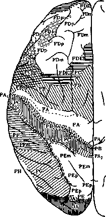
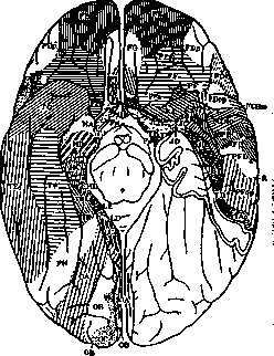
Figure 5. Areal maps of Economo and Koskinas. Regio praerolandica: area praecentralis (FA); area frontalis agranularis (FB); area frontalis intermedia (FC) and others. Regio frontalis: area frontalis granularis (FB); area frontopolaris (FE) and others. Regio orbitalis: area orbitalis (granularis) (FF); area gyri recti (FG); area praefrontalis (FH); area frontoinsularis (FT); area piriformis frontalis (FK); area parolfactoria (FL); area geniculata (FM); area praecommissuralis (FN). Regio limbica sup. ant.: area limb. ant. agr. (LA); area ultracingularis ant. (LB1); area indusiei (LB2). Regio limb. sup. post.: area cingularis post, dorsalis (LC1); area cingularis post, ventral. (LC2); area cingularis lim. post. (LC3). Subregio retrosplenialis: area retrosplenial. agranularis (LD); area retrosplenialis granulosa (LE); area ultracingularis post. (LF1); area (ultracingularis) obtecta (LF2). Lobus insulae: area insulae praecentralis (JA); area insulae postcentralis (JB); area orbito-insularis (JC); area insularis piriformis (JD). Regio postcentralis (parietalis ant.): area postcentralis gigantopyramidalis (PA); area postcentralis oralis simplex (PB); area postcentr. intermedia (PC); area postcentr. caudalis (PD). Regio parietalis sup.: area parietal, sup. (PE). Regio parietal, inf.: area supramarginalis (PF); area angularis (PG). Regio parietalis basalis: area pariet. (temporo-occipitalis) basalis (PH). Lobus occipitalis: area peristriata (OA); area parastriata (OB); area striata (granulosa) (OC). Regio supratemporalis: area temp. sup. (TA); area supratempor. magnocellularis simplex (TB); area supratemporalis granulosa (TC); area supratemp. intercalata (TD). Regio tempor. propria: area tempor. propria (TE). Regio fusiformis: area fusiformis (TF); area hippocampotemporalis (TH). Regio temporopolaris: area temporopolaris (TG); area piriformis tempor. (TG); area subst. pert. post. (TH). Lobus limbicus inferior: area uncinata (HA); area parauncinata (HB); area rhinalis limitans (HC); area praesubicular. granul. (HD); area pyramidalis (HE); area fasciae dentatae (HF).
Architectonics and higher nervous (psychic) activity. The architectonics of the cerebral cortex as an organ of higher nervous (psychic) activity is also of great importance for the study of the latter. Thus, architectonics provides guiding indications for the reflexologist engaged in the formation of conditioned reflexes and for the psychologist studying animal behavior: both, by additionally experimenting with irritation or destruction of the cortex in animals, can base their work not only on the topography of the sulci and gyri, but also on the fine areal structure. Furthermore, the architectonics of the cerebral cortex introduces a new element into the study of the natural experiment on man that is mental illness. The pathological anatomy of the so-called defective psychoses (feeble-mindedness) in the light of topistics, patoclisis, and pathoarchitectonics (S. and O. Foerster) has already received a new understanding. In this context, the anatomical substrate of idiocy and acquired intellectual defects has become clearly evident. The value of architectural research must also be considered proven in relation to the healthy psyche. There are a number of microscopic studies establishing the features of individual architectonics. Racial architectonics, architectonics of criminals, etc. are being outlined. The study of the architectural features of the brains of highly gifted people acquires particular importance. In these studies, not only the new morphological data obtained are important, but even more so the establishment of relationships between them and the various mental peculiarities of these individuals. The task of the future is not only to establish such relationships accurately but also to prove their regularity, i.e., that they satisfy the mathematical formula of correlations. For this, obviously, a large number of studies will need to be conducted. In general, it can be said that all the data from architectural studies conducted so far on both humans and animals confirm the following general biological position: in the process of organic development, differentiation of form corresponds to differentiation of function; mental activity, representing one of the functions of the brain, is no exception to this position. Areal atlas and maps. Above, mention was made of the difficulty of describing the laminar structure of an area without a drawing. This need is met by photographs of the cyto- and myeloarchitectonic pictures of each area. With them, everyone engaged in the study of the architectonics of the cerebral cortex can compare their preparations when studying individual, racial, age-related, and pathological features of cortical structure. An atlas is composed from such photographs (see atlas and exact description of each area by Economo-Koskinas). Once the boundaries of individual areas are determined, it is easy to compile a map of the brain surface (see figures 3, 4, 5). Pathoarchitectonics and patoclisis. The application of architectural methods to the study of nervous and mental diseases has led S. and O. Foerster to the doctrine of pathoarchitectonics and patoclisis. According to S. and O. Foerster, one can often diagnose one or another brain suffering on the basis of pathoarchitectonics alone. They have proven this in relation to diseases of the corpus striatum, and they also try to extend this principle to diseases of the brain. In the literature, there are a number of pathoarchitectonic studies of various demented disease forms (defect-psychoses), lateral sclerosis, Huntington's chorea, cerebral palsy with integrity of pyramidal pathways, early dementia, epilepsy, progressive paralysis, manic-depressive psychosis, etc. It is interesting that changes in pathoarchitectonics follow certain laws. Many harmful agents have an affinity for individual neuronal complexes (patoclisis), affecting them more than others. For example, layers III and V are affected or only layer IV. This is laminar patoclisis. To a lesser extent, it manifests in individual areas (areal patoclisis).
Related articles
Mentioned in
Cite this page
“ARCHITECTONICS OF THE CEREBRAL CORTEX.” Soviet Medical Encyclopedia. English translation of Bolshaya Meditsinskaya Entsiklopediya, 1st ed. (Moscow, 1928–1936), ed. N. A. Semashko. https://sovietmedicalencyclopedia.pages.dev/article/architectonics-of-the-cerebral-cortex/