Cerebro-Cranial Topography
Historical document, translated for reference. It reflects medical knowledge of the 1920s–30s and is not medical advice.
Summary
This article discusses the projection of brain structures onto the skull surface for surgical planning, explaining the challenges and methods of this technique in the 1930s.
Encyclopedia article (1928–1936)
CEREBRO-CRANIAL TOPOGRAPHY, the projection onto the surface of the skull of various cerebral lobes, gyri, sulci, boundary points between different lobes and gyri of the brain (temporal, parietal, and occipital), and finally the projection of individual cerebral centers. Additionally, the outlines of the cerebral ventricles, the direction of the sinuses and arteries of the dura mater, and the vessels of the brain, as well as the exit points of various cranial nerves, can be marked on the surface of the skull. C.-c. t. from a practical point of view has extremely important significance in surgical intervention, first because it makes possible surgical intervention in the form of a bone-plastic operation at a strictly selected location, and second because it makes possible to limit this intervention and not carry it to the extent of hemi-craniotomy, as proposed to do at one time by Doyen. At the present time, it is known what enormous significance in the development of the picture of shock has bleeding from the coverings of the skull, from the bones of the skull and the meninges of the brain, and therefore the possibility to orient oneself in the more precise location of one or another pathological process is advantageous already from the point of view of prevention of bleeding and consequently of shock. In surgical intervention, one has to search for an abscess, or a tumor, or accumulated blood, or a cyst, or scars, or one or another center, pathological excitation of which causes epileptic seizures. - The certain stability of the relations between the skull and the brain and the certain typical location of the cerebral sulci seemed should make easy the establishment of the relation between the lobes and sulci of the brain and the surface of the skull. However in reality this task is very difficult, and accuracy cannot be guaranteed. The projection onto the surface of the coverings of the skull of the lobes of the brain, gyri, centers, the course of vessels, ventricles, cisterns is possible under the following preconditions: 1) it is necessary to recognize the stability and immovability of the brain in the cranial cavity in various positions of the head; 2) it is necessary to develop a certain technique of research, which as much as possible would guarantee against errors. The brain in the cranial cavity is situated b. or m. freely, thanks to which it performs certain rhythmic movements in connection with the activity of the heart and respiration. These movements can be considered as pendulum-like movements with very small amplitude and do not have essential significance in the sense of C.-c. t. The brain with freedom of physiol. movements preserves in general a stable statics thanks to its location in the fossae of the skull, in the receptacles of the dura mater with its formations, as proc. falciformis, tentorium. Impressiones digitatae are proof of the constancy of certain relations between the gyri and the bony surface of the skull. A certain role in the sense of statics of the brain in relation to the skull is played by the cranial nerves, arterial and venous vessels; finally this role is also partly performed by the Pacchionian granulations. Besides this another circumstance is also interesting. The Pacchionian granulations represent very delicate in their structure formations, and hardly they would be able to follow without violation of their anat. integrity for very large deviations of the brain from its certain average position, to which it returns during physiol. movements. In studying the position of the brain one can see that a significant role belongs to the cerebro-spinal fluid. The brain is not only suspended by the mass of the anatomical formations described above, it floats, in the expression of Hurtle and Plant. But if one takes into account the specific gravity of the brain (1.040) and of the cerebro-spinal fluid (1.003-1.008), then one has to speak of floating in the exact sense of the word with certain reservations. With different specific gravity one can assume the possibility of displacement of the brain in different positions of the head, but the excess weight of the brain will be equalized thanks to the pressure of the fluid (Müller), as this can partly be seen from the enormous material of suboccipital punctures: each position of the brain has its determining the stability of the position of the brain pressure of the cerebro-spinal fluid. All this creates the basis for the projection of one or another areas of the brain onto the surface of the skull. But here it is necessary to point to a whole series of circumstances which force one to evaluate this method as giving not unconditional results. The fact is that there is no strict correspondence between the parts of the skull and the lobes of the brain bearing the same names; os frontale does not correspond to the frontal lobe of the brain; os temporale to the temporal lobe of the brain; os occipitale to the occipital lobe of the brain (figure 1). At this however one cannot deny the significance of the sutures for constructing the projection scheme; for establishing many points they have enormous significance, but the sutures themselves are subject to various deviations from the norm: cases of obliterating ossification of the sutures, their incorrect location, the presence of accessory bones, which can give a completely wrong impression about the direction of the suture line (figures 2, 3 and 4). Of course at the present time with the help of roentgenograms one can to a large extent guarantee oneself against errors. Further it is necessary to take into account a whole series of deformations of the skull, which go far beyond the limits of biol. oscillation; here it is necessary to include deformations caused by premature ossification of the sutures and compensatory increase at the same time of the size of the cranial box in the area of normally functioning sutures, as well as asymmetries of the cranial box with uneven growth of the bones of the skull in the area of different sutures. From pathol. changes of the skull one most often has to meet with rachitic changes of the skulls, and from deformations of the oscillation type it is necessary to point to changes in the external outlines of the skull depending on the development of the musculature, attaching to the bones of the skull both in the temporal and occipital areas as well as on the base of the skull. The brain itself in the construction of its hemispheres, individual lobes and gyri gives very large variations. Cases are observed of uneven development of the right and left hemispheres with displacement of proc. falciformis and sometimes tentorium. Cases are observed of uneven development of the anterior and posterior parts of the hemispheres. Such asymmetries of course do not remain without influence on the location of the most important sulci and gyri, as
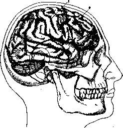
Figure 1. Relation between the sutures of the skull and
the lobes of the brain: 1- sulcus centralis; 2-fissura Sylvii (from Tandler).
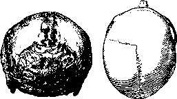
Figure 2.
Figure 3.
Figure 2. The winding and numerous fissures sut. lambdoideae. Figure 3. Absence of sut. coronal, on the right side (from Megkery). e.g. of the central sulcus and corresponding convolutions. Particularly demonstrative in proving the significance of various types for C.-c. t. was Froriep (1897), who established two types: frontopetal and occipitopetal. At present, this doctrine has been developed in more detail, and the basioparietal and parietopetal types have been established. The significance of these forms is evident from the accompanying figures (figs. 5, 6, 7, 8 and 9). In the surgical literature, the extreme importance of these types in performing operations is noted. Above, methods for studying the fundamentals of C.-c. t. were indicated. In this respect, the methodology cannot be considered flawless; all methods applied, when dealing with cadaveric material, do not guarantee accuracy of research: after death, the statics sharply changes due to the cessation of cerebrospinal fluid function; furthermore, in the brain, phenomena of edema appear extremely sharply, both general, occurring during the agonal period and immediately after death, and partial in the lower-lying parts of the brain depending on the position of the corpse. The use of preserving fluids and freezing cannot be considered a reliable method for craniocerebral research. In the latter case, as well as in the use of preserving fluids causing swelling of brain tissue, there first occurs a change in the ventricles and communication pathways, namely their reduction, flattening of convolutions, and a backward shift of the brainstem toward the foramen occipitale magnum. The application of preserving agents causing shrinkage of the brain also changes the relationship of the lobes and convolutions to the surface of the skull due to the expansion of subdural spaces, the system of ventricular cavities and communication pathways. They are filled partly with cerebrospinal fluid, partly with the seeping liquid used for preservation. In view of this, research using radiograms must be considered of little value, when it comes to the method of Taylor and Haughton, who on the corpse opened the skull, inserted lead plates into the sulci, closed the skull and took pictures. In this respect, at present, mention should be made of Sicard's original method of injecting the cerebral vessels in living subjects to determine the location of tumors or aneurysms, of course, with exceptional indications for this. Despite such a critical attitude toward the methodology of craniocerebral research, its data are still widely used in practice. The large number of proposals on the methodology of C.-c. t. characterizes both the difficulty of the practical resolution of the question and the lack so far of the best way to solve the problem.
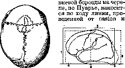
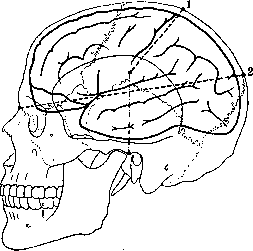
For projecting areas of the brain onto the surface of the skull, many schemes and apparatuses have been proposed, constructed on the basis of a thorough study of the anatomy of the skull and brain, as well as on the basis of clinical observations. To date, up to 20 schemes have been counted, and there is no reason to think that new proposals will not be made. Some authors offer schemes and models for determining only the main convolutions and sulci, while others aim to represent the details of projections of all sulci and convolutions. The methods of Poirier, Kocher, Kronlein, and Tandler are the most popular. Poirier gives in his scheme the determination of the Sylvian and Rolandic sulci. The projection of the Sylvian sulcus is determined by the line named by him lin. nasolambdoidea (fig. 10). Nasion - the point of connection of the suture between both nasal bones and the frontal bone (linea naso-frontalis). Lambda (the name of the Greek letter) - the point of connection of both parietal bones with the apex of the occipital squama. This line intersects the base of the frontal bone, the apex of the greater wing of the sphenoid bone, the apex of the temporal squama, and the lower part of the parietal bone. Poirier calls this line the Sylvian line, characterizing by this the coincidence of this line with the Sylvian sulcus. However, the coincidence is not the rule; only certain parts of this line coincide with the Sylvian sulcus: the part of the line intersecting the apex of the greater wing of the sphenoid bone coincides with the beginning of the Sylvian sulcus, but later the line deviates from the course of the Sylvian sulcus, runs along the first temporal convolution, and finally coincides with the first temporal sulcus. Furthermore, a weak point of this scheme is the variability of lambda. First, it is sometimes very difficult to establish the suture line of the parietal bones with the occipital bone. Second, in the area of lambda, accessory bones (ossa interparietalia) are sometimes observed; in this case, it is completely impossible to determine the suture line of these bones with the occipital bone, and it is easy to mistake the upper part of the accessory bones for Lambda. The projection of the central sulcus is determined by a line whose direction is determined by two points. One point, the upper one, is established on the median sagittal line 48 mm behind the bregma (the point of connection of the coronal and sagittal sutures). The lower guiding point is determined as follows: a vertical line is drawn through the end of the articular process of the mandible, so-called praeauricular vertical, a line perpendicular to the plane in which the French horizontal line passes - the line passing through the alveolar process of the maxilla between the incisors and the lowest point of the articular surface of the skull. Along this vertical line, 7 cm are measured, counting the external auditory meatus as the starting point. The designated point will be the second point determining the direction of the Rolandic sulcus. A weak point of this scheme is the absolute figures. Since in different types of skulls both the size of the bones and the position of the sutures vary, this method does not actually guarantee the accuracy of the projection. Despite the weak points of Poirier's scheme, it is very often used, especially in France; its particular significance lies in the fact that Poirier's principle has been adopted in a number of other schemes: in the schemes of Keler, Kocher, Kronlein; in Kocher's scheme, Poirier's line is adopted for the Sylvian sulcus, and in Keler's, Kronlein's, etc. schemes - the vertical preauricular (praeauricularis verticalis).
Kronlein's method (see vol. VII, art. 577, fig. 3). Kronlein proposed on the basis of Froriep's research the following system of lines: 1) a horizontal line, so-called German horizontal, auriculo-orbital; it passes through the lower edge of the orbit and the upper edge of the external auditory meatus; 2) a horizontal line, supraorbital (linea supra-orbitalis); this line passes through the upper edge of the orbit and runs parallel to the first line; 3) a vertical line, zygomatic, anterior vertical line (linea verticalis zygomatica), which goes from the middle of the zygomatic bone perpendicular to the first line; 4) a vertical line, auricular, middle vertical (verticalis auricularis), going from the head of the articular process of the mandible vertically to the first line; 5) a vertical line behind the mastoid process, posterior vertical line (verticalis retromastoidea), starting from the most posterior point of the base of the mastoid process and running perpendicular to the first line. To determine the central sulcus using this scheme, a line must be drawn connecting two points: one point lies at the intersection of the anterior vertical with the supraorbital horizontal line, and the other point lies at the intersection of the posterior vertical with the median sagittal in the parietal region. The connection of the mentioned points gives a line that corresponds to the position of the central sulcus. The determination of the Sylvian sulcus is carried out as follows: the angle formed by the Rolandic sulcus line and the upper horizontal line is bisected. The direction of this line will correspond to the Sylvian sulcus. Kronlein's scheme is the most popular. To determine the lines, Kronlein proposed a very simple and convenient apparatus,
Figure 6. Frontopetal type of skull. Figure 7. Occipitopetal type. Figure 8. Basiopetal type. Figure 9. Parietopetal type.
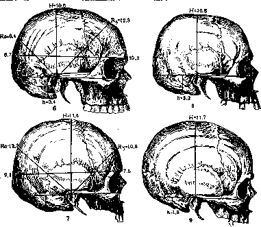
Figure 10. Poirier's method: 1 - point located
The projection of the central sulcus is determined by a line whose direction is determined by two points. One point, the upper one, is established on the median sagittal line 48 mm behind the bregma (the point of connection of the coronal and sagittal sutures). The lower guiding point is determined as follows: a vertical line is drawn through the end of the articular process of the mandible, so-called praeauricular vertical, a line perpendicular to the plane in which the French horizontal line passes - the line passing through the alveolar process of the maxilla between the incisors and the lowest point of the articular surface of the skull. Along this vertical line, 7 cm are measured, counting the external auditory meatus as the starting point. The designated point will be the second point determining the direction of the Rolandic sulcus. A weak point of this scheme is the absolute figures. Since in different types of skulls both the size of the bones and the position of the sutures vary, this method does not actually guarantee the accuracy of the projection. Despite the weak points of Poirier's scheme, it is very often used, especially in France; its particular significance lies in the fact that Poirier's principle has been adopted in a number of other schemes: in the schemes of Keler, Kocher, Kronlein; in Kocher's scheme, Poirier's line is adopted for the Sylvian sulcus, and in Keler's, Kronlein's, etc. schemes - the vertical preauricular (praeauricularis verticalis).
Kronlein's method (see vol. VII, art. 577, fig. 3). Kronlein proposed on the basis of Froriep's research the following system of lines: 1) a horizontal line, so-called German horizontal, auriculo-orbital; it passes through the lower edge of the orbit and the upper edge of the external auditory meatus; 2) a horizontal line, supraorbital (linea supra-orbitalis); this line passes through the upper edge of the orbit and runs parallel to the first line; 3) a vertical line, zygomatic, anterior vertical line (linea verticalis zygomatica), which goes from the middle of the zygomatic bone perpendicular to the first line; 4) a vertical line, auricular, middle vertical (verticalis auricularis), going from the head of the articular process of the mandible vertically to the first line; 5) a vertical line behind the mastoid process, posterior vertical line (verticalis retromastoidea), starting from the most posterior point of the base of the mastoid process and running perpendicular to the first line. To determine the central sulcus using this scheme, a line must be drawn connecting two points: one point lies at the intersection of the anterior vertical with the supraorbital horizontal line, and the other point lies at the intersection of the posterior vertical with the median sagittal in the parietal region. The connection of the mentioned points gives a line that corresponds to the position of the central sulcus. The determination of the Sylvian sulcus is carried out as follows: the angle formed by the Rolandic sulcus line and the upper horizontal line is bisected. The direction of this line will correspond to the Sylvian sulcus. Kronlein's scheme is the most popular. To determine the lines, Kronlein proposed a very simple and convenient apparatus,
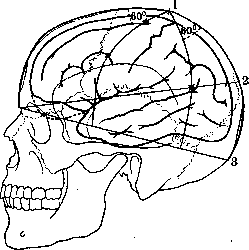
48 mm posterior to bregma along the sagittal suture; 2 - point at the junction of the sagittal suture with the apex of the occipital bone. It has very wide application. The apparatus is constructed of steel bands running along the lower and upper horizontal lines around the circumference of the skull; perpendicular to them are bands reproducing the sagittal meridian, anterior, middle, and posterior perpendicular lines. For drawing the Rolandic and Sylvian sulci, there is a hinged device on which also moves a metal plate. Using this apparatus, one can with extraordinary ease draw all the indicated lines on the surface of the skull. A certain disadvantage of this apparatus is its bulkiness and the impossibility of maintaining strict asepsis when using it. Kocher's method (Fig. 11). Kocher first determines the equatorial line, i.e., the line that crosses the skull from the glabella to the occipital protuberance. The second line is also drawn between the mentioned points, but only in the sagittal direction. Using geographical terminology, this line is called the meridional-sagittal meridional. This line is divided in half and in this way the parietal point is found. From this point, two meridians are laid off - the anterior oblique and posterior oblique, which stand at an angle of 60° to the anterior and posterior parts of the meridional line and extend to the equatorial line. The anterior meridional line corresponds to the direction of the precentral sulcus. Kocher attributes more importance to this line than to determining the central sulcus itself. This line is divided into three equal parts. The resulting division points will correspond to the place where the upper and lower frontal sulci empty into the precentral sulcus. The posterior meridian is called by Kocher the limiting line (linea limitans). This line divides two cerebral convolutions: gyrus supramarginalis and gyrus angularis; the first lies anterior to it, and the second posterior; in its lower part this line separates the temporal lobe from the occipital. On the sagittal-meridional line, Kocher marks a point at the apex of the lambdoid suture. Connecting this point with the glabella gives a line which Kocher calls the temporal line. This line was taken by Kocher from Poirier's scheme and somewhat modified by him. According to Poirier's scheme, this line is known as linea naso-lambdoidea. It somewhat deviates from the course of the Sylvian sulcus, runs along the first temporal convolution and subsequently coincides with the superior temporal sulcus, which gave Kocher reason to call this line the temporal line. Practically, Kocher somewhat changed the point at the occipital bone; he moves it 1 cm upward compared to Poirier's point. In Kocher's, the places where this line intersects with the precentral and limiting lines will correspond: the first - to the beginning of the Sylvian sulcus and the second - to the posterior end of the superior temporal convolution. The portion of this line behind the limiting line to the point of intersection with the median (sagittal) meridian will coincide with the occipito-parietal sulcus. The place where this latter line crosses the precentral line corresponds to the beginning of the Sylvian sulcus, and the place where this line crosses the posterior meridian corresponds to the posterior end of the superior temporal sulcus and convolution. The terminal portion of this line in the
Figure 11. Kocher's method: 1 - midpoint of the distance between glabella and prot. occip. ext. along the sagittal suture; 2 - Poirier's line (Sylvian); 3 - main line, or equatorial line.
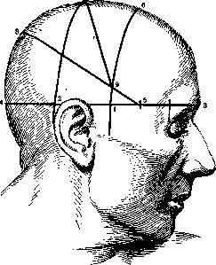
section adjacent to the sagittal meridian corresponds to the occipito-parietal sulcus, i.e., the boundary between the occipital and parietal regions. - For drawing the direction of the convolutions, Kocher proposed an apparatus known as the Kocher craniometer. This instrument, just like Krenlein's instrument, consists of steel plates with divisions marked in centimeters and millimeters. One of the plates passes along the equator of the head and is tightly fixed to the head by screws. Another plate, connected to the first, runs in the sagittal direction through the middle of the skull along the median meridional line. On this plate move 2 or 3 other plates, which are fixed on a movable hinge and can be set at any angle relative to the sagittal plate. On some models, these plates are connected by a common hinge. Due to the mobility of the plates relative to the median meridional line, the above-mentioned lines - precentral, limiting, and temporal 1st - can be easily reproduced. Tandler's scheme. In this scheme, as in Krenlein's scheme, the starting point is the upper horizontal line. On this line five points are marked: one corresponding to the articular head of the mandible, another - to the mastoid process; the third and fourth points correspond to the intersection of the horizontal line with the medial sagittal line on the frontal and occipital parts of the skull. The fifth point lies in the middle between point 1 and the outer edge of the orbit. Through the first and second points, two vertical lines are drawn at right angles to the point where they intersect the median sagittal line at points 6 and 7. The distance between points 7 and 4 is divided in half and then on the median meridional line point 8 is marked. The line connecting the latter point with point 5 will give the projection of the Sylvian sulcus. At the intersection of this line with the anterior vertical line, the ninth point is obtained. Connecting it with the seventh point will give the projection line of the Rolandic sulcus. Applying this scheme to a smoothly shaved skull is usually done with tincture of iodine. One can also use threads glued with kleol. If the direction of the main sulci is known, the approximate orientation in the course of other sulci and centers is significantly facilitated, at least on a gross scale (Fig. 12). The presented schemes sufficiently clearly illustrate what they can provide. Of course, they by no means exhaust all the tasks of cerebro-cranial topography, but they significantly help in solving such questions as the projection of brain lobes, certain convolutions, and even specific centers. - I. Frontal lobe. The projection of its boundary lines - the line of the lower cant anteriorly, the lower cant laterally and posteriorly, the line of the central convolution - is applied according to rules partly indicated above in the schemes of Poirier, Kocher, Krenlein, e.g., for the central convolution. The sagittal line determines the inner boundary of gyr. frontalis sup., while the line of the frontal and temporal cants must be determined by examining the supraorbital area. Here one can quite distinctly palpate the upper edge of the orbital fossa, but its spatial relationship to the frontal cant of the brain is very uncertain, and this instability depends on two circumstances: the development of the frontal sinus and the protrusion of the upper orbital edge above the opening of the orbital fossa. The frontal cavity (sinus frontalis) can vary greatly in both size and shape. Its size can range from 1-2-3 cm3 to 10-15 cm3. The frontal sinus, starting from the supraorbital region, can extend upward to the tubers of the frontal bone and laterally to the proc. zygomaticus, spreading over the upper edge of the orbit. Of course, one or another size of this sinus will affect the position of the frontal lobe of the brain in relation to the anterior surface of both the forehead and the orbit. The varying degree of protrusion of the upper orbital edge determines the size of the orbit in the
Figure 12. Tandler's method.
in the spring direction: with greater protrusion the dimensions will be smaller, with lesser protrusion they will be larger; at the same time the size of the transverse section of the orbit changes accordingly, and the entire cavity takes on a shape resembling a quadrangular pyramid. With such an anatomical structure, the edge of the brain will be farther from the surface of the frontal area than with another type; in the absence of protrusion of the upper orbital edge, when the glabella does not project forward, the position of the brain will be different: its edge will be closer to the surface of the frontal bones. The study of this area has enormous significance for practical surgery, precisely in those cases when the pituitary gland or the chiasm and the central part of the optic nerve are approached through the frontal area. Opening the frontal sinuses during this operation is extremely undesirable, and knowledge of the anatomy and variations is essential for the surgeon. The study of this area is significantly facilitated by X-ray examination. - Further, the lateral border of the frontal lobe, as a rule, goes higher than the zygomatic process of the frontal bone, crosses the junction of the frontal bone and the upper part of the greater wing, or goes near the apex of the upper part of the greater wing. The limiting point of this area of the brain will be the initial part of the Sylvian fissure. Its projection can be determined using models and diagrams, and the posterior border line of the frontal area is defined by the central sulcus. II. The parietal areas have clearly defined border lines: internally - the longitudinal sulcus, in front - the central sulcus, below - the Sylvian line; the posterior boundary does not have sharply defined anatomical structures. The first three boundaries are projected with greater or lesser accuracy, while the last boundary - the place where the parietal area joins the occipital and temporal areas - is established conventionally. Müller describes the posterior boundary of the parietal area as follows: from the occipital lobe, the parietal lobe is bounded by a line that from the parieto-occipital fissure goes obliquely forward and downward to that point on the lower edge of the hemisphere where it passes from the tentorium to the pyramid of the temporal bone. Here also lies the boundary between the temporal and occipital lobes. - The projection of the parieto-occipital fissure is determined by various schemes and methods with greater or lesser accuracy in the region of lambda, but with significant variations ranging up to 4 cm (Forrip). The place where the three lobes of the brain (parietal, occipital, and temporal) meet is projected onto the asterion (the posterior end of the parietomastoid suture and the junction of the occipitomastoid suture). III. The occipital lobe is defined by the boundary points just described, while the middle and lower boundaries are the superior longitudinal fissure of the cerebrum and the transverse fissure of the cerebrum. IV. The temporal lobe has sharply defined boundaries in front, above, and below, but its posterior part merges into adjacent areas, as already described, without sharp anatomical boundaries. The position of the anterior pole of the temporal lobe is subject to significant variations, which is determined by the variation in its outline. The lowest point of the temporal lobe in its position relative to both the sagittal and frontal planes is also subject to various variations. More often it stands at the upper edge of the zygomatic bone or even higher and near the jaw joint. The lower edge of the temporal lobe is projected either according to Kraus - a line connecting the outer angle of the orbital fissure and the supramastoid crest, or according to Müller - a line passing directly above the jaw joint and the external auditory meatus to the asterion. V. Cerebellum. The projection of its upper part is determined by the position of the transverse fissure (fissura transv. cerebri). Both hemispheres of the cerebellum are located in the cerebellar fossae of the occipital bone (fossae cerebellaris ossis occipitalis). The surface of the bone in these areas is usually smooth, although sometimes a elevation is described, running along the entire bone and corresponding to the transverse fissure of the cerebellum between the upper and lower halves of the cerebellar hemisphere. The lower surfaces of the hemispheres pass onto the dorsal side of the medulla oblongata, whereby the cerebellum reaches the foramen occipitale magnum. Usually this refers to the tonsilla cerebelli and adjacent parts. In this same area lies the largest in volume cistern (cisterna cerebello-medullaris). Projection of individual sulci. Already in the schemes presented, this question was partially touched upon, but a particularly detailed study of it was undertaken by Neisser and Pollak. On the basis of cerebro-cranial topography, they proposed brain puncture as a diagnostic method (Fig. 13). - In the scheme of these authors, light circles indicate the sites for puncture of abscesses, while black circles indicate punctures of the lobes and gyri of the brain. The authors give precise numerical indications for each point, both regarding the site of puncture and the depth. - 1. In the topography of the frontal lobes, Neisser and Pollak orient themselves along a line passing through the highest point of the orbital edge parallel to the sagittal meridian. On this line two points are marked: Frontalis 1 and Frontalis 2. The first is laid off 4 cm from the edge of the orbit, and the second - 4 cm from the first; this point will correspond to the pole of the frontal lobe. Frontalis 2 corresponds to the middle of the frontal lobe and lies on the second frontal gyrus. These points are used for diagnosing pathological processes in the frontal lobes; from here one can also puncture the anterior horns of the lateral ventricles. - 2. Area of the central gyri and sulcus. The anterior oblique meridian determines the precentral sulcus. According to its triple division, Neisser and Pollak determine the location of the centers for the leg (C), hand (C2), and face (C3). The first center is placed somewhat above the first division of the oblique meridian; the second point is 1-11/2 cm posterior to the oblique meridian in its middle third; the third point is posterior to the division of the oblique meridian. - 3. Temporal lobe. Neisser and Pollak mark the center of the temporal lobe with the letter Tx and place it 1-1x/2 cm above the attachment of the auricle. The second point, T%, they place 1-11/2 cm anterior to the first point; this point is also used by them for the projection of the center of the temporal area. The point T3, determining the position of an abscess, lies 0.5-0.75 cm above the attachment of the auricle. - 4. The temporal and occipital areas are punctured relatively rarely; the puncture sites are determined according to Kocher's scheme. - 5. Cerebellum. The point Kg (Kleinhirn-cerebellum) marks the center of the cerebellar hemisphere. It lies midway along the line connecting the occipital protuberance with the apex of the mastoid process. This point will correspond to the lobulus gracilis or lobus quadrangularis. The points K2 and K3 marked on the diagram refer to punctures for abscess. These points are chosen as follows: K3 corresponds to the posterior point of the base of the mastoid process. The midpoint of the line connecting point K3 with point Kg will give point K2. The precise determination of this point has exceptional importance: this point will lie within the knee of the sigmoid sinus, but so far from the sinus that needle punctures will not injure the sinus. - All the designated points are chosen not only according to the location of the brain lobes, gyri, and specific areas of gyri, but also
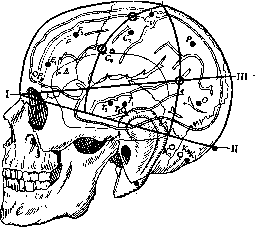
Figure 13. Puncture sites according to Kocher's scheme.
in places that are least safe in terms of the possibility of damaging blood vessels; for example, in the area of the Sylvian fissure, punctures are generally not recommended due to the possibility of injury to the middle artery and vein of the brain. At the present time, following the work of Brodmann, Foerster, Economo, and Koskinas, which has been devoted to the study of the architectonics of the cerebral cortex, the doctrine of localization and, in particular, of the motor area of the brain, has received a different interpretation. It is increasingly entering into clinical practice, and the surgeon often has to deal in practice with the doctrine of cytoarchitectonics of the cerebral cortex. Therefore, in this case, it appears necessary to indicate in the most essential features how the doctrine of cytoarchitectonic fields and areas (see Architectonics of the cerebral cortex) should reflect on the tasks of C.-c. t. On the basis of architectural features, the cerebral cortex is divided by C. and O. Foersts into no less than 200 fields; Brodmann and Economo have distinguished a smaller number. From a practical point of view, for the clarification of the question under discussion, it is necessary to emphasize three facts: 1) the morphological boundaries of each field have linear forms, which is explained by the segmental character of the arrangement of the fields. The boundaries of the fields do not coincide with the fissures of the brain and in most cases run along the convex surface of the gyri. 2) Fields similar in structure have provided the basis for distinguishing certain groups of them into common complexes of areas. Thus, the entire cortex breaks down into several areas, and various researchers describe the areas differently (Brodmann-13 areas, Koskinas-7 areas); but in any case, here also there is no exact coincidence with the division of the brain into frontal, parietal, temporal, occipital lobes. 3) Fields and areas have certain physiological functions, and the localization of individual functions corresponds to a greater or lesser degree to the division of the cortex into fields and areas. This creates new tasks for practical surgery in terms of re-examining cerebro-cranial relations. Specifically, one most often has to deal with this in the surgical treatment of epilepsy, when the focus of irritation is located not in the area of the Rolandic fissure, which, since the time of Bergmann, for a long time was considered the predominant object of brain surgery. At the present time, the following areas are referred to the motor area of the cerebral cortex, which can be easily understood from a comparison of the accompanying drawings [Fig. 14 and t. VII (pp. 535-536), Fig. 1 and 2].- The brief outline of the modern doctrine of the motor area of the cerebral cortex given above gives an idea of the imperfection of cerebro-cranial schemes and apparatuses; they by no means cover the details of the boundaries of fields and areas. Nevertheless, with their help, one can still orient oneself in the location of the fields, and in regard to certain fields that coincide with the data of descriptive anatomy (for example, fields 4 and 6 of Brodmann, fields 22, 19), one can also establish the boundaries exactly. Of course, during an operation, orientation in the boundaries of the fields occurs mainly not on the basis of the data of C.-c. t. alone, but on the basis
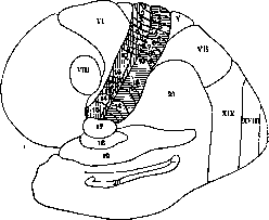
Figure 14. Diagram of Brodmann's fields and their physiological functions (vertically and horizontally hatched are the motor and sensory spheres): 1-foot; 2-leg; 3-thigh; 4-abdomen; 5-chest; 6-upper shoulder; 7-shoulder; 8-forearm; 9-hand; 10-fingers; 11-neck; 12 and 13-face; 14-tongue; 15-jaw, pharynx, larynx; 16-thumb; 17-chewing, licking, swallowing, sneezing; 18-hiccups; 19-turning of the head, eyes, and body to the opposite side, clonic convulsions in the opposite limbs; 20-gustatory aura; 21-sensory aura; clonic twitching of individual muscle groups in sequence. Field V and VII-posterior adversive field: tonic-clonic convulsions in the opposite limbs; VI-anterior adversive field; VIII-clonic twitching of the eyeballs to the opposite side without aura; XVIII-optical aura; XIX-tonic convulsions in the opposite half of the body (according to Foerster).
of the results of electrical irritation of various areas of the cortex according to the data of O. Vogt and Foerster. In hemorrhages, one has to deal either with damage to the branches of art. mening. mediae or with damage to the sinuses and finally with hemorrhages from the vessels of the cerebral cortex or vessels passing in the depth of the brain substance. The projection of art. mening. med. and its most important branches is established according to the scheme of Krenlein [t. VII (pp. 576-577), Fig. 2 and 3].-The topographical location of the sinuses of the dura mater has recently acquired special importance in view of proposals to inject contrast substances into the sinuses in their thrombosis. The location of the sinuses in relation to the bones of the frontal, parietal, and occipital bones, while maintaining a more or less stable type, is nevertheless subject to significant fluctuations in their position; displacements of sin. longitudin. from the median meridian have been described; cases of its doubling either over the entire length or only on certain sections have been described; cases of asymmetrical arrangement in relation to the median-1.
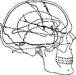
Figure 15. Thane's method-lateral projection of the ventricles: 1 and 2-Rolandic fissure; 3-Sylvian fissure; 4-basic line; 5-parallel line.
line of the place of confluence of sin. longitud. with sin. transversus (confluens sinuum).-The location of the sagittal sinus, asymmetrical in relation to the median line, also presupposes a displacement of proc. falciformis, and consequently a displacement of fissurae longitudinalis cerebri, and thus a displacement of the hemispheres. It is necessary to note here an extremely important circumstance from a practical point of view: cases have been described in which the sinus longitudinalis deviated from the line of attachment of proc. falciformis, the latter remaining on the median line (sagittal meridian), and sin. longitudinalis running to the side and flowing into the transverse sinus several centimeters from the median line. In these anomalies, which can have extremely important significance in practical surgery, it is only sometimes possible to sort things out by careful study of the skull and precisely the median line. The identifying points for the latter are glabella, sometimes the frontal suture or its derivatives. The suture, if it has not ossified, can be felt very easily; sometimes after its closure there remain bony longitudinal protrusions with a groove in the middle; also
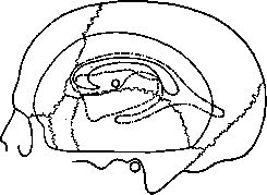
Figure 16. Projection of the lateral ventricles and ins. Reili according to Jenkins.
the sagittal suture, located between the frontal tubercles (if it can be palpated), then lambda, protuberantia occipitalis. These points identifying the median line are not always easily palpated-the sutures may ossify; further, the sutures can be easily palpated, but the results of palpation are inaccurate: along the course of the sutures there may be accessory bones; lambda and protuberantia occipitalis are sometimes not palpated at all, especially in women (Müller). Transverse sinus. Its location and course are determined on the outer surface of the occipital part of the skull, protuberantia occipitalis and lin. superior nuchae, if it can be palpated; if it cannot be palpated, then the identifying line may be the upper edge of the occipital musculature, it being considered a rule that lin. nuchae superior corresponds to the lower edge of the transverse sinus. Establishing the line of passage of the transverse sinus simultaneously characterizes the attachment of tentorium and the posterior cerebral fissure (fissura transversa cerebri). In this area there is no tendency to oscillation on such a wide scale as in sin. longitudinalis superior. It is necessary, however, to note here 1) variants in the area of confluens sinuum, which in part
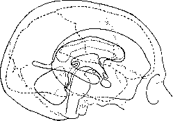
Figure 17. Projection of the cerebrum and the ventricular system according to Symington.
depends on the variants of the superior longitudinal sinus described above; 2) a very marked asymmetry between the right and left halves of the transverse sinus; 3) significant variations in the thickness of the occipital bone above the passage of the transverse sinus, above the point where the longitudinal and transverse sinuses merge. The second point may be of great importance in case of occlusion or the need to ligate the right or left half of the transverse sinus: ligation of the more developed half can have a catastrophic effect on cerebral circulation.-The outlines of the normal cerebral ventricles exhibit a certain stability, but their projection onto the surface of the skull cannot be clearly delineated due to the peculiar shape of the ventricles and the convexity of the skull. For practical purposes, it is extremely important to know the projections of the ventricular contours from the side, front, and back. This is also important for studying the deformations of the ventricles, for puncturing them, and for evaluating X-ray images obtained during encephalography. The study of ventricular projection is possible only through the examination of models of ventricles from various brain sections and specimens of whole brains. The most well-known schemes are those of Thane, Jenkins, Symington, and Dandy, which provide profile contours; among other schemes, mention should be made of the one presented by Corning, Tandler, and Ranzi; these schemes show the projection onto the parietal, occipital, and temporal bones. The primary material for studying ventricular projection for the modern clinician must, of course, be X-ray images of both normal and pathological forms of the ventricles, as can be seen from the accompanying diagrams (Figs. 15, 16, and 17).
Related articles
Mentioned in
Cite this page
“Cerebro-Cranial Topography.” Soviet Medical Encyclopedia. English translation of Bolshaya Meditsinskaya Entsiklopediya, 1st ed. (Moscow, 1928–1936), ed. N. A. Semashko. https://sovietmedicalencyclopedia.pages.dev/article/cerebro-cranial-topography/