Dura Mater
Historical document, translated for reference. It reflects medical knowledge of the 1920s–30s and is not medical advice.
Summary
The dura mater serves as the inner periosteum of the skull and vertebrae and the outer membrane of the brain and spinal cord. It forms various folds and partitions within the skull and creates a cylindrical sac around the spinal cord, with pathological conditions including inflammation, hemorrhages, and tumors.
Encyclopedia article (1928–1936)
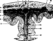
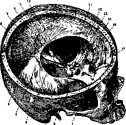
DURA MATER, the hard cerebral membrane, serves simultaneously as the inner periosteum of the bones of the skull and spine and the outer membrane of the brain and spinal cord. In the skull, the inner surface of the dura mater connects with the other membranes through various cerebral veins and arachnoid villi or Pacchionian granulations (Fig. 1) and gives off sagittal and transverse processes: 1. The great falx, falx cerebri, which attaches to the sagittal sulcus of the cranial vault, extends from the crista galli to the internal occipital protuberance, and penetrates accordingly into the longitudinal fissure of the brain. 2. The small falx, falx cerebelli, constitutes a lesser continuation of the great falx and penetrates between the hemispheres of the cerebellum, descending from the internal occipital protuberance to the foramen magnum. 3. The tentorium cerebelli, representing a tightly stretched transverse septum between the inferior surface of the occipital lobe of the cerebral hemisphere and the superior surface of the cerebellum. The tentorial incisure gives the tentorium a semilunar shape; one distinguishes the cerebral and cerebellar surfaces of the tentorium, the anterior free edge, and the posterior edge; the latter attaches to the transverse sulci of the occipital and parietal bones and to the superior border of the pyramid of the temporal bone. At the junction of the tentorium with the falx cerebri is the straight sinus, into which through the tentorial opening enters the great vein of Galen. 4. The diaphragm of the sella turcica, diaphragma sellae turcicae, surrounds the hypophysis cerebri, leaving an opening for the infundibulum (Fig. 2).
Figure 1. Frontal section of the skull and brain; 1-subst. cortic. cerebri; 2 blood vessel; 3 pia mater; 4-arachnoidea; 5-dura mater; 6-os pariet.; 7-venous lacunae; 8-arachnoid processes; 9-sut. sagitt.; 10 sin. sagitt. sup.; 11-Pacchionian granulations; 13-depressions in bone; 13-subarachnoid space; 14-falx cerebri; 15-subdural space; 26-fiss. longitud. cerebri. (After Spalteholz.)
In the spine, the dura mater forms a cylindrical sac, somewhat wider than the spinal cord it encloses, and attaching superiorly to the margin of the foramen magnum and inferiorly to the sacral bone. Together with the outer surface of the arachnoid membrane, the dura mater forms the subdural space; the space located between the two layers of the dura mater and filled with loose connective tissue is called the epidural or interdural cavity. The blood supply to the dura mater in the brain occurs through the middle meningeal, anterior meningeal, and posterior meningeal arteries, which branch from the main cerebral arteries and pass in the outer layer, while in the spinal cord-through branches of the corresponding arteries (intercostal, lumbar, etc.). The venous circulation is carried out by a system of sinuses located in the dura mater (see Brain-anatomy). The dura mater is innervated by meningeal branches arising from the trigeminal, vagus, hypoglossal, and sympathetic nerves, and in the spinal cord-by fibers of the corresponding nerve roots. In addition, there are also vasoconstrictor nerves and the proper nerves of the dura mater.
Figure 2. The dura mater from the right and above: 1 and 2-fossa cranii ant. et med.; 3-incisura tentorii; 4-tentor. cerebelli; 5-attachment of falx cerebri to tent. cerebelli; 6-n. abducens; 7-nn. facialis et acusticus; 8-n. trigeminus; 9-n. trochlearis; 10-n. oculomotorius; 11-proc. clinoid. post.; 12-infundibulum; 13-diaphragma sellae; 14-n. opticus; 15-falx cerebri; 16-crista galli. (After Spalteholz.)
The pathology of the dura mater is expressed by inflammations (see Meningitis), hemorrhages, and tumors. Hemorrhages are of three types: extra- and epidural, occurring from traumatic causes and manifesting as effusion of blood either between the bone and dura mater or between its two layers, and subdural (see Hematoma); the clinical picture is characterized by a series of general symptoms-loss of consciousness, headaches, dizziness, vomiting, slowing of the pulse, elevation of temperature, convulsions, rigidity of the neck and extremities, disturbance of sphincters, etc.; in addition, depending on the simultaneous involvement of one or another part of brain tissue, symptoms of involvement of these parts may be observed. The course depends on the intensity of the trauma. Therapy: rest, ice on the head, low position of the head, and symptomatic remedies. To traumatic hemorrhages of the dura mater also belong hemorrhages in the fetus during a prolonged labor complicated by the application of forceps; twisting of the umbilical cord around the neck can also be the cause of hemorrhage into the dura mater; in this case, the hemorrhages are usually subdural, and the child is either born dead or dies shortly after birth. Tumors of the dura mater either originate from the dura mater itself, developing in various directions, or, arising from brain tissue, also involve the dura mater; most commonly encountered are sarcomas and fibromas. (Symptomatology, course, etc.-see Brain-pathology, tumors.) Isolated tuberculosis of the dura mater is observed very rarely and occurs only simultaneously with lesions of the soft cerebral membranes. In syphilis, gummatous nodes form in the dura mater, sometimes penetrating into the bone and eroding it, and sometimes growing into the brain tissue.
Figure 3. The dura mater from the right and above: 1 and 2-fossa cranii ant. et med.; 3-incisura tentorii; 4-tentor. cerebelli; 5-attachment of falx cerebri to tent. cerebelli; 6-n. abducens; 7-nn. facialis et acusticus; 8-n. trigeminus; 9-n. trochlearis; 10-n. oculomotorius; 11-proc. clinoid. post.; 12-infundibulum; 13-diaphragma sellae; 14-n. opticus; 15-falx cerebri; 16-crista galli. (After Spalteholz.)
Related articles
Mentioned in
- Abducens Nerve
- Amyelia
- Arachnoidea
- Caput Succedaneum
- Cavum
- Cephalhematoma
- Cerebellopontine Angle
- Cerebro-Cranial Topography
- Corpora Arenacea
- Cretinism
- Dermovaccine
- DRAINAGE (is used for removing fluids from body cavities,)
- Dural Sinuses
- Ethmoid Bone
- Fungous
- Galen
- Gasserian Ganglion
- Graduated Cylinder
- Hydrencephalomeningocele
- Infundibulum
Cite this page
“Dura Mater.” Soviet Medical Encyclopedia. English translation of Bolshaya Meditsinskaya Entsiklopediya, 1st ed. (Moscow, 1928–1936), ed. N. A. Semashko. https://sovietmedicalencyclopedia.pages.dev/article/dura-mater/